Alanine for Immune Function
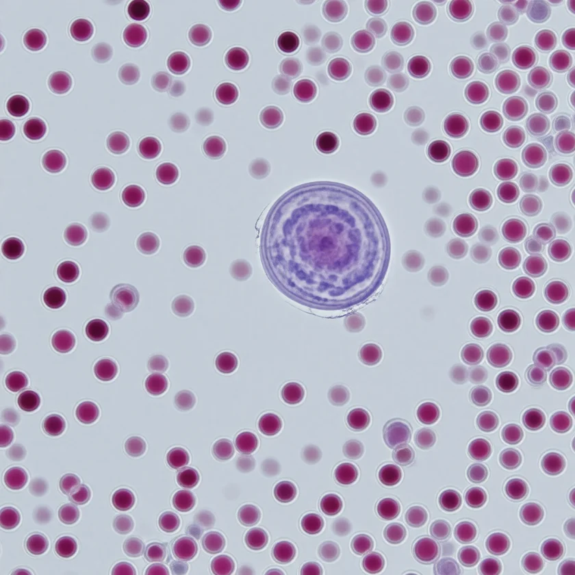
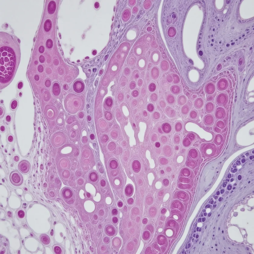
Immune cells — lymphocytes, macrophages, neutrophils, and dendritic cells — are among the most metabolically active cells in the body, with energy and biosynthetic demands that rival those of skeletal muscle and exceed those of most other tissue types. The classic work of Eric Newsholme and his Oxford collaborators in the 1980s established that activated lymphocytes consume glutamine at extraordinary rates and excrete the nitrogen as alanine in a tightly coupled glutamine-alanine relay. The same circuitry was later shown to underlie immune cell function in sepsis, trauma, major surgery, and critical illness — conditions in which the body's endogenous glutamine pool is consumed faster than the muscle can synthesize it, and clinical outcomes deteriorate as the relay fails. This is the biochemical foundation of the modern intravenous alanyl-glutamine dipeptide (L-alanyl-L-glutamine) used in critical-care parenteral nutrition. This page traces the biochemistry of immune-cell amino acid metabolism, the glutamine-alanine relay, the concept of conditional essentiality during critical illness, and the practical clinical applications of amino-acid support in immunocompromised patients.
Table of Contents
- Immune Cells as Metabolically Demanding Tissue
- The Glutamine-Alanine Relay Inside Immune Cells
- Alanine and Lymphocyte Proliferation
- Conditional Essentiality During Critical Illness
- Sepsis and Critical Care Nutrition
- The Intravenous Alanyl-Glutamine Dipeptide
- Exercise-Induced Transient Immunosuppression
- Wound Healing and Post-Surgical Recovery
- Gut Barrier Function and Mucosal Immunity
- Cautions (Including the REDOXS Critical-Care Signal)
- Key Research Papers
- Connections
- Featured Videos
Immune Cells as Metabolically Demanding Tissue
An activated T lymphocyte is one of the most metabolically active cells in the body. When a naive T cell encounters antigen and undergoes clonal expansion, it goes from a quiescent doubling time of months (or never, in a true naive cell) to a doubling time of approximately 6-12 hours. This requires building a complete new daughter cell — all of its proteins, lipids, nucleic acids, and organelles — from scratch in less than half a day. The metabolic demand for substrate is enormous and the substrate requirements are highly amino-acid-skewed.
The Newsholme group's seminal experiments in the 1980s measured the rates at which different amino acids are consumed by activated lymphocytes. Two amino acids stand out:
- Glutamine — consumed at rates 10-100 times higher than any other amino acid. Lymphocytes use glutamine for energy production (via glutaminolysis, the conversion of glutamine to glutamate to alpha-ketoglutarate to the tricarboxylic acid (TCA) cycle), for nucleotide biosynthesis (the amino group of glutamine donates to purine and pyrimidine synthesis), and for redox homeostasis (the glutathione precursor cysteine is in part regenerated from glutamine-derived carbon).
- Glucose — consumed at high rates via glycolysis, primarily for ribose-5-phosphate production in the pentose phosphate pathway (also for nucleotide synthesis) and for anabolic biosynthesis of fatty acids and cholesterol.
The combination of high glutamine and glucose consumption is sometimes called the "Warburg-like" phenotype of activated immune cells (after the German biochemist Otto Warburg, who first described the same metabolic phenotype in tumor cells). The two cell populations share a fundamental biochemical strategy: maximize biosynthetic substrate availability at the expense of complete oxidation, with the metabolic cost paid by elevated glycolytic and glutaminolytic flux.
The dependence on glutamine is so extreme that immune function collapses if glutamine is depleted. In vitro, removing glutamine from culture media stops lymphocyte proliferation, IL-2 production, and antibody secretion within hours. In vivo, the body normally maintains plasma glutamine concentrations of 500-800 micromolar — the highest of any amino acid — specifically to ensure that immune cells have access to this critical substrate even during fasting and stress.
The Glutamine-Alanine Relay Inside Immune Cells
Here is where alanine enters the immune story. When activated lymphocytes consume glutamine, they do not fully oxidize it. The reaction proceeds as follows:
- Glutamine enters the cell via the ASCT2 transporter (SLC1A5), one of the highest-expression amino acid transporters on activated lymphocytes.
- Glutaminase converts glutamine to glutamate, releasing ammonia as a byproduct.
- Glutamate is transaminated to alpha-ketoglutarate, donating its amino group either to pyruvate (forming alanine via alanine aminotransferase, ALT) or to oxaloacetate (forming aspartate via aspartate aminotransferase, AST).
- Alpha-ketoglutarate enters the TCA cycle, where it is partially oxidized for adenosine triphosphate (ATP) production.
- The TCA intermediates are siphoned off for biosynthesis — citrate exits the mitochondria to support fatty acid synthesis, oxaloacetate provides aspartate for nucleotide synthesis, alpha-ketoglutarate provides 2-hydroxyglutarate and succinate for epigenetic modification of transcription factors.
- Alanine (and lactate) are excreted as the metabolic end products. Alanine carries away one nitrogen per glutamine consumed; lactate carries away the partially-oxidized carbon when glycolytic flux exceeds the TCA cycle's capacity to accept pyruvate.
This is the glutamine-alanine relay: the cell consumes glutamine, uses its carbon and nitrogen for biosynthesis, and exports the metabolic waste nitrogen as alanine. The relay is quantitatively important — activated lymphocytes can excrete alanine at rates similar to their glutamine uptake rates. What is not settled is how this fits newer work: the relay picture comes from older lymphocyte cultures (the Newsholme studies), while a 2019 laboratory study found that T cells leaving their resting state take alanine up from outside the cell and use it to build proteins (see the next section). The exported alanine returns to the liver and feeds the same hepatic gluconeogenic and urea-cycle disposal pathway that handles muscle-derived alanine (see the companion Gluconeogenesis page).
The implication is that alanine and glutamine are not independent variables in immune physiology — they are coupled inputs and outputs of the same lymphocyte metabolic engine. Researchers who target immune function through amino acid supplementation therefore consider the pair together; the dipeptide used in modern parenteral nutrition supplies both, although its main practical advantage is that free glutamine is unstable in solution (see the alanyl-glutamine dipeptide section below).
Alanine and Lymphocyte Proliferation
The direct role of alanine in lymphocyte proliferation is more limited than that of glutamine, but several specific contributions matter:
- Alanine is a non-essential substrate for protein synthesis. Roughly 8% of the amino acid residues in human proteins are alanine, so the daughter lymphocyte built during a 6-12 hour proliferation cycle requires alanine for its protein content. While alanine can be synthesized de novo from pyruvate plus a nitrogen donor, the de novo synthesis costs adenosine triphosphate and competes with the nitrogen demands elsewhere in the cell. Exogenous alanine availability spares this biosynthetic burden.
- Alanine is required for the alanine-aspartate-glutamate axis that feeds purine and pyrimidine synthesis. Aspartate (donated by aspartate aminotransferase transamination from glutamate) provides nitrogen atoms for both adenine and thymidine; glutamate (donated by glutamine deamidation) provides nitrogen for both guanine and cytosine. The transamination flux through alanine aminotransferase helps maintain the cytosolic alanine-pyruvate balance that keeps this axis running.
- Alanine in the immunological synapse — the contact zone between an antigen-presenting cell and a T cell. Local amino acid concentrations in the immunological synapse modulate signaling through the mTORC1 nutrient-sensing pathway. mTORC1 integrates amino acid availability, growth factor signaling, and cellular energy status to license clonal expansion. Severe amino acid deprivation (including alanine) inactivates mTORC1 and blocks T cell proliferation.
- T cells need alanine from outside the cell. A 2019 laboratory study (Ron-Harel and colleagues) found that alanine in the surrounding fluid is required for resting (naive) T cells to switch on efficiently and for memory T cells to respond again, even though alanine can be made from pyruvate in a single step. Resting T cells make little alanine aminotransferase, the enzyme that interconverts pyruvate and alanine, so activated T cells switch on alanine transporters instead. Tracer experiments showed the imported alanine was used to build proteins rather than burned for fuel, and removing alanine impaired the T cells' metabolism and function in the dish.
The clinical implication is that severe protein malnutrition impairs lymphocyte proliferation. Patients with kwashiorkor (protein malnutrition with caloric adequacy) and marasmus (combined protein-calorie malnutrition) exhibit profound lymphopenia and reduced T-cell mitogen responsiveness, with secondary increases in opportunistic infection rates. The reciprocal observation — that nutritional repletion restores T-cell function — is the basis for the modern emphasis on adequate enteral and parenteral amino acid provision in hospitalized patients.
For patients in the developed world, frank protein malnutrition is rare but subclinical inadequacy is common in elderly patients, patients with chronic illness, and patients who have recently undergone major surgery. Each of these populations shows reduced immune function that improves with amino acid supplementation.
Conditional Essentiality During Critical Illness
Alanine is conventionally classified as a non-essential amino acid — the body can synthesize it from pyruvate plus a nitrogen donor, so dietary intake is not strictly required. However, the concept of conditional essentiality applies: under certain pathological states, endogenous synthesis cannot keep up with demand and exogenous supply becomes necessary for normal physiological function.
Glutamine is the canonical conditionally essential amino acid — well-established to drop precipitously in critical illness, with associated mortality. Alanine's conditional essentiality status is less well-defined but is increasingly recognized in several specific clinical settings:
- Sepsis — plasma alanine paradoxically remains stable or even rises early in sepsis (due to accelerated muscle catabolism), but tissue-specific alanine availability in the liver and immune compartments becomes inadequate as the cytokine storm and inflammatory state proceed. The hepatic gluconeogenic demand exceeds the alanine supply, contributing to the lactic acidosis of severe sepsis.
- Major trauma — the catabolic response to multiple injury creates a large nitrogen-disposal load that exceeds normal alanine shuttle capacity. Plasma alanine can rise to 600-800 micromolar transiently as the muscle exports nitrogen faster than the liver can clear it.
- Burns — the largest sustained hypermetabolic state in clinical medicine. A patient with 40% total body surface area burns can sustain resting metabolic rates of 200% of predicted for weeks. Alanine flux becomes a major nutritional consideration.
- Liver failure — the alanine cycle collapses from the liver-failure side rather than the muscle side. Plasma alanine rises sharply because the liver cannot clear it, but the cycle no longer delivers useful glucose or safely disposed nitrogen. Branched-chain amino acid-enriched formulations support muscle alanine synthesis without overloading the failing liver.
- Cancer cachexia — the cachectic muscle loss in advanced cancer reflects a sustained shift to net catabolism, with alanine flux elevated for months on end. Nutritional intervention with full amino acid replacement remains controversial as it may also feed the tumor.
The practical clinical inference is that critical illness requires aggressive nutritional support of the amino acid pool, with formulations that provide complete coverage of both essential and conditionally essential amino acids. The trend in modern critical care nutrition has been toward earlier enteral feeding, more complete amino acid profiles, and targeted use of alanine-glutamine dipeptides for the sickest patients.
Sepsis and Critical Care Nutrition
The metabolic state of sepsis is biochemically distinctive: a hypermetabolic, hypercatabolic, insulin-resistant state in which the body is simultaneously trying to fuel an enormous immune response while losing the ability to use its conventional energy substrates efficiently. Several specific features matter for the alanine story:
- Skeletal muscle proteolysis accelerates — net muscle protein breakdown can exceed 100 g/day in severe sepsis, releasing branched-chain amino acids, alanine, and glutamine into the circulation.
- Insulin resistance develops — cytokines (tumor necrosis factor-alpha, interleukin-6, interleukin-1) impair muscle glucose uptake. Hyperglycemia coexists with elevated lactate (the lactate-pyruvate ratio rises).
- Plasma glutamine drops — this is one of the most reliable metabolic markers of severe sepsis. Normal plasma glutamine 500-800 micromolar drops to 200-400 micromolar in severe sepsis. The drop reflects consumption by activated immune cells outstripping muscle synthesis.
- Plasma alanine paradoxically may rise early then fall — reflecting first the proteolytic phase then the failure-to-clear phase as multi-organ dysfunction develops.
- Hepatic gluconeogenesis from alanine is impaired — both because of relative substrate insufficiency and because cytokine-mediated hepatic dysfunction reduces gluconeogenic enzyme activity. The resulting glucose-lactate-alanine imbalance contributes to the lactic acidosis that prognosticates severe sepsis.
Modern critical care nutrition guidelines (the ASPEN/SCCM 2016 update and the ESPEN 2019 guidelines, which ESPEN shortened and partially revised as a practical guideline in 2023) emphasize:
- Early enteral nutrition (within 24-48 hours of intensive care unit (ICU) admission) whenever feasible
- Adequate protein provision (1.2-2.0 g/kg/day in critical illness, higher than the 0.8 g/kg/day Recommended Dietary Allowance (RDA) for healthy adults)
- Selective use of supplemental glutamine in burn patients and patients with major trauma (the ESPEN guideline) — the largest burn trial since, RE-ENERGIZE (2022), found no benefit of enteral glutamine in burns (see the alanyl-glutamine dipeptide section)
- Caution against supplemental glutamine in patients with multi-organ failure (the REDOXS trial signal — see Cautions section)
The era of high-dose glutamine supplementation for all critically ill patients has ended; modern practice individualizes the approach based on illness severity and organ function. Alanyl-glutamine dipeptide formulations remain in use in many European intensive care units but are less commonly used in North America.
The Intravenous Alanyl-Glutamine Dipeptide
Free L-glutamine in solution is unstable — it spontaneously degrades to pyroglutamate and ammonia at a rate that increases with temperature and pH. This makes pure glutamine difficult to formulate for parenteral administration: a TPN bag containing free glutamine would generate substantial ammonia by the end of the infusion. The pharmaceutical solution to this problem is to use a dipeptide form — L-alanyl-L-glutamine — which is heat-stable, soluble at high concentration, and rapidly cleaved by plasma and tissue dipeptidases to release free glutamine and free alanine after infusion.
The intravenous alanyl-glutamine dipeptide solution contains 20 g of L-alanyl-L-glutamine per 100 mL, corresponding to 13.5 g of free glutamine plus 8.2 g of free alanine after enzymatic cleavage. The typical clinical dose is 1.5-2.0 mL/kg/day added to standard parenteral nutrition, providing approximately 0.3-0.4 g/kg/day of the alanyl-glutamine dipeptide (not of free glutamine). A 2017 meta-analysis states the guideline dose as 0.3-0.5 g/kg/day of glutamine dipeptide by vein, at most 30% of the prescribed nitrogen supply.
The clinical rationale combines two effects:
- Replenishes the depleted glutamine pool during critical illness, supporting immune cell function, gut barrier integrity, and antioxidant capacity (glutamine is a glutathione precursor).
- Provides the alanine partner for the glutamine-alanine relay, ensuring that the metabolic engine has both inputs and a viable nitrogen-export route. This is a more theoretical benefit but underlies the formulation choice.
The clinical evidence for intravenous glutamine, including the dipeptide, is mixed. Wischmeyer 2014 pooled 26 trials of intravenous glutamine in 2,484 intensive care unit patients and found a significant reduction in hospital mortality (relative risk 0.68) and a hospital stay 2.56 days shorter, with only a trend toward fewer infections (relative risk 0.86, not statistically significant) and lower overall mortality (relative risk 0.88, not significant). Bollhalder 2013 pooled 40 trials of intravenous glutamine and found significantly fewer infections (relative risk 0.83) and a stay 2.35 days shorter, but no significant reduction in mortality, and noted that one recent trial strongly influenced the results. A 2017 meta-analysis by Stehle and colleagues (five of its eight authors worked for the maker of an alanyl-glutamine product) included 15 trials with 842 critically ill patients without liver or kidney failure given the dipeptide at guideline doses, and found fewer infections, a shorter hospital stay and 45% lower hospital mortality (relative risk 0.55), with no effect on intensive care unit mortality. The 2013 REDOXS trial in patients with multi-organ failure found increased mortality with high-dose glutamine, raising concerns about routine use in the sickest patients. In Europe the dipeptide has been used mainly for patients with severe trauma, burns, and abdominal surgery; in North America, the same formulation is less commonly used and many intensive care unit teams have moved toward enteral immunonutrition formulas instead.
For burns, the largest trial so far, RE-ENERGIZE (2022), gave 1,200 patients with severe burns (on average 33% of the body surface) 0.5 g/kg/day of glutamine through a feeding tube or by mouth, or placebo. Glutamine did not shorten the time to leaving hospital alive (median 40 days vs 38 days with placebo), and deaths at 6 months were similar (17.2% vs 16.2%). This trial used enteral glutamine, not the intravenous dipeptide.
For non-ICU oral alanine-glutamine support, the dipeptide form has limited bioavailability advantage over free amino acids (the gut breaks both down before absorption). Oral L-alanine and L-glutamine sold separately as nutritional supplements perform similarly to combined dipeptide formulations for the rare indications where they are used.
Exercise-Induced Transient Immunosuppression
Heavy endurance exercise produces a transient immunosuppressive state in the hours following the workout, characterized by reduced natural killer cell activity, reduced salivary immunoglobulin A (IgA) secretion, and elevated risk of upper respiratory tract infection. The phenomenon is well documented in marathon runners, ultra-endurance athletes, and high-volume training in Olympic-level swimmers and rowers. The mechanism is multifactorial, but amino acid shifts contribute meaningfully:
- Plasma glutamine falls after heavy endurance exercise — from a baseline of 600 micromolar to 400-500 micromolar after a marathon, and the decline persists for hours. The drop has been hypothesized to contribute to the post-exercise immunosuppression by depriving lymphocytes of their preferred substrate. This is the basis for the "glutamine hypothesis" of exercise immunology popularized by Newsholme in the 1990s.
- Plasma alanine peaks during exercise and falls post-exercise — mirroring the glutamine shift. The alanine pool is being mobilized for hepatic gluconeogenesis during the workout and then is replenished from muscle protein breakdown during recovery.
- The glutamine hypothesis has been partially walked back — subsequent intervention trials with oral glutamine supplementation in endurance athletes have shown only modest effects on immune markers and no consistent reduction in upper respiratory infection rates. The mechanism is more complex than substrate depletion alone (cortisol, beta-adrenergic activation, and direct exercise-induced lymphocyte apoptosis all contribute).
- What athletes commonly do — sports-nutrition practice describes protein intakes of 1.2-1.6 g/kg/day for active individuals and a recovery meal containing both protein and carbohydrate within 1-2 hours of completing a heavy session. Whether dedicated glutamine or alanine-glutamine supplementation provides additional benefit remains unclear; because of their modest cost, some athletes use them preventively without strong evidence.
Wound Healing and Post-Surgical Recovery
Tissue repair after surgery, trauma, or burn injury depends on the rapid proliferation of multiple cell types — fibroblasts depositing collagen, endothelial cells building new capillaries, keratinocytes migrating to close the wound, and immune cells mounting the inflammatory and remodeling response. All of these cell populations share the metabolic profile of activated immune cells: high glutamine and glucose consumption, elevated alanine output, and dependence on adequate amino acid supply.
The clinical correlate is that protein-malnourished patients heal wounds slowly and have higher rates of wound dehiscence, surgical site infection, and anastomotic leak. Modern preoperative nutritional optimization (the "prehab" approach used in major colorectal and pancreatic surgery) emphasizes:
- Preoperative protein supplementation (1.2-1.5 g/kg/day) for 2-4 weeks before elective surgery
- Carbohydrate loading the night before and the morning of surgery (when not contraindicated)
- Early postoperative enteral nutrition (within 24 hours)
- Selective use of immunonutrition formulas (containing arginine, omega-3 fatty acids, and nucleotides) in high-risk surgical patients
For more on wound healing nutrition, see our pages on Glutamine and Arginine.
Gut Barrier Function and Mucosal Immunity
The small intestinal enterocyte is one of the most glutamine-dependent cells in the body, and it shares the alanine-export pattern of activated lymphocytes. Enterocytes consume approximately 30% of total dietary glutamine on the first pass, using it for energy (the small intestine uses glutamine as its preferred fuel, ahead of glucose) and for nucleotide synthesis (the small intestinal epithelium turns over completely every 3-5 days, requiring continuous DNA synthesis). The metabolic waste is excreted as alanine into the portal circulation.
This is the biochemical basis for the well-documented gut-barrier failure that occurs during prolonged fasting, parenteral nutrition without enteral support, and critical illness. When the enterocyte glutamine supply fails, the tight junctions between enterocytes become leaky, bacterial translocation across the gut wall increases, and the gut becomes a source of inflammation and infection. This phenomenon — sometimes called "the gut as the motor of multi-organ failure" underlies the modern emphasis on early enteral nutrition in the intensive care unit even when parenteral nutrition is also being given.
The clinical implication is that gut barrier integrity depends on adequate luminal substrate, with glutamine being the most important substrate and alanine being the secondary export. Patients who must take nothing by mouth (NPO) for extended periods (severe pancreatitis, post-bariatric leaks, complex gut surgery) are given trophic enteral feeding (small-volume drip feeds of formula directly into the small intestine) in hospital practice, even when calorically inadequate, to maintain the gut barrier function that the enterocytes provide.
Cautions (Including the REDOXS Critical-Care Signal)
- REDOXS trial signal in critical illness — the 2013 NEJM REDOXS trial (Heyland and colleagues) was a blinded 2-by-2 factorial trial: 1,223 critically ill adults on mechanical ventilation with multi-organ failure, in 40 intensive care units, were randomly assigned to glutamine, antioxidants, both, or placebo, given by vein and by feeding tube. At 28 days, deaths were 32.4% among patients who received glutamine vs 27.2% among those who did not (P=0.05, reported as a trend because the trial required P below 0.044); in-hospital and 6-month deaths were significantly higher with glutamine, and glutamine had no effect on organ failure or infections. Antioxidants had no effect on 28-day mortality. The mechanism is debated but may relate to amino acid loading in patients with severe renal and hepatic dysfunction. The signal has substantially cooled enthusiasm for routine glutamine supplementation in the sickest intensive care unit patients in North America. The evidence runs both ways: the Wischmeyer 2014 meta-analysis of intravenous glutamine found lower hospital mortality (relative risk 0.68), while REDOXS found higher mortality. For burns, the 2022 RE-ENERGIZE trial found no benefit (see above). Glutamine remains in use for trauma and abdominal surgery (where the evidence is described as more favorable) but is no longer recommended for multi-organ failure.
- Chronic kidney disease — advanced chronic kidney disease (CKD) limits the capacity to handle nitrogen loads. In kidney-failure care, amino acid supplementation (including alanine and glutamine) is kept conservative, and renal-failure-specific essential amino acid formulations are used.
- Hepatic encephalopathy — in cirrhotic encephalopathy, clinical practice withholds supplemental aromatic amino acids and methionine and uses branched-chain amino acid-enriched formulations. There is no established role for routine alanine supplementation.
- Cancer cachexia — the balance between nourishing the patient and feeding the tumor is complex. Whether extra amino acids in advanced cancer help the patient more than the tumor has not been settled.
- Inborn errors of urea cycle — carbamoyl phosphate synthetase deficiency, ornithine transcarbamylase deficiency, and similar disorders impair nitrogen disposal. High-protein meals or amino acid supplements trigger acute hyperammonemia. Protein restriction is the standard management.
- Healthy adults — the clinical applications of alanine for immune function in well-nourished healthy adults are limited. The conditions where alanine genuinely helps immune function (severe protein malnutrition, sepsis, burns) are mostly hospital settings. The evidence does not support routine over-the-counter alanine supplementation for "immune support" in otherwise healthy adults.
- Pregnant and lactating women — standard prenatal nutrition with adequate dietary protein provides sufficient alanine. There is no established role for targeted alanine supplementation in pregnancy or breastfeeding.
Key Research Papers
- Newsholme P, Curi R, Pithon-Curi TC, et al. (1999). Glutamine metabolism by lymphocytes, macrophages, and neutrophils: its importance in health and disease. Journal of Nutritional Biochemistry 10(6):316-324. — DOI: 10.1016/s0955-2863(99)00022-4. — doi:10.1016/s0955-2863(99)00022-4
- Newsholme EA, Crabtree B, Ardawi MS (1985). Glutamine metabolism in lymphocytes: its biochemical, physiological, and clinical importance. Quarterly Journal of Experimental Physiology 70(4):473-489. — PubMed PMID: 3909197
- Wischmeyer PE (2007). Glutamine: mode of action in critical illness. Critical Care Medicine 35(Suppl):S541-S544. — DOI: 10.1097/01.ccm.0000278064.32780.d3. — doi:10.1097/01.ccm.0000278064.32780.d3
- Heyland D, Muscedere J, Wischmeyer PE, Cook D, Jones G, Albert M, Elke G, Berger MM, Day AG (2013). A Randomized Trial of Glutamine and Antioxidants in Critically Ill Patients (REDOXS). NEJM 368(16):1489-1497. — DOI: 10.1056/NEJMoa1212722. — doi:10.1056/NEJMoa1212722
- Calder PC (2006). Branched-chain amino acids and immunity. Journal of Nutrition 136(1 Suppl):288S-293S. — DOI: 10.1093/jn/136.1.288S. — doi:10.1093/jn/136.1.288S
- Wischmeyer PE, Dhaliwal R, McCall M, Ziegler TR, Heyland DK (2014). Parenteral glutamine supplementation in critical illness: a systematic review. Critical Care 18(2):R76. — DOI: 10.1186/cc13836. — doi:10.1186/cc13836
- Mittendorfer B, Volpi E, Wolfe RR (2001). Whole body and skeletal muscle glutamine metabolism in healthy subjects. American Journal of Physiology-Endocrinology and Metabolism 280(2):E323-E333. — DOI: 10.1152/ajpendo.2001.280.2.E323. — doi:10.1152/ajpendo.2001.280.2.E323
- Karinch AM, Pan M, Lin CM, Strange R, Souba WW (2001). Glutamine metabolism in sepsis and infection. Journal of Nutrition 131(9 Suppl):2535S-2538S. — DOI: 10.1093/jn/131.9.2535S. — doi:10.1093/jn/131.9.2535S
- Curi R, Newsholme P, Procopio J, et al. (2007). Glutamine, gene expression, and cell function. Frontiers in Bioscience 12:344-357. — DOI: 10.2741/2068. — doi:10.2741/2068
- Bollhalder L, Pfeil AM, Tomonaga Y, Schwenkglenks M (2013). A systematic literature review and meta-analysis of randomized clinical trials of parenteral glutamine supplementation. Clinical Nutrition 32(2):213-223. — DOI: 10.1016/j.clnu.2012.11.003. — doi:10.1016/j.clnu.2012.11.003
- Stehle P, Ellger B, Kojic D, et al. (2017). Glutamine dipeptide-supplemented parenteral nutrition improves outcomes in critically ill patients: meta-analysis. Clinical Nutrition ESPEN 17:75-85. — DOI: 10.1016/j.clnesp.2016.09.007. — doi:10.1016/j.clnesp.2016.09.007
- Singer P, Blaser AR, Berger MM, et al. (2019). ESPEN guideline on clinical nutrition in the intensive care unit. Clinical Nutrition 38(1):48-79. — DOI: 10.1016/j.clnu.2018.08.037. — doi:10.1016/j.clnu.2018.08.037
- Reeds PJ, Burrin DG (2001). Glutamine and the bowel. Journal of Nutrition 131(9 Suppl):2505S-2508S. — DOI: 10.1093/jn/131.9.2505S. — doi:10.1093/jn/131.9.2505S
- Ron-Harel N, Ghergurovich JM, Notarangelo G, LaFleur MW, Tsubosaka Y, Sharpe AH, Rabinowitz JD, Haigis MC (2019). T cell activation depends on extracellular alanine. Cell Reports 28(12):3011-3021.e4. — DOI: 10.1016/j.celrep.2019.08.034. — PubMed PMID: 31533027
- Heyland DK, Wibbenmeyer L, Pollack J, et al.; RE-ENERGIZE Trial Team (2022). A randomized trial of enteral glutamine for treatment of burn injuries. New England Journal of Medicine 387(11):1001-1010. — DOI: 10.1056/NEJMoa2203364. — PubMed PMID: 36082909
- Singer P, Blaser AR, Berger MM, et al. (2023). ESPEN practical and partially revised guideline: clinical nutrition in the intensive care unit. Clinical Nutrition 42(9):1671-1689. — DOI: 10.1016/j.clnu.2023.07.011. — PubMed PMID: 37517372
PubMed Topic Searches
Connections
- All Amino Acids
- Alanine Overview
- Alanine Benefits Hub
- Alanine for Gluconeogenesis & Blood Sugar
- Beta-Alanine for Endurance
- Alanine for Liver Function (Alanine Aminotransferase)
- Glutamine (Relay Partner)
- Arginine (Immunonutrition)
- Leucine
- Isoleucine
- Valine
- Glutamic Acid
- Immune Boosting
- Sepsis
- Liver Disease
- Small Intestinal Bacterial Overgrowth (Gut Barrier)
- Athletic Performance
- Endogenous Glutamine Synthesis
- History
- Sources