Iron Overload & Hidden Iron Toxicity
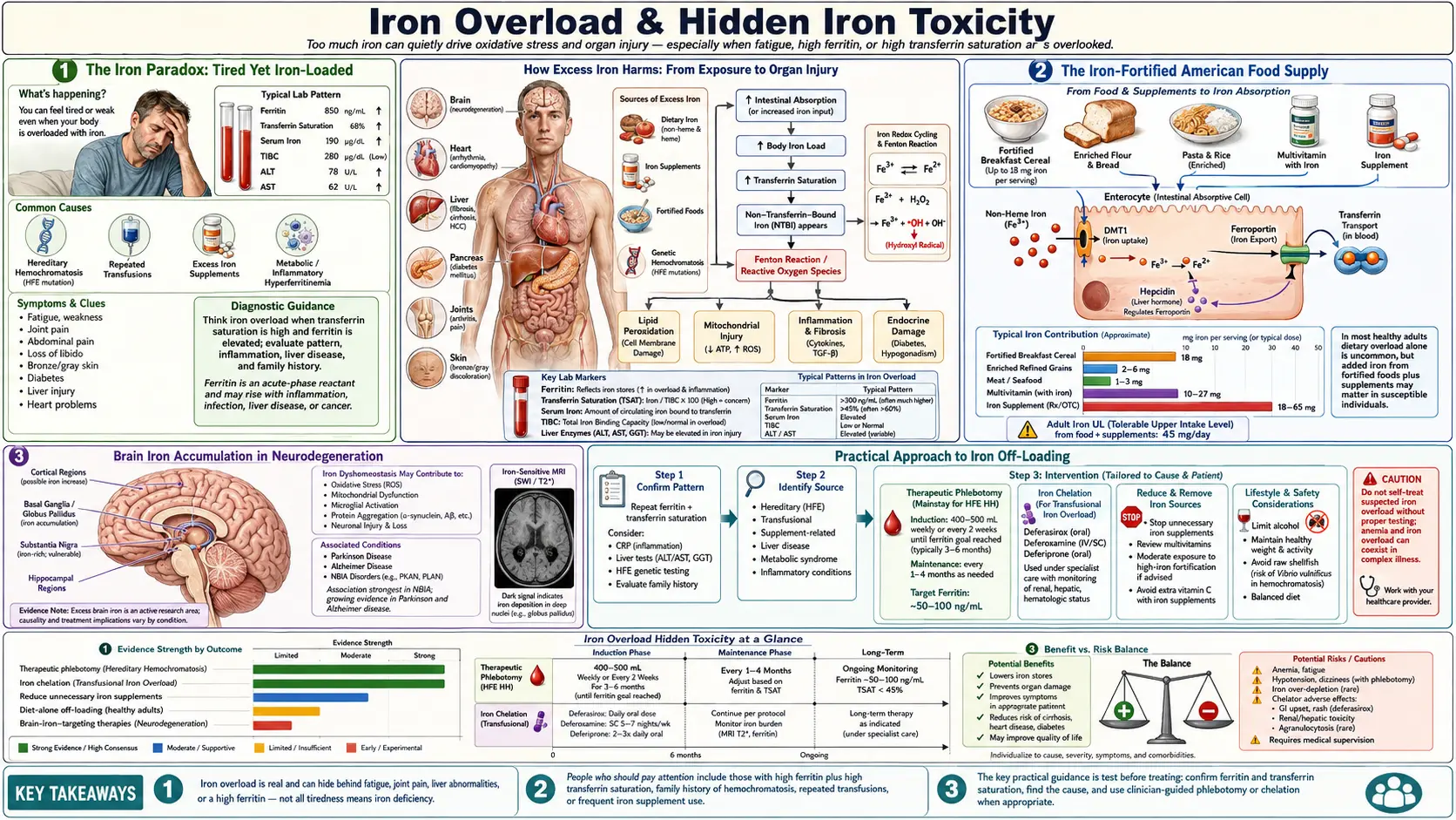
Few claims in functional nutrition are as counter-intuitive as the one Morley Robbins makes about iron. Mainstream medicine and the lay public share the same mental model: iron is a scarce, hard-to-absorb nutrient; low ferritin or hemoglobin is “low iron”; the answer is to take more. The Root Cause Protocol turns that on its head. Robbins argues that the typical American adult is not iron-deficient at all — the typical American adult is iron-loaded in the wrong tissues while the iron in the right tissues fails to function because ceruloplasmin’s ferroxidase activity has collapsed. Adding more iron makes the picture worse, not better. This article walks through that thesis — the iron paradox, the difference between bound and free iron, what ferritin actually measures, the role of mandatory food fortification, the spectrum of hereditary hemochromatosis, brain iron in neurodegeneration, why blood donation is a therapy and not just a charitable act, when iron supplementation is genuinely warranted, and the practical RCP off-loading protocol.
Table of Contents
- The Iron Paradox: Tired Yet Iron-Loaded
- Free Iron vs. Bound Iron
- Ferritin: Storage Marker or Inflammation Marker?
- The Iron-Fortified American Food Supply
- Hereditary Hemochromatosis & The Spectrum
- Brain Iron Accumulation in Neurodegeneration
- Therapeutic Phlebotomy: Donating Blood as Medicine
- When Iron Supplementation IS Genuinely Needed
- Practical Approach to Iron Off-Loading
- Key Research Papers
- Connections
- Featured Videos
1. The Iron Paradox: Tired Yet Iron-Loaded
Robbins’s central observation is one most clinicians can confirm if they look. A patient walks in exhausted. Hemoglobin is 11.8 g/dL — flagged low. Ferritin is 22 ng/mL — flagged low. The standard lab printout pushes both numbers into the “iron-deficient” column, and the standard prescription is ferrous sulfate or a multivitamin with 18 mg of iron. The patient takes it for six months. Hemoglobin barely moves. The fatigue gets worse. GI symptoms appear. Inflammatory markers (CRP, ferritin itself) climb. Eventually a liver MRI is ordered for a different reason — perhaps elevated AST/ALT — and the radiology report mentions increased hepatic iron. The patient is, somehow, iron-deficient by lab cutoffs and iron-loaded on imaging at the same time.
This is the iron paradox. Hemoglobin falls because iron isn’t getting to the bone marrow in usable form — the ferroxidase step that loads iron onto transferrin requires copper-bearing ceruloplasmin, and when ceruloplasmin is functionally low, iron piles up in storage tissues (liver, spleen, brain, heart, pancreas) instead of being shuttled into red-cell production. In Robbins’s account, ferritin in the bloodstream may read low because what little iron is moving is sequestered intracellularly as hemosiderin and as tissue ferritin that the standard serum ferritin assay does not capture. Meanwhile, supplemental iron poured into this broken system has nowhere to go but the wrong places — the very tissues that least need it — where it accelerates Fenton-reaction free-radical generation, deepens oxidative stress, drives more inflammation, and worsens fatigue. The patient is treated harder for “iron deficiency” while the underlying problem — a copper–ceruloplasmin lesion — goes untouched.
Robbins reframes the standard CBC and iron panel through this lens. Anemia in the chronically inflamed adult is rarely true storage-iron deficiency; it is an iron-handling failure. The corrective is to restore copper, magnesium, and retinol so ceruloplasmin can be made — not to throw more iron into a system that cannot use it.
The daily iron cycle in numbers. Mainstream physiology agrees with Robbins on one big point: the body runs mainly on recycled iron, not on the iron in today’s meals. An adult’s bone marrow needs roughly 25 mg of iron a day to build new red cells. Red cells live about 120 days; macrophages in the spleen and liver then break the old ones down and hand about 24 mg of iron a day back to plasma transferrin. The gut adds only 1–2 mg a day, matching the 1–2 mg lost through shed skin and gut cells and minor bleeding (more with menstruation). There is no active way to excrete extra iron, so absorption, controlled by the liver hormone hepcidin acting on the iron exporter ferroportin, is the only regulated step (Nemeth et al., 2004; Camaschella, 2015). Both exits depend on copper enzymes: hephaestin moves iron out of the gut lining (shown in mice; Vulpe et al., 1999) and ceruloplasmin moves it out of macrophages and liver cells (Harris et al., 1999, in mice).
Evidence check. Well established: copper ferroxidases are required for iron export. People born without ceruloplasmin (aceruloplasminemia) build up iron in the liver and the basal ganglia of the brain (Harris et al., 1995), and severe copper deficiency, most often after bariatric surgery or from long-term excess zinc, causes an anemia that iron does not fix but copper does, usually within 4–12 weeks (Myint et al., 2018). Contradicted: the idea that a ceruloplasmin lesion shows up as a low ferritin. When ceruloplasmin is missing, iron is trapped in storage cells and serum ferritin rises, in mice and in the human disorder alike (Harris et al., 1999). Plausible but unproven: that ordinary adults with normal-range ceruloplasmin have a hidden ferroxidase failure. The commonest anemia in chronically ill adults is anemia of inflammation, an iron-handling failure as Robbins says, but one driven by hepcidin blocking ferroportin, with ferritin normal or high (Weiss, Ganz & Goodnough, 2019).
2. Free Iron vs. Bound Iron
Iron biology is, at its heart, about containment. Healthy iron is sequestered. In plasma it is bound to transferrin (which delivers it to cells). Inside cells, what isn’t immediately used is locked into ferritin nanocages or built into heme proteins — hemoglobin, myoglobin, the cytochromes of the electron transport chain. Under normal conditions, less than 0.1% of total body iron exists as free or “labile” iron at any moment. The body works very hard to keep that fraction tiny.
The reason it works so hard is the Fenton reaction. Free Fe²⁺ reacts with hydrogen peroxide to generate the hydroxyl radical (·OH) — the most damaging reactive oxygen species in biology, capable of attacking DNA bases, lipids, and protein thiols indiscriminately. Even nanomolar concentrations of free iron in the wrong cellular compartment cause measurable oxidative damage. Ferroptosis — an iron-dependent form of regulated cell death distinct from apoptosis or necrosis — was characterized in the 2010s and has since been implicated in stroke, heart attack, neurodegeneration, and chemotherapy response. The take-home is simple: iron in the wrong place, at the wrong time, in the wrong oxidation state, is a poison.
Ceruloplasmin’s ferroxidase activity exists precisely to keep this from happening. By oxidizing Fe²⁺ back to Fe³⁺ for transferrin loading, ceruloplasmin keeps the labile-iron pool small and keeps iron moving in regulated channels. When ceruloplasmin function falls — whether through copper deficiency, retinol deficiency, magnesium-dependent enzyme failure, or rare aceruloplasminemia — the labile pool grows. In aceruloplasminemia, the one human condition in which ceruloplasmin is truly absent, the picture is tissue iron accumulation, low serum iron and anemia, but with a high serum ferritin. Robbins extends the same mechanism to a partial, acquired loss of ceruloplasmin function from retinol or magnesium shortfalls; that extension has not been tested in people.
3. Ferritin: Storage Marker or Inflammation Marker?
Ferritin is two things at once, and the failure to remember that is responsible for a great deal of misdiagnosis. Ferritin is, first, a real iron-storage protein — a 24-subunit nanocage that sequesters up to ~4,500 iron atoms per molecule inside cells. Serum ferritin (a small fraction of total ferritin, secreted by hepatocytes and macrophages) does correlate with body iron stores in healthy people. But ferritin is also, second, an acute-phase reactant — an inflammation-induced protein whose serum level rises in response to IL-6 and other inflammatory cytokines, independent of actual iron stores. In a patient with autoimmune disease, fatty liver, metabolic syndrome, chronic infection, or active inflammation of any origin, serum ferritin rises because the liver is making more of it as part of the host-defense program of sequestering iron away from invading pathogens. That ferritin reading no longer represents iron stores.
Standard reference ranges (typically 30–300 ng/mL for adults) reflect a population that is heavily inflamed by modern standards. Robbins, drawing on the Iron Disorders Institute’s work, argues that the healthy ferritin range is much narrower — 50–150 ng/mL — and that ferritin above ~100 ng/mL in a patient with any inflammatory condition is far more likely to be reading inflammation than reading iron. His clinical target is ferritin under 80 ng/mL in adult men and under 50 ng/mL in women. This is not actually radical: mainstream hepatology already adjusts the interpretation of elevated ferritin downward when CRP is elevated, and the literature on hyperferritinemia in metabolic syndrome (where ferritin is elevated without iron overload on liver biopsy) supports the inflammation-marker reading.
Robbins also warns against the mirror-image error: reading low ferritin in a non-bleeding adult as definitive iron deficiency. In a menstruating woman with heavy periods, low ferritin plus low transferrin saturation plus a low reticulocyte count truly is iron deficiency and may need iron. In an adult man with fatigue and a “low” ferritin (say, 25 ng/mL), Robbins reads functional copper deficiency and would order ceruloplasmin and serum copper before reaching for ferrous sulfate. Evidence check: that reading runs against the physiology. A ferritin below 30 ng/mL is the most specific blood marker of empty iron stores, and when copper ferroxidases fail, iron is trapped in storage and ferritin goes up, not down. In a man or a woman past menopause, true iron deficiency calls first for a search for hidden blood loss, most often from the gut (Camaschella, 2015); testing copper and ceruloplasmin alongside is reasonable, but it does not replace that search. Copper-deficiency anemia is real but uncommon (Myint et al., 2018).
Reading ferritin with transferrin saturation. Mainstream hematology reads ferritin together with transferrin saturation (TSAT: serum iron divided by total iron-binding capacity), because the pair separates states either number alone confuses. Ferritin in ng/mL is the same number as in µg/L. The cut-offs below are approximate and vary by laboratory, age, sex and guideline. A ferritin below 30 ng/mL with TSAT below 20% means stores are genuinely empty: absolute iron deficiency (Camaschella, 2015). A ferritin above 100 ng/mL with TSAT below 20% is the pattern of anemia of inflammation: inflammatory signals such as IL-6 raise hepcidin, which traps iron inside macrophages, so stores exist but the marrow cannot reach them (functional iron deficiency); a ferritin between 30 and 100 ng/mL with a low TSAT in an inflamed patient can be both at once (Weiss, Ganz & Goodnough, 2019). A ferritin within the laboratory range with TSAT between 20% and 45% is the ordinary pattern. Iron overload is signalled by TSAT above 45% together with ferritin above 200 ng/mL in women or 300 ng/mL in men (the European liver society’s haemochromatosis criteria, which use a TSAT above 50% for men and postmenopausal women; EASL, 2022). A high ferritin with a normal TSAT usually means inflammation, fatty liver or metabolic syndrome rather than overload.
4. The Iron-Fortified American Food Supply
Most Americans dramatically underestimate how much iron they consume each day, because most of it is added without their knowledge. In 1941 the United States set a federal standard for “enriched” white flour, restoring iron, niacin, riboflavin and thiamin lost in milling (folic acid was added in 1998). The federal standard defines what “enriched” means rather than forcing every miller to use it, but enrichment was made compulsory for white bread during the Second World War, many states require it, and it is near-universal in refined flour today. The federal standard for enriched flour calls for 20 mg of iron per pound, which works out to roughly 1 mg in a typical slice of enriched white bread. Ready-to-eat breakfast cereals are fortified voluntarily, and some deliver 18 mg per serving, 100% of the Daily Value, in a single bowl. Infant formulas are iron-fortified by design, and that is not risk-free in every infant: in a Chilean randomised trial, babies who began with high hemoglobin and were fed iron-fortified formula (12.7 mg/L) from 6 to 12 months scored lower on several developmental tests at age 10 than those given low-iron formula, while babies who started anemic did better on the fortified formula (Lozoff et al., 2012). Pasta is enriched. Rice is enriched. The bagel, the pizza crust, the breakfast sandwich, the kid’s cereal, the after-school crackers, and the dinner pasta are all delivering added inorganic iron that the body cannot easily refuse.
Add to this a typical American diet that includes red meat (rich in heme iron, which is absorbed more efficiently and less tightly regulated than non-heme iron), and add to that the routine use of multivitamins and prenatal vitamins containing 18–27 mg of iron, and supplement users can take in well over what food supplies. US survey data put average intake from food alone at about 16–18 mg a day for men and 12.6–13.5 mg for women (NIH Office of Dietary Supplements). The physiological daily loss in a non-menstruating adult is closer to 1–2 mg, and only a small fraction of intake is absorbed. Robbins argues that the cumulative imbalance over decades is substantial and links rising fortification to rising metabolic syndrome, type 2 diabetes, fatty liver disease and cardiovascular disease. Evidence check: prospective cohorts do find more type 2 diabetes in people with higher ferritin, but ferritin also rises with fatty liver and inflammation, so cause and effect are unsettled, and no controlled comparison has shown that fortifying countries have more of these diseases once diet is accounted for. Tier: hypothesis.
Robbins’s practical advice is straightforward: read the ingredient panel of every packaged food. Look for the words iron, ferrous sulfate, ferrous gluconate, ferrous fumarate, ferric pyrophosphate, or reduced iron. If they appear, the food is delivering added inorganic iron. That iron is not exempt from regulation: it enters through the same gut transporters as other non-heme iron and is still subject to hepcidin control. The point is volume, not a loophole. For an adult attempting iron off-loading, this is the single largest controllable input.
5. Hereditary Hemochromatosis & The Spectrum
Classical hereditary hemochromatosis (HH) is the genetic disorder of uncontrolled iron absorption. The most common form is HFE-related: homozygosity for the C282Y mutation produces a defective HFE protein that fails to signal hepcidin properly, leaving the gut continuously absorbing iron as if the body were always deficient. The result, untreated, is progressive iron loading of the liver (cirrhosis, hepatocellular carcinoma), heart (cardiomyopathy, arrhythmia), pancreas (“bronze diabetes”), pituitary (hypogonadism), joints (arthropathy), and skin (the classic bronze pigmentation). The treatment is therapeutic phlebotomy — weekly to monthly blood removal until ferritin and transferrin saturation normalize, then maintenance phlebotomy two to four times a year for life.
What is less widely recognized is the breadth of the hemochromatosis spectrum. C282Y homozygotes are roughly 1 in 200 in populations of Northern European descent — the highest frequency of any clinically significant Mendelian disorder in those populations. C282Y heterozygotes (1 in 8 to 1 in 10 in the same populations) accumulate iron at lower rates but, in combination with other factors (alcohol intake, hepatitis C infection, metabolic syndrome), can develop clinically meaningful overload. The H63D mutation is even more common; C282Y/H63D compound heterozygotes have intermediate risk. Beyond HFE, juvenile hemochromatosis caused by hemojuvelin (HJV) or hepcidin (HAMP) mutations produces severe, early-onset overload. Mutations in transferrin receptor 2 (TfR2) and ferroportin (SLC40A1) cause additional forms. Carrying a variant is not the same as having disease, though. In a cohort of 31,192 people of northern European descent followed for an average of 12 years, iron-overload-related disease developed in 28.4% of male C282Y homozygotes and 1.2% of female ones, and in only one person who was not a C282Y homozygote (Allen et al., 2008). Current European criteria diagnose haemochromatosis in a C282Y homozygote from a TSAT above 45% with ferritin above 200 ng/mL in women, or a TSAT above 50% with ferritin above 300 ng/mL in men and postmenopausal women (EASL, 2022).
Robbins extends this further. He argues that even non-HFE patients accumulate iron in the modern environment because the regulatory system was never designed for the iron load now being delivered. Whether or not one accepts that broader extension, the clinical implication is the same: any patient with persistently elevated ferritin, especially with a Northern European background, deserves an iron-panel workup including transferrin saturation and (if indicated) HFE genotyping. Iron loading is far more common than the textbooks suggest, and missing it has decade-long consequences.
6. Brain Iron Accumulation in Neurodegeneration
The brain handles iron with extraordinary care — and when that handling fails, the consequences are devastating. Iron is required for myelin synthesis, neurotransmitter production (dopamine synthesis depends on iron-containing tyrosine hydroxylase), and oxidative phosphorylation in mitochondria-rich neurons. But iron in the wrong neuronal compartment, in the wrong oxidation state, is catastrophic. The substantia nigra accumulates iron progressively in Parkinson’s disease, visible on T2*-weighted and R2* MRI sequences and correlating with disease severity. The hippocampus and cortex accumulate iron in Alzheimer’s, where it co-localizes with amyloid-β plaques. The globus pallidus accumulates iron in pantothenate-kinase-associated neurodegeneration (PKAN); Friedreich’s ataxia involves iron mishandling in mitochondria of the heart and the cerebellum’s dentate nucleus. And the basal ganglia accumulate iron in aceruloplasminemia, the inherited loss of ceruloplasmin (Harris et al., 1995).
The mechanism converges on the same chemistry: iron-driven Fenton-reaction free-radical generation, propagated by ferroptosis, in cells that already have high baseline oxidative metabolism and limited regenerative capacity. Iron also accelerates α-synuclein and amyloid-β aggregation in vitro, suggesting it is not just an innocent bystander but a co-pathogen. Iron disrupts the blood-brain barrier in animal models, allowing further peripheral inflammation and immune-cell infiltration into the parenchyma. A 2014 pilot study of the chelator deferiprone in Parkinson’s disease (Devos et al.) raised hopes, but the large follow-up trial reversed them: in 372 people with newly diagnosed Parkinson’s, 36 weeks of deferiprone lowered iron in the nigrostriatal region yet worsened movement scores compared with placebo (a 15.6-point versus 6.3-point rise), and caused agranulocytosis in 2 participants (Devos et al., 2022). Lowering brain iron with a drug is not, on current evidence, a treatment.
From the RCP perspective, the prevention story is more important than the treatment story. Brain iron accumulation does not start in the seventh decade — it accumulates over a lifetime of dietary iron loading and copper depletion. Anyone with a family history of neurodegenerative disease has a strong rationale to think about iron load decades before symptoms appear, and to think about ceruloplasmin function as the upstream protective variable. The brain has very limited iron off-loading capacity once accumulated; the lever that moves easily is the input side — what comes in via diet and supplements, and how well the ferroxidase system can keep iron sequestered.
7. Therapeutic Phlebotomy: Donating Blood as Medicine
Therapeutic phlebotomy is the most efficient iron-removal method available, by a wide margin. A single 500 mL whole-blood donation removes roughly 250 mg of iron — the iron contained in the hemoglobin of the removed red cells. Oral chelation drugs, by comparison, remove far less iron per dose and carry kidney, liver and blood-count side effects, which is why they are reserved for people who cannot be bled, such as those with transfusion-dependent anemias. Three or four blood donations per year off-load 750–1,000 mg of iron, which can shift ferritin from the high triple digits down into Robbins’s target range of 50–80 over twelve to eighteen months in an adult man whose dietary inputs have also been reduced.
The American Red Cross requires at least 56 days (8 weeks) between whole-blood donations for men and women alike, with a hemoglobin check at each visit. For patients with diagnosed hemochromatosis, formal therapeutic phlebotomy under medical supervision proceeds on a more aggressive schedule — weekly or biweekly during the de-iron phase, then maintenance every two to four months; European guidance aims for ferritin below 50 ng/mL during the induction phase and below 100 ng/mL during maintenance (EASL, 2022). A standing order from a treating physician converts what would otherwise be a regular blood donation into a coded therapeutic procedure, often covered by insurance.
For the broader Robbins audience — adults without classical hemochromatosis but with elevated ferritin, metabolic syndrome, fatty liver, or family history of neurodegenerative disease — routine blood donation through the Red Cross or a local blood bank is a no-cost, well-tolerated, and powerful iron-management tool. Eligibility, screening, and aftercare are all standardized. Anyone with elevated ferritin and no contraindication can use it, ideally once the cause of the high ferritin (inflammation, fatty liver or true overload; see the ferritin section) has been looked into. The cost-benefit ratio is unusually favorable: donate two to four times per year, lower body iron stores, contribute to the blood supply, and check ferritin annually to monitor trajectory.
8. When Iron Supplementation IS Genuinely Needed
Robbins is sometimes mischaracterized as an absolute opponent of iron supplementation. He is not. The RCP framework recognizes specific clinical situations where iron supplementation is both appropriate and essential:
- Active gastrointestinal bleeding — visible bleeding (melena, hematochezia, hematemesis) or occult bleeding identified by stool testing. Iron loss exceeds replacement and supplementation is required while the source is identified and treated.
- Heavy menstrual losses with documented true iron deficiency — all three of low ferritin, low transferrin saturation, and low reticulocyte count, ideally with low retic-Hb content. Heavy menstrual bleeding is the most common cause of true iron-deficiency anemia in adult women.
- Pregnancy with confirmed deficiency — expanded blood volume and fetal demand can outpace dietary intake even in well-nourished women. Confirmed deficiency on labs warrants supplementation.
- Post-bariatric surgery malabsorption — gastric bypass patients lose acid-dependent iron absorption and frequently need supplemental iron lifelong.
- Iron-deficient infants — rare in iron-fortified countries, common in low-income global settings where the public-health calculus is different. The WHO supplementation programs exist for clear reasons in those populations, but even there iron is not harmless when given to everyone: in a randomised trial of about 24,000 preschool children in a high-malaria area of Zanzibar, routine iron plus folic acid raised the risk of death or hospital admission by 12%, and the trial arms were stopped early (Sazawal et al., 2006).
Even in these legitimate cases, Robbins would emphasize co-supplementation with copper, retinol (vitamin A), and the cofactors needed for ceruloplasmin synthesis so that the supplemental iron can actually be loaded and used safely. He would prefer food-form iron — red meat, organ meats (especially beef liver), blackstrap molasses — over isolated ferrous-salt tablets, because food-form iron arrives packaged with the cofactors that help direct it to the right places. And he would re-test in three to four months and de-escalate as soon as the deficiency is corrected, rather than continuing supplementation indefinitely.
9. Practical Approach to Iron Off-Loading
The RCP iron off-loading protocol is layered — reduce inputs, increase outputs, and restore the regulatory machinery in parallel.
- Stop ADDED iron. Read every label. Remove iron-fortified breakfast cereals, multivitamins with iron, prenatal vitamins (if not pregnant), iron-containing protein powders, and iron-fortified breads where possible. Choose unfortified whole-grain or sourdough bread, oatmeal, and whole-food breakfasts (eggs, fruit, real cheese) instead of fortified cereal.
- Donate blood every 8 to 16 weeks for men, or after menses cease for women. Check ferritin annually to monitor progress. Document the trajectory.
- Drink coffee with iron-containing meals. Coffee’s polyphenols, chiefly chlorogenic acid, reduce non-heme iron absorption: in isotope studies a cup of coffee cut iron absorption from a hamburger meal by 39%, and tea by 64% (Morck et al., 1983). Coffee with the meal, or up to an hour after it, blunts iron uptake; coffee drunk an hour before the meal had no effect.
- Drink black or green tea with meals. Tea tannins reduce iron absorption similarly to coffee polyphenols. Tea with lunch or dinner, every day, is a passive iron-reduction lever.
- Calcium with meals (food calcium, not supplemental). Calcium competes with iron for absorption at the brush-border level. A glass of milk, a serving of yogurt, or sardines (calcium plus omega-3) at the same meal as iron-containing foods reduces uptake. Note that this conflicts with the RCP’s general recommendation to stop calcium supplements, so use food calcium specifically — dairy, sardines with bones, leafy greens.
- Separate vitamin C from iron-containing meals. High-dose ascorbic acid increases non-heme iron absorption substantially — the opposite of what the iron-loaded patient wants. The RCP already discourages synthetic ascorbic acid for other reasons; this is an additional one. If using whole-food vitamin C (acerola, camu camu), take it on an empty stomach, away from iron-containing meals.
- Restore ceruloplasmin. The off-loading effort is incomplete without this. Bioavailable copper from beef liver, oysters, and cacao; retinol from cod-liver oil and liver; magnesium glycinate; whole-food vitamin C; and removal of the depleting inputs (fluoride, glyphosate, synthetic D3 at high doses) are the inputs to ceruloplasmin synthesis. Safety beside the dose: the US Tolerable Upper Intake Level for copper is 10 mg a day for adults from all sources (Trumbo et al., 2001); people with Wilson’s disease must not add copper at all; and preformed vitamin A (retinol) from liver and cod-liver oil can cause birth defects in high doses, so pregnant women and those trying to conceive should stay within prenatal guidance. See the Ceruloplasmin and Whole-Food Copper articles for details.
- Re-test ferritin every four to six months. Track the trajectory. Adjust phlebotomy frequency, food-iron intake, and ceruloplasmin support based on the numbers. Aim for ferritin in the healthy range — 50–80 ng/mL for adult men, lower for women — with a stable transferrin saturation of 20–35%.
This protocol is not aggressive. It is patient. Iron took years or decades to load, and it will take twelve to twenty-four months of consistent off-loading to bring stores into the healthy range while simultaneously rebuilding the ferroxidase system. For most adults, the changes are tolerable — many were going to drink the coffee, donate the blood, and skip the fortified cereal anyway. What changes is the framing: these are not random lifestyle choices, they are an iron-management system, and they are the upstream lever for a great deal of metabolic, cardiovascular, and neurological resilience over a lifetime.
Key Research Papers
- Nemeth E, Tuttle MS, Powelson J, Vaughn MB, Donovan A, Ward DM, Ganz T, Kaplan J (2004). Hepcidin regulates cellular iron efflux by binding to ferroportin and inducing its internalization. Science. — PubMed PMID: 15514116
- Vulpe CD, Kuo YM, Murphy TL, Cowley L, Askwith C, Libina N, Gitschier J, Anderson GJ (1999). Hephaestin, a ceruloplasmin homologue implicated in intestinal iron transport, is defective in the sla mouse. Nat Genet. — PubMed PMID: 9988272
- Harris ZL, Durley AP, Man TK, Gitlin JD (1999). Targeted gene disruption reveals an essential role for ceruloplasmin in cellular iron efflux. Proc Natl Acad Sci U S A. — PubMed PMID: 10485908
- Harris ZL, Takahashi Y, Miyajima H, Serizawa M, MacGillivray RT, Gitlin JD (1995). Aceruloplasminemia: molecular characterization of this disorder of iron metabolism. Proc Natl Acad Sci U S A. — PubMed PMID: 7708681
- Myint ZW, Oo TH, Thein KZ, Tun AM, Saeed H (2018). Copper deficiency anemia: review article. Ann Hematol. — PubMed PMID: 29959467
- Camaschella C (2015). Iron-deficiency anemia. N Engl J Med. — PubMed PMID: 25946282
- Weiss G, Ganz T, Goodnough LT (2019). Anemia of inflammation. Blood. — PubMed PMID: 30401705
- European Association for the Study of the Liver (2022). EASL Clinical Practice Guidelines on haemochromatosis. J Hepatol. — PubMed PMID: 35662478
- Allen KJ, Gurrin LC, Constantine CC, Osborne NJ, Delatycki MB, Nicoll AJ, et al. (2008). Iron-overload-related disease in HFE hereditary hemochromatosis. N Engl J Med. — PubMed PMID: 18199861
- Devos D, Labreuche J, Rascol O, Corvol JC, Duhamel A, Guyon Delannoy P, et al. (2022). Trial of Deferiprone in Parkinson’s Disease. N Engl J Med. — PubMed PMID: 36449420
- Lozoff B, Castillo M, Clark KM, Smith JB (2012). Iron-fortified vs low-iron infant formula: developmental outcome at 10 years. Arch Pediatr Adolesc Med. — PubMed PMID: 22064877
- Sazawal S, Black RE, Ramsan M, Chwaya HM, Stoltzfus RJ, Dutta A, et al. (2006). Effects of routine prophylactic supplementation with iron and folic acid on admission to hospital and mortality in preschool children in a high malaria transmission setting: community-based, randomised, placebo-controlled trial. Lancet. — PubMed PMID: 16413877
- Morck TA, Lynch SR, Cook JD (1983). Inhibition of food iron absorption by coffee. Am J Clin Nutr. — PubMed PMID: 6402915
- Trumbo P, Yates AA, Schlicker S, Poos M (2001). Dietary reference intakes: vitamin A, vitamin K, arsenic, boron, chromium, copper, iodine, iron, manganese, molybdenum, nickel, silicon, vanadium, and zinc. J Am Diet Assoc. — PubMed PMID: 11269606
PubMed Topic Searches
- PubMed: Hereditary hemochromatosis (HFE)
- PubMed: Ferritin as an acute-phase reactant
- PubMed: Brain iron in neurodegeneration (MRI)
- PubMed: Parkinson’s disease & substantia nigra iron
- PubMed: Alzheimer’s, iron & amyloid
- PubMed: Therapeutic phlebotomy & ferritin
- PubMed: Iron fortification & disease risk
- PubMed: Non-HFE hemochromatosis
- PubMed: Fenton reaction, iron & ROS
- PubMed: Deferiprone in Parkinson’s disease
- PubMed: Hepcidin & iron regulation
- PubMed: Ferroptosis: iron-dependent cell death
Connections
- All Remedies
- Morley Robbins
- Ceruloplasmin and Bioavailable Copper
- Relationship Between Hemoglobin and Ceruloplasmin — why ceruloplasmin (the copper ferroxidase) is required to load iron into hemoglobin, and why "iron-deficiency anemia" is often functional copper deficiency.
- Whole Food Copper Sources
- Copper-Iron Dysregulation
- Root Cause Protocol
- Magnesium Replenishment
- Vitamin D Controversy
- Adrenal Cortisol and Mineral Connection
- Cure Your Fatigue
- Iron
- Copper
- Magnesium
- Hemochromatosis
- Berberine and Iron Overload
- Liver Cleansing
- Iron Panel
- Metabolic Syndrome
- Glyphosate Copper Chelation
- Lab Tests