Manganism (Manganese Toxicity): Tremor and Parkinsonism
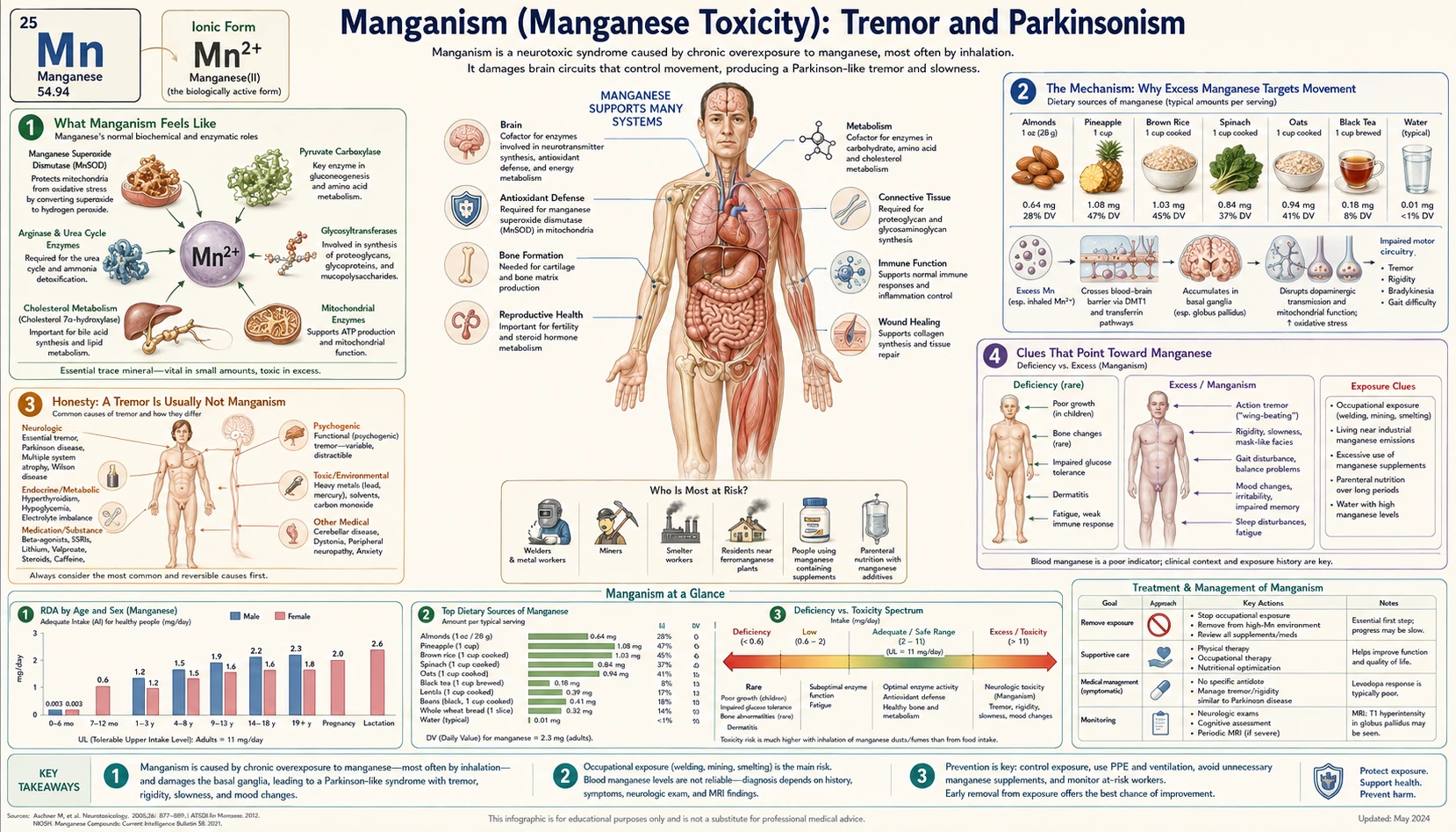
When the body is overloaded with manganese over months or years, the metal settles in a deep brain region called the globus pallidus and can produce a movement disorder known as manganism — slow, stiff, clumsy movements, a shuffling or rooster-like walk, and sometimes a tremor. Because it looks so much like Parkinson’s disease, manganism is often called “manganese-induced parkinsonism.” But two honest points come first. Most people will never develop it: ordinary diet does not cause it, and even most workers exposed to manganese do not get the full syndrome. And it is not the same disease as Parkinson’s — it injures a different part of the brain, responds poorly to the standard Parkinson’s drug, and follows a different course. This page explains what manganism feels like, why excess manganese targets movement, and — just as importantly — why a tremor is far more likely to be something else.
Table of Contents
- What Manganism Feels Like
- The Mechanism: Why Excess Manganese Targets Movement
- Honesty: A Tremor Is Usually Not Manganism
- Clues That Point Toward Manganese
- What Actually Causes Manganese Overload
- Getting Checked
- How Manganese Overload Is Managed
- Red Flags and Warning Signs
- Key Research Papers
- Connections
- Featured Videos
What Manganism Feels Like
Manganism comes on slowly, usually after months to years of heavy manganese exposure, and it tends to unfold in stages. Early on, the symptoms are easy to dismiss: vague tiredness, irritability, trouble sleeping, leg cramps, or a subtle clumsiness. These early changes overlap heavily with mood and thinking problems, which is why they are covered on the companion page on mood and cognitive changes. As exposure continues, the movement problem — the focus of this page — becomes the dominant feature.
The established movement picture of manganism has a recognizable shape:
- Slowness and stiffness (bradykinesia and rigidity). Movements become slow and effortful; muscles feel rigid, and the face can lose its expressiveness (a “masked” look). This slowing — not the tremor — is usually the most prominent and disabling feature.
- A characteristic gait. People with advanced manganism often develop a distinctive walk: high-stepping on the toes, sometimes described as a “cock walk” or rooster-like gait, with a tendency to fall backward. This gait is unusual in ordinary Parkinson’s disease and is one of the syndrome’s clinical fingerprints.
- Tremor — but a different kind. When tremor appears in manganism, it is more often an action or postural tremor (worse when holding a posture or reaching) rather than the classic resting “pill-rolling” tremor of Parkinson’s disease, which is most visible when the hand is at rest. A pronounced resting tremor is actually relatively uncommon in manganism, and its absence can be a clue.
- Dystonia and balance trouble. Sustained muscle contractions that twist a limb or the trunk (dystonia), poor balance, and a stooped posture are common, and difficulty with balance often appears earlier and more severely than in Parkinson’s disease.
- Speech and writing changes. Speech may become soft or monotonous, and handwriting can shrink — features shared with parkinsonism in general.
The combined effect is a person who moves slowly and stiffly, walks oddly, and may have a tremor when they use their hands — a presentation close enough to Parkinson’s disease that the two are genuinely hard to tell apart at the bedside. The differences, which matter a great deal for diagnosis and treatment, are explained in the sections below.
The Mechanism: Why Excess Manganese Targets Movement
Manganese is an essential trace mineral — the body needs small amounts for enzymes that handle antioxidant defense, bone formation, and metabolism (see the Manganese overview). The problem is purely one of excess. In overload, manganese does not spread evenly through the brain; it concentrates in the deep gray-matter nuclei known as the basal ganglia, and especially in one of them, the globus pallidus — a hub that helps make movement smooth and well-timed.
An analogy helps. Picture the brain’s movement system as an orchestra. The basal ganglia are the conductor, keeping every section in time, neither too loud nor too soft. When manganese piles up in the globus pallidus, it is as if heavy mud has been poured over the conductor’s podium — the conductor can still wave the baton, but the cues come late and clumsy, so the whole orchestra plays slowly and out of step. That is bradykinesia and rigidity: not a problem of the instruments (the muscles) but of the timing center that coordinates them.
At the cellular level, several injuries pile up. Manganese accumulates inside neurons and disrupts the mitochondria — the cell’s power plants — impairing energy production and triggering a flood of damaging oxidative stress (reactive molecules that injure cell membranes and proteins). It interferes with dopamine handling and signaling in the basal ganglia, and it promotes protein misfolding and neuroinflammation. Animal and laboratory studies tie this triad — mitochondrial failure, oxidative damage, and inflammation — directly to the movement disturbances of manganese excess.
This is also where manganism quietly parts ways with Parkinson’s disease. In Parkinson’s, the damage centers on dopamine-producing neurons in a different structure, the substantia nigra pars compacta, which feed dopamine to the basal ganglia. Manganism injures the receiving station downstream — the globus pallidus — while the dopamine-producing neurons themselves are relatively spared. Same orchestra, but the fault sits in a different chair. That single anatomical difference, explored next, explains why the standard Parkinson’s drug often disappoints in manganism.
Honesty: A Tremor Is Usually Not Manganism
This is the most important section on the page. If you have a tremor, manganism is one of the least likely explanations. Tremor is extraordinarily common and has many causes; manganese overload is a rare one, seen almost entirely in specific high-exposure situations described later. It is not caused by eating manganese-rich foods, by a typical multivitamin, or by living an ordinary life. Naming the far more common causes is not hedging — it is the honest and useful thing to do.
The usual reasons for a tremor include:
- Essential tremor — by far the most common tremor disorder. It is typically an action tremor (most visible when holding a cup, writing, or reaching), often runs in families, frequently affects the hands and head, and sometimes eases briefly with a small amount of alcohol. See Essential Tremor.
- Parkinson’s disease — the classic neurodegenerative cause, with a slow resting tremor (often “pill-rolling” in the hand), slowness, and stiffness. Far more common than manganism and a completely separate condition. See Parkinson’s Disease.
- Too much caffeine or stimulants, and a normal physiologic tremor exaggerated by fatigue, stress, or low blood sugar.
- Medications — certain asthma inhalers, some antidepressants, lithium, valproate, thyroid hormone, and others can all produce a fine tremor.
- An overactive thyroid (hyperthyroidism), which causes a fine, fast tremor along with weight loss, a racing heart, and heat intolerance.
- Alcohol withdrawal, which produces a prominent tremor (the “shakes”) within a day or two of stopping heavy drinking.
- Anxiety, low blood sugar, and simply being cold — ordinary, reversible causes of shaking.
Because this list is long and manganism sits near the bottom of it, a tremor is usually sorted out on its own terms first — by its type (resting vs. action), what makes it better or worse, family history, medications, and thyroid function — before an unusual metal explanation is considered. Manganese becomes a realistic possibility mainly when the specific exposures in the next sections are present.
Clues That Point Toward Manganese
Manganese enters the picture when the story fits, not when symptoms appear in isolation. Several features, taken together, raise the suspicion:
- A real exposure. This is the single biggest clue. Years of welding, mining, smelting, working in dry-cell battery or ferromanganese-alloy plants, long-term reliance on intravenous (parenteral) nutrition, or advanced liver disease — the settings detailed in the causes section, and on the companion page on occupational and water exposure. Without an exposure like one of these, manganism is very unlikely.
- The wrong kind of tremor for Parkinson’s. A prominent action or postural tremor, with relatively little resting tremor, in someone who also has slowness and stiffness, is more in keeping with manganism than with classic Parkinson’s disease.
- The “cock walk” and early falls. The high-stepping, toe-walking gait, a tendency to topple backward, and balance trouble appearing early all point away from ordinary Parkinson’s disease.
- A poor response to levodopa. Levodopa — the drug that replenishes dopamine and is the mainstay of Parkinson’s treatment — usually works well in Parkinson’s disease but tends to help little in manganism, because the dopamine-producing neurons are intact while the downstream target is damaged. A parkinsonism that does not respond to levodopa is a notable clue. A 2015 review describes levodopa as “ineffective in alleviating” manganese-induced movement symptoms and explains why: the damage lies in the basal ganglia’s “direct pathway” (the caudate/putamen, the internal globus pallidus and the substantia nigra pars reticulata), where the release of dopamine is held back while the dopamine nerve endings themselves do not degenerate.
- Symmetry and timing. Manganism usually, though not always, affects both sides of the body fairly evenly from early on (in an 18-year follow-up in Taiwan, the one-sided pattern present at the start in 2 of 4 patients was still there 18 years later), whereas Parkinson’s disease classically begins on one side. Stopping exposure does not always stop the worsening: in a small Taiwan series followed after exposure ended, disability scores on the King’s College Hospital Rating Scale rose from 15.0 in 1987 to 28.3 in 1991 and 38.1 in 1995, and the authors found a plateau only over the following decade.
None of these is proof on its own; a tremor is not evidence of manganese overload, and even all of them together still require objective testing. But the combination of a genuine high-dose exposure plus an atypical, levodopa-unresponsive, symmetric parkinsonism is the pattern that earns a manganese work-up.
What Actually Causes Manganese Overload
Healthy people are well protected from manganese overload. The gut tightly limits how much dietary manganese is absorbed, and the liver clears the excess into bile and out through the stool. Toxicity therefore requires either an exposure that bypasses these defenses or a body that can no longer clear the metal. The recognized causes are:
- Inhalation at work (the classic cause). Breathing manganese-containing dust or fumes is the most important route, because inhaled manganese can reach the brain efficiently and bypasses the gut’s controls. The classic occupations are welding, mining and ore-crushing, smelting and ferroalloy production, and dry-cell battery manufacturing. Studies of welders show parkinsonian signs that worsen with cumulative exposure: a 2017 study of 886 American welders, 398 of them re-examined for up to 9.9 years, found the movement score (the Unified Parkinson Disease Rating Scale motor part, UPDRS3) worsened by 0.24 points a year for each mg Mn/m3-year of cumulative manganese exposure, mostly in slowness of the arms, stiffness of the limbs, and speech and facial expression; the link was most marked in welders who did flux-core arc welding in a confined space. Surveys of mine workers link manganese exposure to parkinsonism and reduced quality of life.
- Liver failure. Because the liver is the body’s main exit route for manganese, severe liver disease lets the metal build up in the blood and brain. People with cirrhosis and other forms of advanced liver disease can develop a manganese-related parkinsonism as part of what is called acquired hepatocerebral degeneration — sometimes described as a “Parkinson’s of cirrhosis.”
- Long-term intravenous (parenteral) nutrition. When all nutrition is delivered into a vein, manganese in the feeding solution skips the gut’s protective gatekeeping and goes straight into the blood. Long-term use — in adults with intestinal failure and in infants — can deposit manganese in the brain, which is why feeding formulas are carefully manganese-limited and monitored.
- Very high manganese in drinking water. Naturally high or contaminated well water is a less common but real route of excess, of particular concern for the developing brain (discussed below and on the water-exposure page).
- Injecting a manganese-containing stimulant. Manganese parkinsonism is also seen in a group of young drug users who inject ephedron, a stimulant that contains manganese. Brain-imaging studies in this group show the same pattern as in manganese-exposed workers: damage in the basal ganglia, while the dopamine nerve endings are not lost.
- Rare inherited transport disorders. A handful of genetic conditions impair the proteins that carry manganese out of the body, causing it to accumulate from childhood — an uncommon but instructive cause that proves the principle that failing to excrete manganese, not just over-consuming it, drives toxicity. People who inherit faults in the manganese-transporter gene SLC30A10 show the same brain-imaging pattern as exposed workers and ephedron users.
A pattern runs through this list: ordinary eating almost never causes manganese toxicity. The danger lies in inhaling it, in losing the liver’s ability to clear it, or in infusing it past the gut. That is the practical reason a tremor in a person with none of these exposures is so unlikely to be manganese.
Getting Checked
There is no single perfect test for manganism; the diagnosis rests on putting together the exposure history, the clinical picture, brain imaging, and blood work — while ruling out the far more common causes of tremor and parkinsonism first.
The exposure history comes first. A careful account of work (welding, mining, smelting, batteries), of any long-term intravenous nutrition, and of liver disease is the most valuable piece of information, because manganism without an exposure is so improbable.
Brain magnetic resonance imaging (MRI) is the most telling test. Manganese is unusual in that it shows up on a routine brain magnetic resonance imaging: deposits in the globus pallidus produce a bright signal on what radiologists call T1-weighted images. This symmetric brightening of the basal ganglia is a recognized fingerprint of manganese accumulation (though it can also appear in liver failure and long-term intravenous nutrition, which fits, since those are themselves causes). Importantly, in classic Parkinson’s disease the routine magnetic resonance imaging is typically normal — so the imaging can help separate the two.
Blood and other tests. A blood manganese level can support the diagnosis when it is high, but it is an imperfect marker: blood largely reflects recent exposure and correlates only loosely with how much has accumulated in the brain, so a normal level does not rule out past overload and a high level alone does not prove brain injury. A Comprehensive Metabolic Panel and dedicated liver function tests help evaluate liver disease as both a cause and a contributor. Where Parkinson’s disease is the real question, a specialized dopamine-transporter brain scan (a DaTscan) is typically abnormal in Parkinson’s but normal in manganism — another way the workup distinguishes the two, since manganism spares the dopamine-producing neurons that such scans measure.
The goal of the evaluation is twofold: confirm that excess manganese is genuinely present and affecting the brain, and — just as important — make sure a more common, sometimes more treatable, diagnosis is not being missed.
How Manganese Overload Is Managed
Management is led by a doctor — usually a neurologist, occupational-medicine physician, or toxicologist — and the single most effective step is also the most obvious: ending the exposure. Beyond that, the realistic aim is to halt further injury and ease symptoms, because brain damage that has already occurred may only partly recover.
- Removing the source. Leaving a high-manganese workplace (or using effective respiratory protection and engineering controls), adjusting a parenteral-nutrition formula to limit or remove manganese, and treating the underlying liver disease are the foundation. Ending exposure does not always halt the illness: in a small Taiwan series, symptoms kept worsening for about 10 years after exposure stopped, even after manganese levels in blood, urine and hair had returned to normal, and then reached a plateau over the following decade.
- Chelation has limited value. Chelating agents (drugs that bind metals so the body can excrete them) can lower blood manganese, but the clinical benefit for established neurological manganism has been disappointing in practice; chelation is not a reliable cure and is used selectively and under specialist supervision, not as a routine fix. A 2025 review lists the treatments that have been studied: chelation with calcium disodium EDTA (CaNa2EDTA), with or without plasma exchange, and para-aminosalicylic acid (PAS), along with natural compounds such as vinpocetine, punicalagin, niacin, vitamin E, curcumin and sesame oil. The review presents these as interventions under study and points to future research for better treatments; none is an established cure.
- Levodopa usually helps little. Because the dopamine-producing neurons are intact while their downstream target is damaged, the standard Parkinson’s drug levodopa generally provides only modest, if any, benefit in manganism — one of the clearest practical differences from Parkinson’s disease. In the Taiwan follow-up of six people with manganese parkinsonism, three received levodopa and improved at first, but the response faded after 2 to 3 years; during treatment they did not develop the on-off swings or extra involuntary movements (dyskinesia), and the authors concluded that their response to levodopa differs from that of people with Parkinson’s disease. A clinician may still trial it, partly to confirm the diagnosis.
- Supportive care matters. Physical therapy, occupational therapy, speech therapy, balance and fall-prevention work, and treatment of associated mood and cognitive symptoms all improve day-to-day function and safety, even when the movement disorder cannot be reversed.
For people in at-risk occupations, the real victory is prevention: workplace exposure limits, ventilation and fume extraction, respiratory protection, and periodic monitoring so that excess is caught long before any tremor or stiffness appears. As with most metal toxicities, not getting overloaded in the first place beats every treatment that follows.
Red Flags and Warning Signs
The cause of a new tremor or movement change is usually a common, manageable one. Certain situations are the ones doctors treat as calling for prompt evaluation:
- A new or worsening tremor or slowness — especially with stiffness, a change in walking or balance, falls, smaller handwriting, or a softer voice. These are the signs a neurological assessment looks at, whatever the suspected cause.
- Movement symptoms in someone with a real manganese exposure — years of welding, mining, smelting, or battery work; long-term intravenous nutrition; or known cirrhosis or advanced liver disease. This is the combination in which doctors specifically consider manganese.
- Signs of advanced liver disease alongside confusion, personality change, or movement problems — yellowing of the skin or eyes, abdominal swelling, or easy bruising — which can reflect cirrhosis driving both the liver failure and the manganese accumulation. Hospitals treat this combination as urgent.
- A tremor with a racing heart, weight loss, and heat intolerance, which suggests an overactive thyroid — a common, treatable cause that is far more likely than a metal problem.
Movement changes together with sudden severe confusion, marked drowsiness, or loss of consciousness are signs hospitals treat as an emergency — in someone with liver disease they can signal hepatic encephalopathy. An ordinary, slowly developing tremor without these features is not an emergency: as the sections above explain, the overwhelming majority of tremors turn out to have a common cause rather than manganism.
Key Research Papers
- Khindri NM, Maj MC (2025). Manganese-Induced Parkinsonism: A Review of Etiologies and Treatments. Degenerative Neurological and Neuromuscular Disease;15:65-79. — DOI: 10.2147/DNND.S482018. — doi:10.2147/DNND.S482018
- Kwakye GF, Paoliello MMB, Mukhopadhyay S, Bowman AB, Aschner M (2015). Manganese-Induced Parkinsonism and Parkinson’s Disease: Shared and Distinguishable Features. International Journal of Environmental Research and Public Health;12(7):7519-7540. — DOI: 10.3390/ijerph120707519. — doi:10.3390/ijerph120707519
- Guilarte TR, Gonzales KK (2015). Manganese-Induced Parkinsonism Is Not Idiopathic Parkinson’s Disease: Environmental and Genetic Evidence. Toxicological Sciences;146(2):204-212. — DOI: 10.1093/toxsci/kfv099. — doi:10.1093/toxsci/kfv099
- Guilarte TR (2010). Manganese and Parkinson’s Disease: A Critical Review and New Findings. Environmental Health Perspectives;118(8):1071-1080. — DOI: 10.1289/ehp.0901748. — doi:10.1289/ehp.0901748
- Lucchini RG, Tieu K (2023). Manganese-Induced Parkinsonism: Evidence from Epidemiological and Experimental Studies. Biomolecules;13(8):1190. — DOI: 10.3390/biom13081190. — doi:10.3390/biom13081190
- Harischandra DS, Ghaisas S, Zenitsky G, et al. (2019). Manganese-Induced Neurotoxicity: New Insights Into the Triad of Protein Misfolding, Mitochondrial Impairment, and Neuroinflammation. Frontiers in Neuroscience;13:654. — DOI: 10.3389/fnins.2019.00654. — doi:10.3389/fnins.2019.00654
- Milatovic D, Zaja-Milatovic S, Gupta RC, Yu Y, Aschner M (2009). Oxidative damage and neurodegeneration in manganese-induced neurotoxicity. Toxicology and Applied Pharmacology;240(2):219-225. — DOI: 10.1016/j.taap.2009.07.004. — doi:10.1016/j.taap.2009.07.004
- Bouabid S, Tinakoua A, Lakhdar-Ghazal N, Benazzouz A (2016). Manganese neurotoxicity: behavioral disorders associated with dysfunctions in the basal ganglia and neurochemical transmission. Journal of Neurochemistry;136(4):677-691. — DOI: 10.1111/jnc.13442. — doi:10.1111/jnc.13442
- Racette BA, Searles Nielsen S, Criswell SR, et al. (2017). Dose-dependent progression of parkinsonism in manganese-exposed welders. Neurology;88(4):344-351. — DOI: 10.1212/WNL.0000000000003533. — doi:10.1212/wnl.0000000000003533
- Mehkari Z, Mohammed L, Javed M, et al. (2020). Manganese, a Likely Cause of ‘Parkinson’s in Cirrhosis’, a Unique Clinical Entity of Acquired Hepatocerebral Degeneration. Cureus;12(9):e10448. — DOI: 10.7759/cureus.10448. — doi:10.7759/cureus.10448
- Zaitout Z, Romanowski C, Karunasaagarar K, Connolly D, Batty R (2014). A review of pathologies associated with high T1W signal intensity in the basal ganglia on Magnetic Resonance Imaging. Polish Journal of Radiology;79:126-130. — DOI: 10.12659/PJR.890043. — doi:10.12659/PJR.890043
- Huang CC, Lu CS, Chu NS, Hochberg F, Lilienfeld D, Olanow W, Calne DB (1993). Progression after chronic manganese exposure. Neurology;43(8):1479-1483. — DOI: 10.1212/wnl.43.8.1479. — doi:10.1212/wnl.43.8.1479
- Huang CC, Chu NS, Lu CS, Chen RS, Calne DB (1998). Long-term progression in chronic manganism: ten years of follow-up. Neurology;50(3):698-700. — DOI: 10.1212/wnl.50.3.698. — doi:10.1212/wnl.50.3.698
- Huang CC, Chu NS, Lu CS, Chen RS, Schulzer M, Calne DB (2007). The natural history of neurological manganism over 18 years. Parkinsonism & Related Disorders;13(3):143-145. — DOI: 10.1016/j.parkreldis.2006.09.002. — doi:10.1016/j.parkreldis.2006.09.002
- National Institutes of Health, Office of Dietary Supplements. Manganese — Fact Sheet for Health Professionals (adult Tolerable Upper Intake Level 11 mg/day; toxicity and manganism). — NIH Office of Dietary Supplements