Iron Overload (High Iron): Bronze Skin and Diabetes
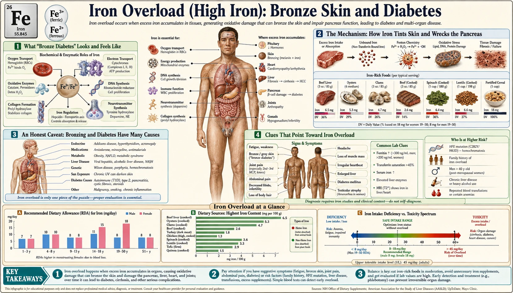
When too much iron builds up in the body over many years — a condition called iron overload, most often from the inherited disorder hereditary hemochromatosis — it can leave two striking marks: a gradual bronze or slate-gray tint to the skin, and diabetes caused by iron damaging the pancreas. Together these gave the old textbook name “bronze diabetes.” It is worth being honest from the start: this dramatic pairing is a late picture that most people with high iron never reach, and a bronze tan or new diabetes has dozens of far more common explanations that have nothing to do with iron. Still, when bronzing and diabetes appear together — especially alongside fatigue, joint pain, or liver trouble — iron overload is a diagnosis worth ruling out, because it is one of the few causes of diabetes that is partly reversible if caught early. This page explains how iron tints the skin and injures the pancreas, why these signs are unreliable on their own, and the pattern in which a simple blood test is used to check for it.
Table of Contents
- What “Bronze Diabetes” Looks and Feels Like
- The Mechanism: How Iron Tints Skin and Wrecks the Pancreas
- An Honest Caveat: Bronzing and Diabetes Have Many Causes
- Clues That Point Toward Iron Overload
- What Causes Iron to Build Up
- Getting Checked
- How Iron Overload Is Treated
- Red Flags and When the Blood Tests Are Used
- Key Research Papers
- Connections
- Featured Videos
What “Bronze Diabetes” Looks and Feels Like
The two signs in the name appear slowly, usually over years, and they often arrive together because both reflect the same underlying problem: iron that has quietly piled up in tissues that were never meant to store it. By the time the skin has changed and blood sugar has risen, the body has typically been accumulating excess iron for a long time.
The skin change. The classic description is a deepening bronze, tan, or grayish-bronze color, as if the person has a permanent suntan that will not fade in winter. Two things actually drive it:
- A genuine tan-like darkening. Iron overload stimulates the skin's pigment cells to make more melanin, the same brown pigment responsible for ordinary tanning. This is what produces most of the visible bronze color.
- A slate-gray tint from iron itself. Iron also deposits directly in the skin, adding a grayish or metallic undertone on top of the melanin. The mix of the two is what gives the skin its distinctive “bronze” quality.
The darkening is usually most obvious on sun-exposed areas — the face, neck, forearms, and the backs of the hands — but it can also show in skin folds, old scars, and, classically, the gums and mouth. It comes on so gradually that people often do not notice it themselves; a relative looking at an old photo, or a doctor, may be the first to point it out.
The diabetes. When iron damages the pancreas, blood sugar rises, producing the familiar symptoms of diabetes: increased thirst, frequent urination, unexplained weight loss, blurred vision, and fatigue. In iron overload this often develops in someone who does not fit the usual diabetes picture — sometimes leaner than expected, or without the typical family history of type 2 diabetes — which is one of the quiet hints that something else is going on.
It is important to be clear that this full “bronze diabetes” presentation is the advanced form, and it is now uncommon, precisely because iron overload is increasingly caught early on blood tests before it ever gets this far. Many people with high iron have only vague fatigue and joint pain — or no symptoms at all — and never develop visible bronzing or diabetes.
The Mechanism: How Iron Tints Skin and Wrecks the Pancreas
To understand both signs you only need one idea: iron in excess is chemically reactive, and the body has no fast way to get rid of it. We are very good at absorbing iron and very poor at excreting it — there is no regulated route to dump a surplus. So when more iron comes in than the body needs, year after year, the excess is shunted into storage in organs throughout the body, where it slowly causes damage.
The damage itself is a chemistry problem. Iron readily flips between two forms (ferrous and ferric), and in doing so it can spark the formation of free radicals — unstable molecules that attack cell membranes, proteins, and DNA. This is called oxidative stress. In small, tightly bound amounts iron is essential and harmless; in large, loosely held amounts it becomes a slow-burning source of cellular injury wherever it settles.
An analogy. Think of iron as firewood for the body's chemistry. The right amount, stacked safely in the hearth, gives steady warmth — this is iron doing its essential jobs, like carrying oxygen. But keep hauling in wood with no way to throw any out, and it piles up against the walls and under the floorboards. Now a stray spark — the free-radical reaction — doesn't just warm the room; it can smolder in the woodpile and char whatever it is stacked against. In iron overload the “woodpiles” build up in specific rooms of the house: the skin, the pancreas, the liver, the heart, and the joints.
Why the skin bronzes. Iron deposits in the deeper layers of the skin and, alongside that, drives the pigment cells (melanocytes) to ramp up melanin production. The result is the blend described above: a tan-like browning from extra melanin, plus a gray-bronze metallic note from the iron itself. The color is essentially the visible surface of a body-wide storage problem.
Why the pancreas fails — the heart of “bronze diabetes.” The pancreas contains the beta cells in the islets of Langerhans, the cells that make insulin, the hormone that lets the body use sugar for fuel. Beta cells are unusually vulnerable to iron: they take it up readily and have relatively weak defenses against oxidative stress. As iron accumulates in the islets, the free-radical damage gradually kills beta cells, so the pancreas makes less insulin. On top of that, iron loading in the liver and muscle worsens insulin resistance — the body responds less well to whatever insulin remains. The combination — falling insulin supply plus rising insulin demand — is what tips blood sugar into the diabetic range. Laboratory and clinical work on this “cross-talk” between iron and glucose control is a well-studied area of metabolism.
This two-hit mechanism explains a hopeful feature of the disease: if iron is removed before too many beta cells have died, glucose control can improve, and the diabetes may become easier to manage or, occasionally in early cases, resolve. Once the beta cells are gone, however, the diabetes is permanent — which is why early detection matters so much.
An Honest Caveat: Bronzing and Diabetes Have Many Causes
This is the most important section for keeping perspective. Both halves of “bronze diabetes” are, on their own, extremely common and almost never caused by iron. Seeing one — or even both — is not proof of iron overload.
Skin darkening has dozens of ordinary causes. By far the most common is simple sun exposure — a real tan. Other frequent, unrelated causes of generalized or patchy darkening include:
- Ethnic skin tone — naturally deeper complexions are normal variation, not a sign of disease.
- Addison's disease (adrenal insufficiency), which classically darkens the skin and the creases of the palms.
- Medications — amiodarone, minocycline, certain chemotherapy drugs, and others can tint or darken the skin.
- Other liver conditions and chronic kidney disease, which can dull or darken the complexion.
- Melasma and post-inflammatory pigmentation — common, usually patchy rather than a uniform bronze.
Diabetes overwhelmingly is not caused by iron. The vast majority of diabetes is ordinary type 2 diabetes (linked to genetics, weight, and inactivity) or autoimmune type 1 diabetes. Iron overload is a rare secondary cause — important to recognize, but a small slice of all diabetes. A new diagnosis of diabetes, by itself, is almost never a reason to suspect hemochromatosis.
So the honest bottom line is this: a tan plus type 2 diabetes is an everyday combination with an everyday explanation. Iron overload climbs the list of suspects only when the pattern is unusual — the bronzing is unexplained and not just sun, the diabetes is atypical, and there are other clues pointing the same way. Those clues are the subject of the next section.
Clues That Point Toward Iron Overload
Iron overload becomes a stronger suspect — one checked with a simple blood test — when bronzing and/or atypical diabetes appear together with one or more of the following:
- The classic companion symptoms. Persistent fatigue and joint pain — especially aching in the knuckles of the index and middle fingers — are among the most common early features of hemochromatosis and often precede the skin and pancreas changes.
- Liver signals. Mildly elevated liver enzymes, an enlarged liver, or signs of liver problems alongside the other findings raise the suspicion, because the liver is usually the first organ to load with iron.
- Heart or hormonal hints. Unexplained heart problems (a weakened heart muscle or rhythm disturbance) or loss of libido and sexual function — iron can also injure the pituitary and gonads.
- Family history. A relative with hemochromatosis, unexplained cirrhosis, or “iron problems” is a strong clue, because the common form is inherited. People of Northern European ancestry are at higher risk.
- Atypical diabetes. Diabetes in someone who is not overweight, has no family history of type 2 diabetes, or whose diabetes is unusually hard to explain — particularly with any of the above — is one of the patterns an iron check is used for.
The takeaway is that no single sign is enough, but a cluster — bronzing, atypical diabetes, fatigue, joint pain, liver changes, and a suggestive family history — is exactly the picture the inexpensive blood tests described below are used to check. The siblings of this page (fatigue & joint pain, liver problems, and heart problems) cover those other organ effects in detail.
What Causes Iron to Build Up
Bronze skin and diabetes are the downstream result of long-standing iron overload; the question is why the iron accumulated in the first place. The causes fall into two broad groups.
- Hereditary hemochromatosis (the usual cause). This is an inherited disorder — most commonly from two copies of a variant in the HFE gene (the C282Y variant) — in which the body absorbs more iron from food than it needs, every day, for life. The controller hormone hepcidin, which normally tells the gut to stop absorbing iron once stores are full, is deficient, so absorption never properly shuts off. It is one of the most common inherited conditions in people of Northern European descent. Crucially, having the genes does not guarantee disease: penetrance is incomplete, and many people who carry two copies never accumulate enough iron to cause organ damage — a key reason the full bronze-diabetes picture is now uncommon.
- Why it shows up later in life, and differently by sex. Because iron builds slowly, symptoms typically emerge in middle age or later. Women are often affected later than men because menstruation and pregnancy shed iron for decades, delaying the buildup until after menopause.
- Iron from repeated transfusions or other overload. A separate route to overload is receiving many blood transfusions over time — for example, in some chronic anemias — because each unit of blood delivers a large iron load the body cannot excrete. Certain other inherited anemias also drive excess iron absorption. This transfusion-related (“secondary”) overload can damage the same organs, including the pancreas.
- What does not cause it. For people without one of these conditions, ordinary diet — even iron-rich foods — and standard multivitamins do not cause iron overload, because a healthy body simply absorbs less when stores are full. For supplements, the US Institute of Medicine (2001) set the adult Tolerable Upper Intake Level at 45 mg of iron a day, based on stomach and gut upset (gastrointestinal distress), and noted that people with hereditary hemochromatosis may not be protected by that limit. Everyday eating is not the culprit.
Pinning down the cause matters because it shapes treatment and family screening: hereditary hemochromatosis is treated by removing blood and warrants testing relatives, whereas transfusion-related overload is managed differently (since removing blood from someone who needs transfusions is not an option).
Getting Checked
The reassuring news is that confirming or excluding iron overload is straightforward and inexpensive, and it starts with two simple blood tests.
Step one: iron blood tests. The screening pair is serum ferritin (a measure of how much iron is stored in the body) and transferrin saturation (the percentage of the blood's iron-carrying protein that is loaded with iron). In iron overload, both tend to be high — a fasting transferrin saturation above roughly 45% is a common trigger to investigate further, and a markedly elevated ferritin signals heavy stores. These are reported on an Iron Panel. One caveat: ferritin also rises with inflammation, infection, and liver inflammation, so a single high ferritin is not proof of overload — it is interpreted alongside the transferrin saturation and the clinical picture.
Step two: confirm and assess the organs. If the iron studies are abnormal, the next steps usually include:
- Genetic testing for the HFE variants (chiefly C282Y), which confirms hereditary hemochromatosis and helps decide which relatives are tested.
- A check for organ involvement, since this is what determines urgency. That means assessing the blood sugar — a fasting glucose and a hemoglobin A1c (HbA1c) — to detect diabetes, plus liver function tests for the liver. A Comprehensive Metabolic Panel conveniently reports glucose and several liver values in one routine draw.
- Imaging of liver iron. A specialized magnetic resonance imaging (MRI) can now measure liver iron non-invasively and has largely replaced the older liver biopsy for gauging how much iron has accumulated; biopsy is reserved for assessing liver scarring in selected cases.
Because the skin and diabetes findings are late, the goal of modern practice is to catch overload before this stage — on a routine ferritin and transferrin saturation, or through screening relatives of someone diagnosed. The skin will not tell you in time; the blood test will.
How Iron Overload Is Treated
The central treatment is refreshingly simple, old, and effective: remove iron by removing blood.
- Therapeutic phlebotomy (the mainstay). For hereditary hemochromatosis, the standard treatment is regular phlebotomy — drawing off a unit of blood, much like donating, on a schedule. Because each unit of red cells carries away a substantial amount of iron, the body is forced to pull iron out of storage to make new blood. Sessions are frequent at first (often weekly) until iron stores fall into the target range, then spaced out to a maintenance rhythm (every few months) for life. It is low-cost, well established, and for most people the iron studies normalize within months to a couple of years.
- Iron-chelation drugs (when phlebotomy can't be used). For people who cannot have blood removed — most importantly those with transfusion-related overload and an underlying anemia — medications called iron chelators bind iron so it can be excreted. These are the main tool when phlebotomy is not an option.
- Diet and alcohol facts that sit alongside treatment. The 2001 Institute of Medicine iron report states that vitamin C (ascorbic acid) strongly enhances the absorption of non-heme iron (the iron in plant foods and supplements). Alcohol adds to the liver injury that iron causes. Raw or undercooked seafood can carry Vibrio vulnificus, a bacterium that depends on iron; a 2023 case report describes it as the main cause of seafood-related deaths in the United States and notes that people with high body iron are more susceptible to the infection. The GeneReviews summary of HFE hemochromatosis lists medicinal iron, iron-containing mineral supplements, excess alcohol, excess vitamin C and uncooked seafood among the agents and circumstances to avoid. These points sit alongside phlebotomy, which the guidelines describe as the mainstay of treatment; they do not replace it.
What treatment does for the skin and the diabetes. The effects are encouraging but not symmetric. The bronze skin discoloration often lightens as iron is removed, sometimes substantially over months to years. The diabetes responds only partially: if treatment begins before extensive beta-cell loss, blood sugar control can improve and insulin needs may fall; but once enough insulin-producing cells have been destroyed, the diabetes is permanent and is managed like any other diabetes. This asymmetry is the real lesson of bronze diabetes — the pigment is cosmetic and reversible, while the pancreatic damage is the part you most want to prevent by acting early. Treating overload early also lowers the risk of the most serious complications, including cirrhosis and liver cancer, and improves long-term survival.
Red Flags and When the Blood Tests Are Used
None of this is an emergency in the way a heart attack is — iron overload develops over years. These are the situations in which the simple iron blood tests (serum ferritin and transferrin saturation, reported on an iron panel) are used:
- Unexplained skin bronzing or graying that is not simply sun exposure — particularly if it is accompanied by any other feature on this page.
- New or atypical diabetes in someone who does not fit the usual pattern, especially with fatigue, joint pain, or a family history of “iron problems.”
- The classic cluster: bronzing plus diabetes plus fatigue and joint pain — this combination is the textbook reason for an iron panel.
- A close relative with hemochromatosis or unexplained cirrhosis — relatives are screened with ferritin, transferrin saturation and often HFE genetic testing, even when they feel completely well; GeneReviews describes offering genetic testing to the adult brothers and sisters of a person diagnosed with HFE hemochromatosis.
How the results are read. The 2022 European Association for the Study of the Liver (EASL) guideline states that in people with two copies of the C282Y variant, a transferrin saturation above 45% with ferritin above 200 µg/L in women, or a transferrin saturation above 50% with ferritin above 300 µg/L in men and post-menopausal women, is enough to diagnose haemochromatosis. Its targets for phlebotomy are ferritin below 50 µg/L during the first (induction) phase and below 100 µg/L during maintenance. The 2019 American College of Gastroenterology (ACG) guideline states that a serum ferritin below 1,000 ng/mL at diagnosis identifies people at low risk of advanced liver scarring (fibrosis). (µg/L and ng/mL are the same unit.)
Pronounced symptoms of high blood sugar — intense thirst, frequent urination, marked weight loss, blurred vision, or confusion — are signs hospitals treat as urgent, because they need diabetes treatment whatever the cause. Signs of advanced liver disease — yellowing of the skin or eyes, a swollen abdomen, or confusion — are also treated as urgent; those details are covered on the liver problems page. The encouraging message stands: iron overload is one of the most treatable causes of these problems when found early, and the test that finds it is quick, cheap, and widely available.
Key Research Papers
- Fleming RE, Ponka P (2012). Iron Overload in Human Disease. New England Journal of Medicine;366(4):348-359. — DOI: 10.1056/NEJMra1004967. — doi:10.1056/NEJMra1004967
- Pietrangelo A (2004). Hereditary Hemochromatosis — A New Look at an Old Disease. New England Journal of Medicine;350(23):2383-2397. — DOI: 10.1056/NEJMra031573. — doi:10.1056/NEJMra031573
- Powell LW, Seckington RC, Deugnier Y (2016). Haemochromatosis. The Lancet;388(10045):706-716. — DOI: 10.1016/S0140-6736(15)01315-X. — doi:10.1016/S0140-6736(15)01315-X
- Pietrangelo A (2010). Hereditary Hemochromatosis: Pathogenesis, Diagnosis, and Treatment. Gastroenterology;139(2):393-408. — DOI: 10.1053/j.gastro.2010.06.013. — doi:10.1053/j.gastro.2010.06.013
- European Association for the Study of the Liver (2022). EASL Clinical Practice Guidelines on haemochromatosis. Journal of Hepatology;77(2):479-502. — DOI: 10.1016/j.jhep.2022.03.033. — PubMed PMID: 35662478
- Kowdley KV, Brown KE, Ahn J, Sundaram V (2019). ACG Clinical Guideline: Hereditary Hemochromatosis. American Journal of Gastroenterology;114(8):1202-1218. — DOI: 10.14309/ajg.0000000000000315. — PubMed PMID: 31335359
- Barton JC, Parker CJ (2000, updated 2024). HFE-Related Hemochromatosis. In: Adam MP, Bick S, Mirzaa GM, Wallace SE, Amemiya A, editors. GeneReviews. Seattle (WA): University of Washington, Seattle. — PubMed PMID: 20301613
- Institute of Medicine, Panel on Micronutrients (2001). Iron. In: Dietary Reference Intakes for Vitamin A, Vitamin K, Arsenic, Boron, Chromium, Copper, Iodine, Iron, Manganese, Molybdenum, Nickel, Silicon, Vanadium, and Zinc. Washington (DC): National Academies Press. — National Academies Press, chapter 9
- Strubchevska K, Kozyk M, Marijanovich N (2023). Case of Vibrio Vulnificus bacteremia in a patient heterozygous for HFE p.C282Y mutation and alcoholic liver cirrhosis. SAGE Open Medical Case Reports;11:2050313X231172329. — PubMed PMID: 37205158
- European Association for the Study of the Liver (2010). EASL Clinical Practice Guidelines for HFE Hemochromatosis. Journal of Hepatology;53(1):3-22. — DOI: 10.1016/j.jhep.2010.03.001. — doi:10.1016/j.jhep.2010.03.001
- Allen KJ, Gurrin LC, Constantine CC, et al. (2008). Iron-Overload-Related Disease in HFE Hereditary Hemochromatosis. New England Journal of Medicine;358(3):221-230. — DOI: 10.1056/NEJMoa073286. — doi:10.1056/NEJMoa073286
- Niederau C, Fischer R, Sonnenberg A, et al. (1985). Survival and Causes of Death in Cirrhotic and in Noncirrhotic Patients with Primary Hemochromatosis. New England Journal of Medicine;313(20):1256-1262. — DOI: 10.1056/NEJM198511143132004. — doi:10.1056/NEJM198511143132004
- Fernández-Real JM, López-Bermejo A, Ricart W (2002). Cross-Talk Between Iron Metabolism and Diabetes. Diabetes;51(8):2348-2354. — DOI: 10.2337/diabetes.51.8.2348. — doi:10.2337/diabetes.51.8.2348
- Jiang R, Manson JE, Meigs JB, et al. (2004). Body Iron Stores in Relation to Risk of Type 2 Diabetes in Apparently Healthy Women. JAMA;291(6):711-717. — DOI: 10.1001/jama.291.6.711. — doi:10.1001/jama.291.6.711
- Jehn M, Clark JM, Guallar E (2004). Serum Ferritin and Risk of the Metabolic Syndrome in U.S. Adults. Diabetes Care;27(10):2422-2428. — DOI: 10.2337/diacare.27.10.2422. — doi:10.2337/diacare.27.10.2422
- McDonnell SM, Preston BL, Jewell SA, et al. (1999). A survey of 2,851 patients with hemochromatosis: symptoms and response to treatment. American Journal of Medicine;106(6):619-624. — PubMed
PubMed Topic Searches
Connections
- All Minerals
- Iron Overload Symptom Hub
- Iron Overload and Fatigue & Joint Pain
- Iron Overload and Liver Problems
- Iron Overload and Heart Problems
- Iron Deficiency Hub
- Iron Overview
- Hemochromatosis
- Diabetes
- Cirrhosis
- Liver Disease
- Iron Panel
- Liver Function Tests
- Hemoglobin A1C
- Comprehensive Metabolic Panel
- Copper