Lipase Test: Diagnosing Acute Pancreatitis and Pancreatic Disease
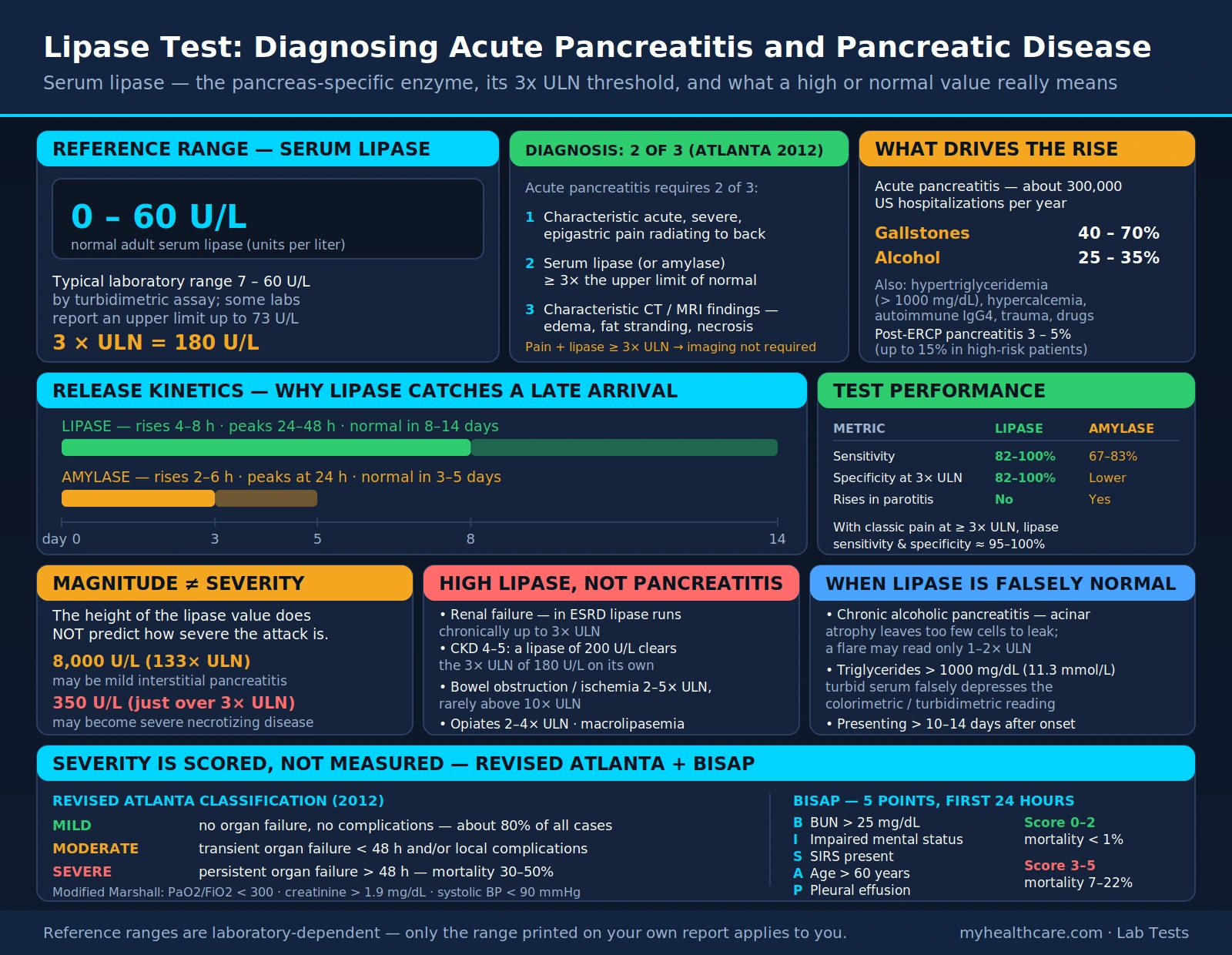
Serum lipase is the blood test that current guidelines prefer for diagnosing acute pancreatitis. Lipase breaks down dietary fats (triglycerides) in the small intestine. Most of the body's lipase is made in the pancreas, with smaller amounts made in the salivary glands and stomach, and its concentration in pancreatic tissue is about 100-fold higher than in tissues such as the duodenum, stomach, fat and lung. When the pancreas is inflamed or injured, lipase leaks into the bloodstream, where it can be measured. A level at least three times the upper limit of normal (ULN) is one of the three diagnostic criteria for acute pancreatitis. In a 2017 Cochrane review of patients seen in emergency departments with abdominal pain, serum lipase above three times normal had a pooled sensitivity of 0.79 and specificity of 0.89 — so roughly a quarter of people with acute pancreatitis were missed by the enzyme tests the review evaluated. The 2024 American College of Gastroenterology (ACG) guideline states that serum amylase alone cannot be used reliably for the diagnosis and that serum lipase is preferred.
Table of Contents
- Overview — What Is Serum Lipase?
- Biochemistry: How Lipase Works
- Reference Ranges and the 3× ULN Threshold
- Lipase vs. Amylase: How the Two Tests Compare
- Pancreatic Causes of Elevated Lipase
- Non-Pancreatic Causes of Elevated Lipase
- When Lipase Is Falsely Normal
- Pancreatitis Severity: Atlanta Classification and BISAP Score
- Clinical Use: What Happens After a High Lipase Result
- Key Research Papers
- Connections
- Featured Videos
Overview — What Is Serum Lipase?
Pancreatic triglyceride lipase and its protein cofactor, colipase, are required for efficient digestion of dietary triglycerides. Pancreatic acinar cells also make two related proteins, pancreatic lipase-related proteins 1 and 2 (PLRP1 and PLRP2), which closely resemble the main enzyme in sequence and structure (Lowe 2002).
In normal physiology, only small amounts of lipase circulate in the blood. Acute pancreatitis is thought to begin with the premature activation of digestive proenzymes inside the pancreas, which starts a local inflammatory response; how each cause (gallstones, alcohol and others) triggers that activation is not well understood (Basnayake and Ratnam 2015). According to the same review, serum lipase typically rises 3–6 hours after the onset of acute pancreatitis, usually peaks at 24 hours, and stays elevated for 8–14 days, which the review attributes to significant reabsorption of lipase in the kidney tubules. Amylase also rises within 3–6 hours but may stay elevated for only up to five days, and the 2024 ACG guideline states that amylase returns to normal within 3–5 days. This longer window is why lipase is described as more useful when a patient presents, or is tested, more than 24 hours after the pain began.
Like amylase, lipase reference ranges vary between laboratories, and there is ongoing debate about the best diagnostic cut-off (Basnayake and Ratnam 2015). Results are expressed against the reporting laboratory's own upper limit of normal.
Biochemistry: How Lipase Works
A 1993 review in Clinical Chemistry describes lipase as a glycoprotein of 420–449 amino acids, with a molecular mass of 46,000–56,000 for pancreatic lipase and 32,000–39,000 for serum lipase. The concentration gradient between pancreatic tissue and serum is about 20,000-fold. Catalysis depends on a serine that forms part of an Asp-His-Ser triad, and lipase differs from other esterases in having a hydrophobic recognition site (Tietz and Shuey 1993).
The Lipase-Colipase Interface
Pancreatic triglyceride lipase has an amino-terminal domain that contains the catalytic site and a carboxyl-terminal domain that carries the main colipase-binding site. In its inactive form, a surface loop called the lid covers the active site. When the enzyme is activated by bile salt micelles and colipase, or by a lipid-water interface, the lid moves to open and shape the active site, and lipase and colipase together form a large water-repelling surface that can attach to the fat droplet (Lowe 2002).
Additional Pancreatic Lipases
Of the two related proteins, PLRP1 has no known activity. The main enzyme and PLRP2 differ in which fats they act on, in how they behave in bile salts, and in how much they depend on colipase (Lowe 2002).
What the Laboratory Test Measures
Assays specific for lipase use a triglyceride substrate together with colipase, a secondary bile salt and calcium, and the 1993 review notes that lipolytic activity in a sample is not always due to pancreatic lipase (Tietz and Shuey 1993). One example: in a 1996 study of hemodialysis patients, measured lipase rose after heparin was given, and the authors traced this mainly to cross-reactivity with lipoprotein lipase and hepatic triglyceride lipase released by heparin (Shibasaki 1996).
The timeline above draws the figures from the 2015 review and the 2024 ACG guideline: a patient who reaches care after amylase's 3–5 day window may still have an elevated lipase, which stays raised for 8–14 days; past that, lipase may be back to normal as well.
Reference Ranges and the 3× ULN Threshold
There is no single universal normal range for serum lipase: reference ranges vary between laboratories, and there is debate about the optimal diagnostic cut-off (Basnayake and Ratnam 2015). The criteria below are expressed as multiples of the reporting laboratory's upper limit of normal. The 2024 ACG guideline adds that a Japanese consensus conference could not agree on appropriate upper limits of normal for amylase and lipase.
The 3× ULN Diagnostic Threshold for Acute Pancreatitis
The 2012 revised Atlanta classification (PubMed PMID: 23100216), as summarized in the 2024 ACG guideline and a 2015 review, establishes the diagnosis of acute pancreatitis when two of these three criteria are present:
- Abdominal pain consistent with the disease (acute onset of persistent, severe epigastric pain, often radiating to the back)
- Serum lipase (or amylase) at least three times the upper limit of normal
- Characteristic findings of acute pancreatitis on abdominal imaging
The 2013 ACG guideline states that because the diagnosis is most often made from symptoms and laboratory testing, contrast-enhanced CT and/or MRI of the pancreas are reserved for patients in whom the diagnosis is unclear or who fail to improve. The cut-off itself is debated: a 2012 review found that cut-offs between 2 and 4 times the upper limit of normal are preferable, without a single definitive threshold, and the 2024 ACG guideline notes that some patients without acute pancreatitis have lipase above three times normal, that a higher multiple (3–5 times) may be needed in some groups such as people with diabetes, and that without abdominal pain consistent with the disease, raised amylase and lipase do not predict acute pancreatitis.
Magnitude Does Not Equal Severity
The height of the lipase value does not predict how severe the pancreatitis will be. The 2024 ACG guideline states that once the diagnosis is established, there is no reason to follow amylase or lipase, because the levels bear no relationship to severity or prognosis and do not guide decisions on refeeding or discharge; a 2002 review likewise found that no enzyme assay predicts severity or cause, and a 2015 review notes that neither enzyme predicts severity in adults. Severity is assessed with clinical scoring systems (such as Ranson, BISAP, APACHE and Glasgow) and, when needed, imaging.
Lipase vs. Amylase: How the Two Tests Compare
For decades, amylase was the default first test for suspected pancreatitis. A 2015 review states that lipase has now replaced amylase as the biochemical test of choice, and the 2024 ACG guideline describes serum lipase as preferred.
Why Lipase Has Replaced Amylase
| Characteristic | Lipase | Amylase |
|---|---|---|
| Primary source | Mostly pancreas (tissue concentration about 100-fold higher than in other tissues); smaller amounts from salivary glands and stomach | Salivary glands and pancreas (separate isoforms); total amylase measured in most laboratories |
| Rises after onset | 3–6 hours; usually peaks at 24 hours | 3–6 hours |
| Stays elevated | 8–14 days | Up to about 5 days (normal within 3–5 days) |
| Sensitivity for acute pancreatitis | 0.79 pooled at >3× normal (Cochrane 2017); 85–100% in studies cited by a 2015 review | 0.72 pooled at >3× normal (Cochrane 2017); 81–95% in studies cited by a 2015 review, as low as 60% at a 2–4× cut-off |
| Specificity | 0.89 pooled at >3× normal (Cochrane 2017); higher than amylase in several studies, and may exceed 95% depending on the cut-off (2015 review) | 0.93 pooled at >3× normal (Cochrane 2017); the review could not formally compare the tests because data were sparse |
| Alcohol-related pancreatitis | Greater sensitivity than amylase | May be normal or less elevated |
| Salivary gland disease | Salivary disease is not listed among causes of raised lipase in the 2015 review's table (mumps is listed) | Raised by salivary gland disease |
When Amylase Still Has a Role
Amylase is still measured in many settings, and the reviews describe situations in which an isolated amylase rise has non-pancreatic explanations:
- Macroamylasemia: The 2024 ACG guideline describes macroamylasemia as a syndrome in which amylase forms large complexes with abnormal immunoglobulins, raising serum amylase without acute pancreatitis; a 2015 review describes it as uncommon and caused by reduced clearance of amylase.
- Salivary gland and other conditions: The 2015 review lists salivary disease, renal failure, ketoacidosis, pneumonia, cerebral trauma, burns, anorexia nervosa and bulimia among the extra-abdominal causes of raised amylase.
On ordering both tests together, a 2015 review states that most guidelines do not advocate it because the gain in sensitivity over a single test is only marginal and not cost-effective; a 2002 review found that measuring both does not improve accuracy, and a 2017 review concluded that co-ordering adds little to no diagnostic sensitivity or specificity, so testing lipase alone can reduce unnecessary testing.
Pancreatic Causes of Elevated Lipase
Acute Pancreatitis
The classic indication for lipase testing. The 2024 ACG guideline reports almost 300,000 admissions for acute pancreatitis each year in the United States and lists the two most common causes:
- Gallstones (40–70% of cases): A 2013 review identifies gallstones as the most common cause of acute pancreatitis and reports that early cholecystectomy (gallbladder removal) eliminates the risk of future attacks.
- Alcohol (25–35% of cases): The 2024 ACG guideline states that alcohol-induced pancreatitis is not considered unless a person has had moderate or heavy alcohol consumption for over 5 years, and a 2006 review describes alcohol-induced acute pancreatitis as an acute toxic insult on a pancreas already damaged by long-standing alcohol injury. Lipase has greater sensitivity than amylase in alcohol-related pancreatitis.
Among other causes, the 2024 ACG guideline states that medications, infectious agents, and metabolic causes such as hypercalcemia and hypertriglyceridemia are rare and more often falsely attributed. Some drugs, such as 6-mercaptopurine, azathioprine and didanosine, clearly can cause acute pancreatitis, but data are limited for most medications. Hypertriglyceridemia accounts for about 5% of cases, and triglycerides are considered the cause when they rise above 1,000 mg/dL. Anatomic variants such as pancreas divisum occur in 10–15% of the population, and it remains unclear whether they cause acute pancreatitis. ERCP is a recognized cause (see below).
Chronic Pancreatitis (Acute Exacerbation)
A 2015 review states that in an acute exacerbation of chronic pancreatitis, neither amylase nor lipase may be elevated. MedlinePlus notes that low lipase levels may reflect permanent damage to the lipase-making cells of the pancreas, as can occur in chronic pancreatitis and cystic fibrosis.
Pancreatic Cancer
MedlinePlus lists pancreatic cancer and blocked pancreatic ducts among the causes of higher-than-normal lipase, and a 2015 review lists malignancy (especially of the oesophagus, stomach, duodenum and pancreas) among the causes of raised lipase. The 2024 ACG guideline reports that 5–14% of patients with benign or malignant pancreatobiliary tumors present with apparently idiopathic acute pancreatitis, that about 1% of acute pancreatitis was due to pancreatic cancer in a recent review, and that in patients older than 40 in whom no cause is established, a pancreatic tumor is to be considered as a possible cause.
Post-ERCP Pancreatitis
The 2024 ACG guideline describes acute pancreatitis as the most common complication of endoscopic retrograde cholangiopancreatography (ERCP), with an incidence that varies widely, from 1% to 30%, depending on the patient, the procedures performed and whether prophylaxis was given. In a 2012 randomized trial of 602 patients at elevated risk, post-ERCP pancreatitis (defined as new upper abdominal pain, pancreatic enzymes at least three times the upper limit of normal 24 hours after the procedure, and hospitalization for at least 2 nights) developed in 9.2% of patients given a single dose of rectal indomethacin immediately after ERCP versus 16.9% given placebo (PubMed PMID: 22494121).
Non-Pancreatic Causes of Elevated Lipase
Lipase is not specific to the pancreas. A 2002 review notes that although lipase was once considered specific for acute pancreatitis, non-specific elevations have been reported in almost as many disorders as for amylase, and a 2015 review of intensive-care patients states that neither amylase nor lipase is specific for pancreatitis.
Renal Failure
Reviews list renal insufficiency among the causes of raised lipase (Matull 2006; Basnayake and Ratnam 2015). In a 2025 retrospective study of 251 people with chronic kidney disease, median serum lipase rose with CKD stage, from 40.50 U/L in stages G1–2 to 100.00 U/L in stage G4 and 155.50 U/L in stage G5, and correlated with serum creatinine and urea. In hemodialysis patients, measured lipase can also rise after heparin through cross-reactivity with other lipases (Shibasaki 1996). The 2024 ACG guideline notes that abdominal imaging may help confirm the diagnosis when doubt exists.
Other Abdominal and Extra-Abdominal Conditions
The table in the 2015 review lists these causes of raised serum lipase:
- Abdominal conditions: acute pancreatitis, pancreatic trauma, perforated viscus, intestinal infarction and obstruction, peritonitis, acute cholecystitis, appendicitis, hepatitis, abdominal aortic aneurysm, and malignancy.
- Extra-abdominal conditions: renal failure, ketoacidosis, fat embolism and bony fractures.
- Others: mumps and hyperlipoproteinaemia.
MedlinePlus additionally lists peptic ulcer, gallbladder disease, blocked intestine, diabetes, salivary gland disorders and alcohol use disorder among the causes of higher-than-normal lipase.
Macrolipasemia
Macrolipasemia is reported in case reports. In a 2000 case report, a woman with lupus in remission had an unexplained increase in serum amylase and lipase, and immunoprecipitation showed lipase bound to an IgA1-kappa immunoglobulin; the authors described it as the fourth reported case of simultaneous macroamylasemia and macrolipasemia, and wrote that macroenzymes are a possibility to consider when raised enzymes come with atypical clinical features.
Medications
The 2015 review lists these drugs as raising serum lipase: adrenocorticotropic hormone, tetracycline, oestrogens, frusemide (furosemide), valproate, thiazides, rifampicin, metronidazole, zalcitabine, opioids, methylprednisolone and indomethacin. It marks most of them as drugs that can cause acute pancreatitis but can also raise pancreatic enzymes without pancreatitis. A 2006 review likewise notes that frusemide can increase serum lipase, and MedlinePlus lists codeine, diuretics, certain cholesterol medicines and birth control pills among medicines that can affect lipase levels. Separately, the 2024 ACG guideline states that 6-mercaptopurine, azathioprine and didanosine clearly can cause acute pancreatitis, while data are limited for most medications.
When Lipase Is Falsely Normal
A normal lipase does not rule out acute pancreatitis in every case. In the 2017 Cochrane review, about a quarter of people with acute pancreatitis were not identified by the enzyme tests evaluated, and the tests performed worse as time passed from symptom onset; the reviewers concluded that clinicians need a low threshold to admit and treat people whose symptoms suggest acute pancreatitis, even when these tests are normal. A 2010 case series described acute pancreatitis with normal serum lipase, which the authors called extremely rare, and concluded that the diagnosis remains possible even with normal amylase and lipase.
Chronic Pancreatitis with Loss of Acinar Tissue
In an acute exacerbation of chronic pancreatitis, a 2015 review notes that neither enzyme may be elevated. A 2006 review states that because pancreatic lipase activity is more than four times that of amylase, lipase is less likely than amylase to be affected by chronic pancreatic insufficiency.
Hypertriglyceridemia-Induced Pancreatitis
Here it is amylase, not lipase, that is most affected. A 2015 review states that in pancreatitis due to hypertriglyceridaemia, serum amylase can be normal in up to 50% of cases, because of assay interference from a circulating inhibitor or the hyperlipidaemia itself, and the 2024 ACG guideline notes that amylase may be normal in hypertriglyceridemia. A 2006 review states that hypertriglyceridaemia does not interfere with the laboratory measurement of lipase. In a 1995 four-hospital review of 70 patients with hyperlipidemic pancreatitis, amylase was raised to two times normal in 54% and lipase in 67%. The 2024 ACG guideline states that triglycerides are considered the cause of acute pancreatitis when they rise above 1,000 mg/dL.
Delayed Presentation
Lipase stays elevated for 8–14 days (Basnayake and Ratnam 2015), so a patient who reaches care after that window may already have a normal level; amylase returns to normal sooner, within 3–5 days. The 2024 ACG guideline notes that abdominal imaging may assist when doubt about the diagnosis exists.
Pancreatitis Severity: Atlanta Classification and BISAP Score
Because lipase magnitude does not predict severity, standardized classification and scoring systems are used for patients with confirmed acute pancreatitis.
Revised Atlanta Classification (2012)
The 2012 revised Atlanta classification (Banks et al., PubMed PMID: 23100216), an international web-based consensus, identifies early and late phases of the disease and three severity categories:
- Mild acute pancreatitis: The most common form. No organ failure and no local or systemic complications; usually resolves in the first week.
- Moderately severe acute pancreatitis: Transient organ failure, local complications, or exacerbation of co-morbid disease. The 2024 ACG guideline defines transient organ failure as organ failure that resolves within 48 hours.
- Severe acute pancreatitis: Persistent organ failure, that is, organ failure lasting more than 48 hours. The 2024 ACG guideline describes organ failure in clinical terms as shock (systolic blood pressure below 90 mm Hg), pulmonary insufficiency (PaO2 below 60 mm Hg), renal failure (creatinine above 2 mg/dL after rehydration) and/or gastrointestinal bleeding (more than 500 mL in 24 hours), or a modified Marshall score of 2 or more in the three accepted organ systems.
The classification lists the local complications as peripancreatic fluid collections, pancreatic and peripancreatic necrosis (sterile or infected), pseudocyst and walled-off necrosis (sterile or infected). The 2024 ACG guideline states that almost one-fifth of patients go on to have complications such as pancreatic necrosis and/or organ failure.
BISAP Score
The Bedside Index for Severity in Acute Pancreatitis (BISAP) was derived from 17,992 cases at 212 hospitals and validated on 18,256 cases at 177 hospitals (Wu et al. 2008, PubMed PMID: 18519429). One point is assigned for each of the following during the first 24 hours:
- Blood urea nitrogen (BUN) >25 mg/dL
- Impaired mental status
- Systemic inflammatory response syndrome (SIRS), which the 2024 ACG guideline defines using pulse >90 per minute, respirations >20 per minute or PaCO2 <32 mm Hg, temperature above 38 °C, and a white-cell count >12,000 or <4,000 cells/mm3 or >10% immature neutrophils
- Age >60 years
- Pleural effusion
In the derivation study, in-hospital mortality ranged from more than 20% in the highest-risk group to less than 1% in the lowest-risk group, and in the validation cohort BISAP predicted mortality about as well as APACHE II (area under the curve 0.82 versus 0.83). A 2012 head-to-head comparison found that existing scoring systems had only modest accuracy for predicting persistent organ failure (areas under the curve 0.62–0.84 at admission in the training cohort and 0.57–0.74 in the validation cohort), with the Glasgow score the best classifier at admission. The 2024 ACG guideline states that BISAP has consistently been shown to be superior to other scores but may be no more accurate than simply monitoring patients for a rising BUN and/or the development of SIRS.
CT Severity Index (Balthazar Score)
A 1990 study of 88 patients developed a CT severity index combining peripancreatic inflammation, phlegmon and the degree of pancreatic necrosis seen on the initial CT. Patients with a high index had 92% morbidity and 17% mortality, while those with a low index had 2% morbidity and none died; patients with necrosis had 23% mortality. The 2024 ACG guideline states that patients with infected necrosis receive antibiotics known to penetrate pancreatic necrosis, largely to delay drainage beyond 4 weeks, that minimally invasive methods are preferred to open surgery for debridement in stable patients with symptomatic necrosis, and it suggests against prophylactic antibiotics in severe acute pancreatitis.
Clinical Use: What Happens After a High Lipase Result
Understanding what comes next helps patients make sense of their diagnosis and the tests and treatments they will encounter. The points below are what published guidelines and reviews describe; decisions about care belong with a clinician.
Initial Workup When Lipase Is ≥3× ULN
When acute pancreatitis is confirmed or strongly suspected, guidelines and reviews describe these steps:
- Abdominal ultrasound: The 2024 ACG guideline calls for abdominal ultrasound in all patients with acute pancreatitis to look for gallstones, with a repeat ultrasound if the first is inconclusive.
- Liver tests: A 2006 review states that a serum alanine transaminase (ALT) above 150 IU/l together with jaundice suggests a gallstone cause.
- Calcium and triglycerides: The same review lists serum calcium and triglycerides for unclear causes, and the 2024 ACG guideline lists triglycerides when there are no gallstones and no significant history of alcohol use, considered the cause when above 1,000 mg/dL.
- Markers of severity: The 2024 ACG guideline lists elevated BUN (≥20 mg/dL), hematocrit (≥44), creatinine (≥2 mg/dL), the presence of obesity, comorbidities and SIRS among risk factors for severe disease.
- CT or MRI: The ACG guidelines reserve these for patients whose diagnosis is unclear or who fail to improve after 48–72 hours, when they are used to assess local complications such as necrosis.
Treatment Principles for Acute Pancreatitis
Management described in the guidelines is largely supportive:
- Intravenous fluids: The 2024 ACG guideline suggests moderately aggressive fluid resuscitation and lactated Ringer solution over normal saline (both conditional recommendations, low quality of evidence), with extra boluses when there is evidence of low blood volume. It describes an initial rate of no more than 1.5 mL/kg per hour in patients without hypovolemia and a 10 mL/kg bolus in patients with hypovolemia. The 2013 guideline states that early intravenous hydration is most beneficial within the first 12–24 hours.
- Feeding: In mild acute pancreatitis, the 2024 ACG guideline suggests early oral feeding (within 24–48 hours) as tolerated, rather than nothing by mouth, starting with a low-fat solid diet rather than a stepwise liquid-to-solid approach. In moderately severe or severe disease, enteral nutrition appears to prevent infectious complications; parenteral nutrition is avoided unless the enteral route is not possible, and nasogastric feeding is preferred to nasojejunal because of comparable safety and efficacy.
- Gallstone pancreatitis: The 2024 ACG guideline states that patients with mild acute biliary pancreatitis undergo cholecystectomy early, preferably before discharge, and the 2013 guideline calls for ERCP within 24 hours of admission in patients with concurrent acute cholangitis.
- Alcohol-induced pancreatitis: A 2013 review states that alcohol abstinence and smoking cessation can alter the progression of pancreatitis and reduce recurrence.
Key Research Papers
- Banks PA, Bollen TL, Dervenis C, et al.; Acute Pancreatitis Classification Working Group. Classification of acute pancreatitis — 2012: revision of the Atlanta classification and definitions by international consensus. Gut. 2013;62(1):102–111. — PubMed PMID: 23100216
- Tenner S, Baillie J, DeWitt J, Vege SS; American College of Gastroenterology. American College of Gastroenterology guideline: management of acute pancreatitis. Am J Gastroenterol. 2013;108(9):1400–1415. — PubMed PMID: 23896955
- Wu BU, Johannes RS, Sun X, Tabak Y, Conwell DL, Banks PA. The early prediction of mortality in acute pancreatitis: a large population-based study. Gut. 2008;57(12):1698–1703. — PubMed PMID: 18519429
- Lippi G, Valentino M, Cervellin G. Laboratory diagnosis of acute pancreatitis: in search of the Holy Grail. Crit Rev Clin Lab Sci. 2012;49(1):18–31. — PubMed PMID: 22339380
- Mounzer R, Langmead CJ, Wu BU, et al. Comparison of existing clinical scoring systems to predict persistent organ failure in patients with acute pancreatitis. Gastroenterology. 2012;142(7):1476–1482. — PubMed PMID: 22425589
- Phillip V, Steiner JM, Algül H. Early phase of acute pancreatitis: assessment and management. World J Gastrointest Pathophysiol. 2014;5(3):158–168. — PubMed PMID: 25133018
- Yadav D, Lowenfels AB. The epidemiology of pancreatitis and pancreatic cancer. Gastroenterology. 2013;144(6):1252–1261. — PubMed PMID: 23622135
- Elmunzer BJ, Scheiman JM, Lehman GA, et al. A randomized trial of rectal indomethacin to prevent post-ERCP pancreatitis. N Engl J Med. 2012;366(15):1414–1422. — PubMed PMID: 22494121
- Petrov MS, Windsor JA. Classification of the severity of acute pancreatitis: how many categories make sense? Am J Gastroenterol. 2010;105(1):74–76. — PubMed PMID: 19844203
- Fortson MR, Freedman SN, Webster PD 3rd. Clinical assessment of hyperlipidemic pancreatitis. Am J Gastroenterol. 1995;90(12):2134–2139. — PubMed PMID: 8540502
- Smotkin J, Tenner S. Laboratory diagnostic tests in acute pancreatitis. J Clin Gastroenterol. 2002;34(4):459–462. — PubMed PMID: 11907364
- Al-Bahrani AZ, Ammori BJ. Clinical laboratory assessment of acute pancreatitis. Clin Chim Acta. 2005;362(1–2):26–48. — PubMed PMID: 16024009
- Tenner S, Vege SS, Sheth SG, Sauer B, Yang A, Conwell DL, Yadlapati RH, Gardner TB. American College of Gastroenterology Guidelines: Management of Acute Pancreatitis. Am J Gastroenterol. 2024;119(3):419–437. — PubMed PMID: 38857482
- Crockett SD, Wani S, Gardner TB, Falck-Ytter Y, Barkun AN; American Gastroenterological Association Institute Clinical Guidelines Committee. American Gastroenterological Association Institute Guideline on Initial Management of Acute Pancreatitis. Gastroenterology. 2018;154(4):1096–1101. (Cited by the 2024 ACG guideline among recent guidelines that emphasize the advantage of serum lipase.) — PubMed PMID: 29409760
- Rompianesi G, Hann A, Komolafe O, Pereira SP, Davidson BR, Gurusamy KS. Serum amylase and lipase and urinary trypsinogen and amylase for diagnosis of acute pancreatitis. Cochrane Database Syst Rev. 2017;4:CD012010. — PubMed PMID: 28431198
- Yadav D, Agarwal N, Pitchumoni CS. A critical evaluation of laboratory tests in acute pancreatitis. Am J Gastroenterol. 2002;97(6):1309–1318. — PubMed PMID: 12094843
- Ismail OZ, Bhayana V. Lipase or amylase for the diagnosis of acute pancreatitis? Clin Biochem. 2017;50(18):1275–1280. — PubMed PMID: 28720341
- Basnayake C, Ratnam D. Blood tests for acute pancreatitis. Aust Prescr. 2015;38(4):128–130. — PubMed PMID: 26648641
- Matull WR, Pereira SP, O'Donohue JW. Biochemical markers of acute pancreatitis. J Clin Pathol. 2006;59(4):340–344. — PubMed PMID: 16567468
- Muniraj T, Dang S, Pitchumoni CS. Pancreatitis or not? Elevated lipase and amylase in ICU patients. J Crit Care. 2015;30(6):1370–1375. — PubMed PMID: 26411523
- Shah AM, Eddi R, Kothari ST, Maksoud C, DiGiacomo WS, Baddoura W. Acute pancreatitis with normal serum lipase: a case series. JOP. 2010;11(4):369–372. — PubMed PMID: 20601812
- Balthazar EJ, Robinson DL, Megibow AJ, Ranson JH. Acute pancreatitis: value of CT in establishing prognosis. Radiology. 1990;174(2):331–336. — PubMed PMID: 2296641
- Zhang G, Li Z, Xu CX. Association between serum lipase levels and chronic kidney disease stages. PLoS One. 2025;20(8):e0330329. — PubMed PMID: 40824906
- Shibasaki T, Matsuda H, Ohno I, et al. Significance of serum lipase in patients undergoing hemodialysis. Am J Nephrol. 1996;16(4):309–314. — PubMed PMID: 8739284
- Goto H, Wakui H, Komatsuda A, Imai H, Miura AB, Fujita K. Simultaneous macroamylasemia and macrolipasemia in a patient with systemic lupus erythematosus in remission. Intern Med. 2000;39(12):1115–1118. — PubMed PMID: 11197804
- Lowe ME. The triglyceride lipases of the pancreas. J Lipid Res. 2002;43(12):2007–2016. — PubMed PMID: 12454260
- Tietz NW, Shuey DF. Lipase in serum — the elusive enzyme: an overview. Clin Chem. 1993;39(5):746–756. — PubMed PMID: 8485865
Also consulted: MedlinePlus (U.S. National Library of Medicine), Lipase Tests.
PubMed Topic Searches
Connections
- All Lab Tests
- Comprehensive Metabolic Panel
- Amylase and Lipase — the plain-language overview of both pancreatic enzymes, and why lipase is now preferred over amylase.
- Liver Function Tests
- Gamma-Glutamyl Transferase (GGT)
- Kidney Function Tests
- Inflammatory Markers Panel
- Lipid Panel
- Iron Panel
- Pancreatitis — the disease this test diagnoses: lipase ≥3× ULN plus epigastric pain meets the Atlanta criteria.