LDH Test: Lactate Dehydrogenase — Tissue Damage and Disease Activity Marker
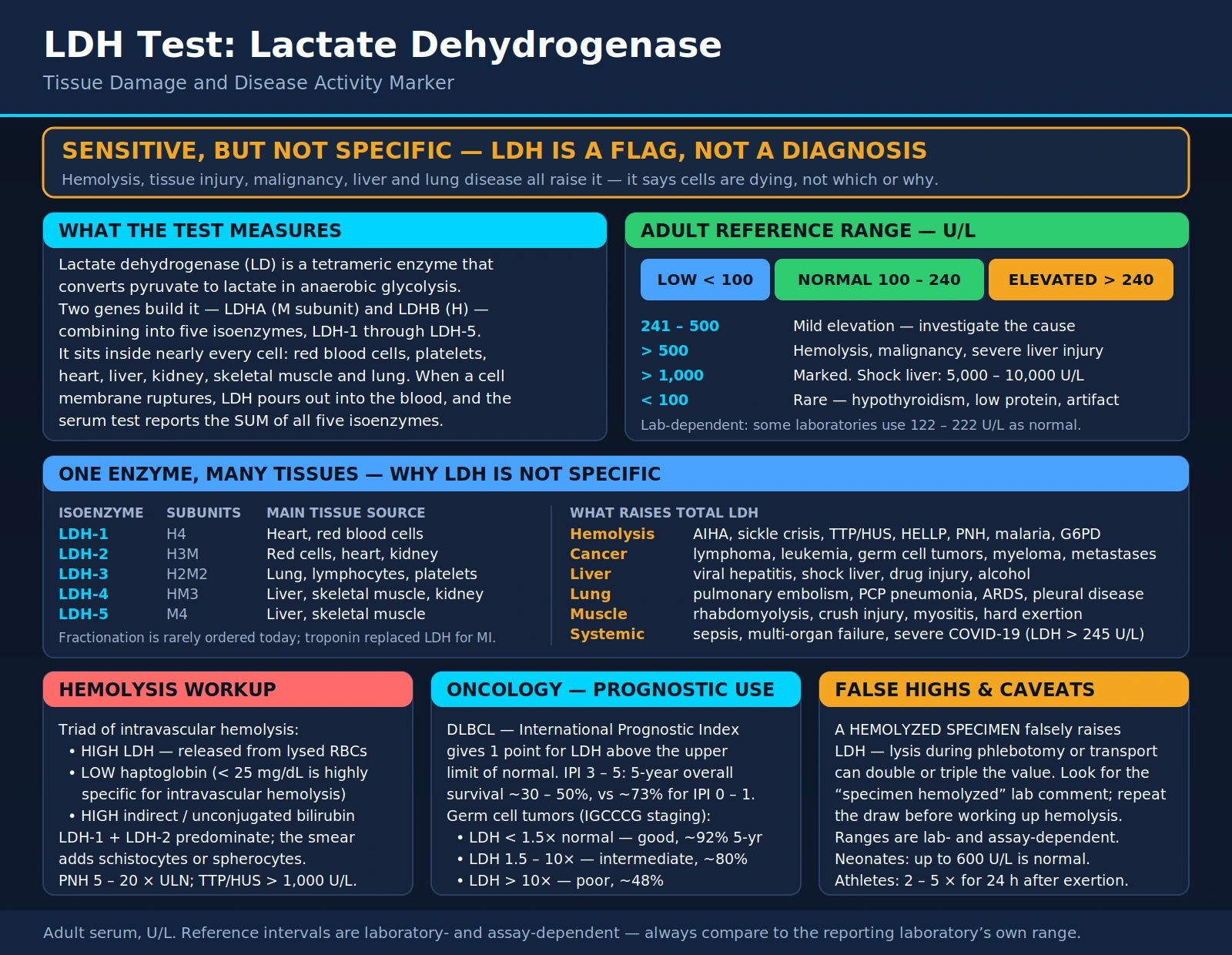
Lactate dehydrogenase (LDH) is an enzyme found in nearly every cell of the human body, where it catalyzes the reversible conversion of pyruvate to lactate — the final step of anaerobic glycolysis. When cells are damaged or die, LDH leaks out of them and into the bloodstream, so a raised serum LDH is a sensitive but non-specific sign of tissue injury: it shows that cells somewhere are breaking down, not which ones or why. The test is used to follow hemolysis, to stage and follow lymphoma, germ-cell tumours and several other cancers, to classify pleural effusions, and as one of many severity markers in critical illness, including COVID-19. Its old role in diagnosing heart attacks has been taken over by cardiac troponin. Which LDH isoenzyme is raised, read together with companion tests, narrows down the source.
Table of Contents
- Overview — What LDH Measures
- LDH Isoenzymes
- Reference Ranges
- Causes of Elevated LDH
- LDH in Hemolysis and Megaloblastic Anaemia
- LDH in Cancer and Lymphoma
- LDH in Lung Disease: Pulmonary Embolism, Pleural Fluid and Pneumocystis
- LDH in Critical Illness and COVID-19
- Key Research Papers
- Connections
- Featured Videos
Overview — What LDH Measures
LDH (also written LD) is a four-subunit enzyme built from two kinds of subunit — M, encoded by the LDHA gene, and H, encoded by LDHB — which combine in different ratios to form five isoenzymes, LDH-1 through LDH-5. The enzyme interconverts pyruvate and lactate, reducing NAD+ to NADH or the reverse, a step that lets cells keep producing energy when oxygen is short, as in intense exercise or hypoxia.
When cell membranes are disrupted by injury, ischemia, inflammation or cell death, LDH passes into the extracellular fluid and the bloodstream. The total serum LDH test measures the sum of all five isoenzymes. Because LDH is present in red blood cells, white blood cells, heart, liver, kidney, skeletal muscle and lung, many kinds of significant cell injury raise the total. That breadth makes LDH useful for showing that something is wrong; isoenzyme testing or the clinical context is needed to show what. MedlinePlus notes that an LDH test alone cannot show what is damaging tissue or where the damage is, so it is usually done alongside other tests.
The main uses today are following hemolytic anaemia, staging and following lymphoma and germ-cell tumours, classifying pleural effusions as exudates or transudates, and, more loosely, as a severity marker in sepsis, organ failure and COVID-19.
LDH Isoenzymes (LDH-1 through LDH-5)
The five isoenzymes differ in their mix of H (heart-type) and M (muscle-type) subunits, and their tissue distributions differ, which is what lets an isoenzyme test point toward the source of cell injury. The tissue sources below are those listed by MedlinePlus (U.S. National Library of Medicine):
- LDH-1 (four H subunits) — heart muscle and red blood cells.
- LDH-2 (three H, one M) — concentrated in white blood cells. In healthy serum LDH-2 normally exceeds LDH-1.
- LDH-3 (two H, two M) — highest in the lung.
- LDH-4 (one H, three M) — highest in the kidney, placenta and pancreas.
- LDH-5 (four M subunits) — highest in the liver and skeletal muscle. Liver congestion raises LDH-5, and a study of isoenzyme profiles after heart attack found that LDH-5 rises could produce false-negative results for one LDH-1 measure (percent LDH-1 of total).
The LDH-1/LDH-2 “flip” and myocardial infarction (historical): a pattern in which LDH-1 rises above LDH-2, reversing the usual order, was once used as a late laboratory sign of acute myocardial infarction. A 1992 clinical review described a raised CK-MB together with a “flipped” LDH isoenzyme pattern as reliable confirmation of a heart attack. That role has ended. The 2000 joint ESC/ACC redefinition of myocardial infarction built its biochemical criterion on troponin (or CK-MB), with no place for LDH, and the 2018 Fourth Universal Definition of Myocardial Infarction rests on cardiac troponin. Isoenzyme testing is now ordered mainly when the source of a raised total LDH is unclear. The cardiac troponin and CK-MB tests are covered on their own pages in the Lab Tests section.
Isoenzymes in red-cell disorders: red blood cells carry mainly LDH-1 and LDH-2; isoenzyme testing in a sickle cell cohort confirmed that these two predominated. A reversed pattern (LDH-1 above LDH-2) is also described in megaloblastic anaemia, where one study found it helped separate megaloblastic from hemolytic anaemia (see the next sections). By contrast, LDH-5 points toward liver or skeletal muscle.
Reference Ranges
LDH — Adults, MedlinePlus example range (IU/L)
The MedlinePlus Medical Encyclopedia gives a normal range of 125 to 220 international units per liter (IU/L) as an example. Reference ranges vary between laboratories and assay methods, so a result is read against the range printed on the reporting laboratory's own report.
What MedlinePlus and the studies cited on this page report about results:
- Higher than normal: usually reflects some type of tissue damage from disease, infection or injury, but a high value does not always mean a condition that needs treatment. Intense exercise and certain medicines, including aspirin, can raise LDH, and so can red blood cells breaking open while the sample is collected and processed.
- Lower than normal: uncommon and usually not considered a health problem. MedlinePlus lists large amounts of vitamin C or vitamin E, and a rare inherited lactate dehydrogenase deficiency, as causes.
- Very high values: in a 2000 study of 75 people with macrocytic anaemia, a total LDH above 3,000 IU/L was diagnostic of megaloblastic anaemia in that series. In acute liver injury, LDH rises far more relative to ALT in ischemic hepatitis and acetaminophen injury than in viral hepatitis (see the Causes section).
Pregnancy: HELLP syndrome (Hemolysis, Elevated Liver enzymes, Low Platelets) is a pregnancy complication in which a raised LDH is one of the laboratory findings.
Causes of Elevated LDH
Because LDH is present in nearly every tissue, the list of conditions that raise it is long. Interpretation rests on grouping a raised LDH with companion findings that narrow the source.
Hemolytic conditions:
- Autoimmune hemolytic anaemia (warm and cold antibody types)
- Sickle cell disease
- Mechanical hemolysis (prosthetic heart valves, ventricular assist devices)
- Microangiopathic hemolytic anaemia: thrombotic thrombocytopenic purpura (TTP), hemolytic uremic syndrome (HUS), HELLP syndrome
- Paroxysmal nocturnal hemoglobinuria (PNH)
- Transfusion reactions
- Malaria
- G6PD deficiency during oxidative stress
Red blood cells carry mainly LDH-1 and LDH-2, so these isoenzymes predominate when red cells break down.
Megaloblastic anaemia: vitamin B12 or folate deficiency causes ineffective red-cell production in the bone marrow, and the destruction of precursor cells there raises LDH, often strikingly (see the Hemolysis and Megaloblastic Anaemia section).
Cancer and hematologic malignancy:
- Non-Hodgkin lymphoma — LDH is one of the five factors in the International Prognostic Index (IPI) for aggressive lymphoma
- Hodgkin lymphoma
- Acute leukaemia (ALL, AML)
- Germ cell tumours (testicular cancer) — LDH is one of three serum markers, with AFP and hCG, in the International Germ Cell Consensus Classification
- Multiple myeloma — LDH is part of the Revised International Staging System
- Metastatic solid tumours, including melanoma and renal cell carcinoma
Myocardial injury (historical role):
- LDH and the LDH-1/LDH-2 ratio were once used as late markers of myocardial infarction.
- The 2000 ESC/ACC redefinition and the 2018 Fourth Universal Definition of Myocardial Infarction base the diagnosis on cardiac troponin; LDH is not part of either definition.
Liver disease:
- Acute viral hepatitis
- Ischemic hepatitis (“shock liver”) and acetaminophen injury — in a 1994 study (51 viral hepatitis, 20 ischemic hepatitis, 26 acetaminophen cases), LDH was higher for a given ALT and AST in ischemic hepatitis and acetaminophen injury than in viral hepatitis. The mean ALT/LDH ratio was 4.65 in viral hepatitis, 0.87 in ischemic hepatitis and 1.46 in acetaminophen injury; a ratio of 1.5 separated viral hepatitis from the other two with 94% sensitivity and 84% specificity.
- Drug-induced liver injury
- Hepatic infiltration by lymphoma
- Alcoholic hepatitis
Pulmonary conditions:
- Pulmonary embolism
- Pneumocystis jirovecii pneumonia (PCP) — see the Lung Disease section
- ARDS (acute respiratory distress syndrome)
- Exudative pleural effusion (pleural fluid LDH, measured with serum LDH, is part of Light's criteria)
Skeletal muscle injury:
- Rhabdomyolysis — creatine kinase (CK) is the companion marker
- Trauma, crush injury, compartment syndrome
- Intense exertion
- Inflammatory myopathies: dermatomyositis, polymyositis
- Statin-associated myopathy
Renal conditions:
- Renal infarction
- Acute tubular necrosis (ATN)
Critical illness and systemic conditions:
- Sepsis and multi-organ dysfunction
- COVID-19 — meta-analyses link a raised LDH with severe disease and death (see the Critical Illness section)
Falsely high LDH from a hemolyzed sample: red cells that break open during blood drawing, transport or processing release their own LDH into the tube, raising the measured value without any disease. In a 2006 laboratory study that lysed blood cells in samples from 12 people on purpose, the false rise in LDH grew roughly in step with the amount of lysate, and clinically meaningful changes in LDH appeared even in samples whose hemolysis was mild or almost invisible to the eye (free hemoglobin below 0.6 g/L). The authors found the response too variable to correct mathematically; for in-vitro hemolysis they suggested measuring free hemoglobin, alerting the clinician and collecting a new sample. A laboratory comment such as “specimen hemolyzed” flags this. MedlinePlus likewise notes that a high LDH can result from red cells breaking open when the sample was collected and tested.
LDH in Hemolysis and Megaloblastic Anaemia
Detecting and following hemolysis is one of the main uses of LDH. When red blood cells break down in the body, they release LDH, mainly LDH-1 and LDH-2. A raised LDH, a raised indirect (unconjugated) bilirubin and a low haptoglobin together form the classic laboratory picture of hemolysis.
Each part of this picture reflects a different consequence of red-cell breakdown:
- LDH rises because the enzyme is released from the broken cells into the circulation.
- Haptoglobin falls because it binds free hemoglobin and the complex is cleared from the blood. In a 1980 study of 100 hospital patients, a haptoglobin of 25 mg/dL or less separated hemolytic from non-hemolytic disorders with 83% sensitivity and 96% specificity, an 87% probability of hemolytic disease when the level fell below that limit. Haptoglobin has its own page in the Lab Tests section.
- Indirect bilirubin rises because the heme from broken red cells is converted to unconjugated bilirubin.
The peripheral blood smear adds morphological evidence: schistocytes (fragmented red cells) in microangiopathic hemolytic anaemia (TTP, HUS, HELLP), spherocytes in autoimmune hemolytic anaemia, and sickled cells in sickle cell disease.
Findings by condition:
- Sickle cell disease: in a 2006 study of 213 patients, steady-state LDH tracked other markers of hemolysis (lower hemoglobin and haptoglobin, higher reticulocytes, bilirubin and plasma hemoglobin), and isoenzyme testing showed LDH-1 and LDH-2 predominating. A higher LDH was associated with pulmonary hypertension, leg ulcers, priapism and risk of death; the authors proposed it as a marker of a hemolysis-related vascular syndrome.
- TTP/HUS: a raised LDH alongside a low platelet count and schistocytes on the smear is part of the picture of microangiopathic hemolysis, and LDH is followed during treatment.
- PNH: LDH is the standard measure of intravascular hemolysis in PNH trials. In the 2006 randomized phase 3 trial of the complement inhibitor eculizumab (87 patients, 26 weeks), the median area under the LDH-over-time curve was 85.8% lower with eculizumab than with placebo.
- Autoimmune hemolytic anaemia (AIHA): LDH is one of the markers used, with haptoglobin and bilirubin, to gauge hemolysis.
Megaloblastic anaemia: in vitamin B12 or folate deficiency, DNA synthesis in developing blood cells is impaired. A 2026 review describes how the resulting ineffective erythropoiesis destroys red-cell precursors inside the bone marrow, raising LDH and indirect bilirubin and lowering haptoglobin, so the picture can mimic a hemolytic anaemia, while the reticulocyte count stays inappropriately low for the degree of anaemia. In a 2000 study of 75 patients with macrocytic anaemia classified by bone marrow examination, a total LDH above 3,000 IU/L was diagnostic of megaloblastic anaemia in that series; for totals between 451 and 3,000 IU/L, a reversed isoenzyme pattern (LDH-1 above LDH-2) helped confirm megaloblastic anaemia and separate it from hemolytic anaemia.
LDH in Cancer and Lymphoma
In oncology LDH is used in two ways: as a factor in established staging and prognostic systems, and as a value followed over the course of treatment.
Aggressive non-Hodgkin lymphoma (International Prognostic Index): the 1993 International Prognostic Index was built from 2,031 patients treated with doxorubicin-containing chemotherapy. Its five factors are age, tumour stage, serum LDH, performance status and number of extranodal sites. The four risk groups had predicted five-year survival of 73%, 51%, 43% and 26%; an age-adjusted index for patients 60 or younger, based on stage, LDH and performance status, gave 83%, 69%, 46% and 32%. A 2014 enhanced index for diffuse large B-cell lymphoma in the rituximab era (NCCN-IPI, 1,650 patients) uses LDH normalized to the upper limit of normal and separated low- and high-risk groups more widely than the IPI (five-year overall survival 96% vs 33%, against 90% vs 54% for the IPI).
Germ cell tumours: the 1997 International Germ Cell Consensus Classification (data on 5,202 patients with non-seminomatous and 660 with seminomatous germ cell tumours) combines the degree of elevation of the three serum markers — alpha-fetoprotein (AFP), human chorionic gonadotropin (hCG) and LDH — with the primary site (mediastinal) and the presence of non-pulmonary visceral metastases in non-seminoma. It defined good, intermediate and poor prognosis groups with 5-year survival of 91%, 79% and 48% across all germ cell tumours (the poor group contained only non-seminomas). A 2021 update in 9,728 men with metastatic non-seminoma treated between 1990 and 2013 found 5-year overall survival of 96%, 89% and 67% in the three groups, and identified an LDH above 2.5 times the upper limit of normal, older age and lung metastases as additional adverse factors.
Multiple myeloma: the 2015 Revised International Staging System (R-ISS) adds chromosomal abnormalities and LDH to the original ISS. Stage I requires a normal LDH, and a high LDH is one of the features that places ISS stage III patients in R-ISS stage III.
Metastatic solid tumours:
- Melanoma: in the AJCC eighth edition (2017), LDH is recorded as a descriptor on each M1 subcategory and no longer upstages a patient to M1c; M1d is a new category for central nervous system metastases. A 2022 meta-analysis of 6,817 patients treated with immune checkpoint inhibitors (anti-CTLA-4 or anti-PD-1/PD-L1 agents) found that a high pretreatment LDH was associated with poorer overall and progression-free survival.
- Renal cell carcinoma: an LDH above 1.5 times the upper limit of normal was one of five adverse factors in the 1999 Memorial Sloan-Kettering (MSKCC) model. The 2009 International Metastatic RCC Database Consortium (IMDC) model kept four of those five factors — anaemia, high calcium, low performance status and less than one year from diagnosis to treatment — and added high neutrophil and platelet counts; LDH is not one of the IMDC factors.
Tumour lysis syndrome (TLS): when treatment kills a large mass of fast-growing cancer cells, their contents flood the blood. An international expert panel defines TLS by its metabolic abnormalities — high uric acid, high phosphate, high potassium and low calcium — which can lead to kidney failure, arrhythmias and seizures. The 2008 evidence-based guidelines describe prevention with hydration and rasburicase in high-risk patients and hydration plus allopurinol or rasburicase at intermediate risk.
LDH in Lung Disease: Pulmonary Embolism, Pleural Fluid and Pneumocystis
Pulmonary embolism: LDH can be raised in pulmonary embolism, but it is not part of current diagnosis or risk assessment. The 2019 ESC guideline on acute pulmonary embolism builds its early risk classification from hemodynamic status, the Pulmonary Embolism Severity Index (PESI or sPESI), right-ventricular dysfunction on imaging and cardiac biomarkers (troponin, with natriuretic peptides as additional markers); LDH is not among them. BNP/NT-proBNP, troponin and D-dimer have their own pages in the Lab Tests section.
Pleural effusion (Light's criteria): LDH measured in pleural fluid, together with serum LDH and protein, is used to classify effusions as transudates or exudates. Under Light's criteria (1972), an effusion is classed as an exudate when any one of the following is present:
- Pleural fluid protein / serum protein > 0.5
- Pleural fluid LDH / serum LDH > 0.6
- Pleural fluid LDH > two-thirds of the upper limit of normal for serum LDH
A 2014 systematic review of 48 studies found that an exudate was unlikely when all of Light's criteria were absent (likelihood ratio 0.04), and that a pleural fluid LDH above 200 U/L (likelihood ratio 18), a pleural fluid cholesterol above 55 mg/dL, or a pleural-to-serum cholesterol ratio above 0.3 were among the most accurate single indicators of an exudate. Exudates arise from causes such as pneumonia, cancer, rheumatoid disease and empyema; transudates from fluid shifts in conditions such as heart failure, cirrhosis and nephrotic syndrome.
Pneumocystis jirovecii pneumonia (PCP): in a 1988 study of 84 HIV-infected patients with lung problems, mean serum LDH was 361 IU in the 54 with PCP versus 224 IU in those with other lung diseases. Individual values overlapped, but levels above 450 IU consistently predicted PCP in that population, and only 7% of PCP patients had a normal LDH. Mean LDH was higher in patients who died (447 IU) than in survivors (340 IU), though the overlap was too great to predict death in an individual. During treatment, 75% of survivors had gradually falling LDH while 75% of non-survivors had rising values. Diagnosis rests on finding the organism, typically in bronchoalveolar lavage fluid.
LDH in Critical Illness and COVID-19
In intensive care, a raised LDH is common and usually has several sources at once — liver ischemia from low blood pressure, hemolysis, muscle breakdown, lung injury and widespread inflammation. A trend over repeated measurements carries more information than a single value.
COVID-19: a 2020 pooled analysis of 9 studies (1,532 patients) found that an elevated LDH at the earliest point in hospital was associated with about a 6-fold increase in the odds of severe disease and about a 16-fold increase in the odds of death; the authors called for larger studies. A later meta-analysis of 21 studies (10,399 patients) found an elevated LDH in 44% of patients and linked it with a composite poor outcome (odds ratio 5.33) and with death (odds ratio 4.22); as a predictor, elevated LDH had a sensitivity of 0.74 and specificity of 0.69. Proposed sources of LDH in severe COVID-19 include direct lung injury and wider inflammatory damage to other organs.
Sepsis: LDH is not part of the 2016 Sepsis-3 definitions. Septic shock is identified by the need for vasopressors to keep mean blood pressure at 65 mm Hg or higher together with a serum lactate above 2 mmol/L (18 mg/dL) after fluid resuscitation; that group had a hospital mortality of 42.3% in the data the task force examined. Serum lactate and LDH are different tests: lactate measures the product of anaerobic metabolism, LDH the enzyme that interconverts lactate and pyruvate.
Ischemic hepatitis (“shock liver”): sudden loss of blood flow to the liver — from cardiac arrest, cardiogenic shock or severe low blood pressure — raises LDH steeply along with the aminotransferases. In the 1994 study described in the Causes section, the mean ALT/LDH ratio was 0.87 in ischemic hepatitis against 4.65 in acute viral hepatitis, so a low ALT/LDH ratio points toward ischemia or acetaminophen injury rather than viral hepatitis.
Key Research Papers
The following are peer-reviewed publications, guidelines and reference chapters behind the statements on this page.
- Wroblewski F, LaDue JS. Lactic dehydrogenase activity in blood. Proc Soc Exp Biol Med. 1955;90(1):210–213. — PubMed PMID: 13273400
- Farhana A, Lappin SL. Biochemistry, Lactate Dehydrogenase. In: StatPearls [Internet]. Treasure Island (FL): StatPearls Publishing; updated 2023 May 1. NCBI Bookshelf NBK557536 — PubMed PMID: 32491468
- The International Non-Hodgkin's Lymphoma Prognostic Factors Project. A predictive model for aggressive non-Hodgkin's lymphoma. N Engl J Med. 1993;329(14):987–994. — PubMed PMID: 8141877
- International Germ Cell Cancer Collaborative Group. International germ cell consensus classification: a prognostic factor-based staging system for metastatic germ cell cancers. J Clin Oncol. 1997;15(2):594–603. — PubMed PMID: 9053482
- Lippi G, Salvagno GL, Montagnana M, Brocco G, Guidi GC. Influence of hemolysis on routine clinical chemistry testing. Clin Chem Lab Med. 2006;44(3):311–316. — PubMed PMID: 16519604
- Henry BM, Aggarwal G, Wong J, Benoit S, Vikse J, Plebani M, Lippi G. Lactate dehydrogenase levels predict coronavirus disease 2019 (COVID-19) severity and mortality: A pooled analysis. Am J Emerg Med. 2020;38(9):1722–1726. — PubMed PMID: 32738466
- Thygesen K, Alpert JS, Jaffe AS, Chaitman BR, Bax JJ, Morrow DA, White HD; Executive Group on behalf of the Joint ESC/ACC/AHA/WHF Task Force for the Universal Definition of Myocardial Infarction. Fourth Universal Definition of Myocardial Infarction (2018). J Am Coll Cardiol. 2018;72(18):2231–2264. — PubMed PMID: 30153967
- Brodsky RA. Paroxysmal nocturnal hemoglobinuria. Blood. 2014;124(18):2804–2811. — PubMed PMID: 25237200
- Shankar-Hari M, et al. Developing a New Definition and Assessing New Clinical Criteria for Septic Shock: For the Third International Consensus Definitions for Sepsis and Septic Shock (Sepsis-3). JAMA. 2016;315(8):775–787. — PubMed PMID: 26903336
- Cassidy WM, Reynolds TB. Serum lactic dehydrogenase in the differential diagnosis of acute hepatocellular injury. J Clin Gastroenterol. 1994;19(2):118–121. — PubMed PMID: 7963356
- Konstantinides SV, et al. 2019 ESC Guidelines for the diagnosis and management of acute pulmonary embolism developed in collaboration with the European Respiratory Society (ERS). Eur Heart J. 2020;41(4):543–603. — PubMed PMID: 31504429
- Zaman MK, White DA. Serum lactate dehydrogenase levels and Pneumocystis carinii pneumonia. Diagnostic and prognostic significance. Am Rev Respir Dis. 1988;137(4):796–800. — PubMed PMID: 3258483
- Light RW, Macgregor MI, Luchsinger PC, Ball WC Jr. Pleural effusions: the diagnostic separation of transudates and exudates. Ann Intern Med. 1972;77(4):507–513. — PubMed PMID: 4642731
- Wilcox ME, Chong CA, Stanbrook MB, Tricco AC, Wong C, Straus SE. Does this patient have an exudative pleural effusion? The Rational Clinical Examination systematic review. JAMA. 2014;311(23):2422–2431. — PubMed PMID: 24938565
- Marchand A, Galen RS, Van Lente F. The predictive value of serum haptoglobin in hemolytic disease. JAMA. 1980;243(19):1909–1911. — PubMed PMID: 7365971
- Kato GJ, McGowan V, Machado RF, et al. Lactate dehydrogenase as a biomarker of hemolysis-associated nitric oxide resistance, priapism, leg ulceration, pulmonary hypertension, and death in patients with sickle cell disease. Blood. 2006;107(6):2279–2285. — PubMed PMID: 16291595
- Hillmen P, Young NS, Schubert J, et al. The complement inhibitor eculizumab in paroxysmal nocturnal hemoglobinuria. N Engl J Med. 2006;355(12):1233–1243. — PubMed PMID: 16990386
- Parakh N, Dewan P. Revisiting the hematological manifestations of vitamin B12 deficiency. Front Nutr. 2026;13:1898533. — PubMed PMID: 42657046
- Jaswal TS, Mehta HC, Gupta V, Singh M, Singh S. Serum lactate dehydrogenase in diagnosis of megaloblastic anaemia. Indian J Pathol Microbiol. 2000;43(3):325–329. — PubMed PMID: 11218680
- Ruzich RS. Cardiac enzymes. How to use serial determinations to confirm acute myocardial infarction. Postgrad Med. 1992;92(7):85–89, 92. — PubMed PMID: 1437917
- Levinson SS, Hobbs GA. Usefulness of various lactate dehydrogenase isoenzyme 1 profiles after myocardial infarction. Ann Clin Lab Sci. 1994;24(4):364–370. — PubMed PMID: 7944273
- Alpert JS, Thygesen K, Antman E, Bassand JP. Myocardial infarction redefined — a consensus document of The Joint European Society of Cardiology/American College of Cardiology Committee for the redefinition of myocardial infarction. J Am Coll Cardiol. 2000;36(3):959–969. — PubMed PMID: 10987628
- Zhou Z, Sehn LH, Rademaker AW, et al. An enhanced International Prognostic Index (NCCN-IPI) for patients with diffuse large B-cell lymphoma treated in the rituximab era. Blood. 2014;123(6):837–842. — PubMed PMID: 24264230
- Gillessen S, Sauvé N, Collette L, et al. Predicting Outcomes in Men With Metastatic Nonseminomatous Germ Cell Tumors (NSGCT): Results From the IGCCCG Update Consortium. J Clin Oncol. 2021;39(14):1563–1574. — PubMed PMID: 33822655
- Palumbo A, Avet-Loiseau H, Oliva S, et al. Revised International Staging System for Multiple Myeloma: A Report From International Myeloma Working Group. J Clin Oncol. 2015;33(26):2863–2869. — PubMed PMID: 26240224
- Gershenwald JE, Scolyer RA, Hess KR, et al. Melanoma staging: Evidence-based changes in the American Joint Committee on Cancer eighth edition cancer staging manual. CA Cancer J Clin. 2017;67(6):472–492. — PubMed PMID: 29028110
- Zhang Y, Liu B, Kotenko S, Li W. Prognostic value of neutrophil-lymphocyte ratio and lactate dehydrogenase in melanoma patients treated with immune checkpoint inhibitors: A systematic review and meta-analysis. Medicine (Baltimore). 2022;101(32):e29536. — PubMed PMID: 35960066
- Motzer RJ, Mazumdar M, Bacik J, Berg W, Amsterdam A, Ferrara J. Survival and prognostic stratification of 670 patients with advanced renal cell carcinoma. J Clin Oncol. 1999;17(8):2530–2540. — PubMed PMID: 10561319
- Heng DY, Xie W, Regan MM, et al. Prognostic factors for overall survival in patients with metastatic renal cell carcinoma treated with vascular endothelial growth factor-targeted agents: results from a large, multicenter study. J Clin Oncol. 2009;27(34):5794–5799. — PubMed PMID: 19826129
- Cairo MS, Coiffier B, Reiter A, Younes A; TLS Expert Panel. Recommendations for the evaluation of risk and prophylaxis of tumour lysis syndrome (TLS) in adults and children with malignant diseases: an expert TLS panel consensus. Br J Haematol. 2010;149(4):578–586. — PubMed PMID: 20331465
- Coiffier B, Altman A, Pui CH, Younes A, Cairo MS. Guidelines for the management of pediatric and adult tumor lysis syndrome: an evidence-based review. J Clin Oncol. 2008;26(16):2767–2778. — PubMed PMID: 18509186
- Martha JW, Wibowo A, Pranata R. Prognostic value of elevated lactate dehydrogenase in patients with COVID-19: a systematic review and meta-analysis. Postgrad Med J. 2022;98(1160):422–427. — PubMed PMID: 33452143
PubMed Topic Searches
External Authoritative Resources
- MedlinePlus — Laboratory Tests Reference
- MedlinePlus — Medical Tests (U.S. National Library of Medicine)
- MedlinePlus — Medical Tests (U.S. National Library of Medicine)
- MedlinePlus — Lactate Dehydrogenase (LDH) Test
- MedlinePlus Medical Encyclopedia — Lactate dehydrogenase test
- MedlinePlus Medical Encyclopedia — LDH isoenzyme blood test
- StatPearls — Pleural Effusion (NCBI Bookshelf NBK448189)