Black Ginger, Blood Sugar and Brown Fat: What a Small Apple Buys You
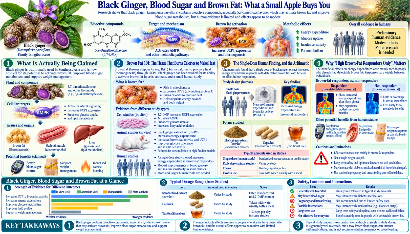
Of the three claims covered across this Benefits leg, the metabolic one has the most genuinely interesting human physiology behind it — a real positron-emission-tomography measurement of a real tissue doing a real, measurable thing in real people. It is also the claim where the honest arithmetic matters most, because the actual number, once converted out of a laboratory unit into something a reader can picture, is genuinely small.
Table of Contents
- What Is Actually Being Claimed
- Brown Fat 101: The Tissue That Burns Calories to Make Heat
- The Single-Dose Human Finding, and the Arithmetic
- Why "High Brown-Fat Responders Only" Matters
- The Blood-Glucose Signal: A Meta-Analysis of Small Trials
- How Black Ginger’s AMP-Activated Protein Kinase Story Compares With Metformin
- A Second Mechanism: Blocking New Fat-Cell Formation
- What a Trial That Actually Measured Weight Loss Would Look Like
- Pharmacokinetics: How Much of an Oral Dose Reaches Blood and Tissue
- Numbers This Page Refuses to Give
- Evidence Ledger for This Claim
- Practical Bottom Line
- Key Research Papers
- Connections
- Featured Videos
What Is Actually Being Claimed
The metabolic marketing around black ginger runs from a modest, real finding to a far less tested one across three claims:
- A single dose measurably increases whole-body energy expenditure via brown fat activation. True, measured directly in humans by positron emission tomography (PET) scan, and covered in detail below — along with exactly how small the effect is.
- It lowers fasting blood glucose. A real signal from a meta-analysis of small trials, with a human confidence interval that comes close to crossing zero.
- It produces meaningful weight loss. Three 12-week placebo-controlled trials reported less abdominal or body fat with black ginger extract or its purified polymethoxyflavones, and one of them also reported lower body weight; none ran for the 52 weeks or more used in obesity-drug trials, and the abstracts do not give the size of the weight change. The step from those short fat-loss results to lasting weight loss is the leap this page spends the most time examining.
Brown Fat 101: The Tissue That Burns Calories to Make Heat
Most body fat is white adipose tissue, built to store energy. A separate, smaller tissue type, brown adipose tissue (BAT), is built to do almost the opposite: it burns fuel specifically to generate heat, via a protein called UCP1 that uncouples mitochondrial respiration from adenosine triphosphate (ATP) production, releasing the energy as heat instead of storing it. Infants have substantial BAT deposits (it is why babies do not shiver much), and for years it was assumed adults lost nearly all of theirs.
Cypess and colleagues' landmark 2009 study, scanning 3,640 clinical positron emission tomography/computed tomography (PET-CT) scans from 1,972 patients, established that functionally active brown fat persists in some adults, in a region extending from the front of the neck to the chest: positive scans were seen in 76 of 1013 women (7.5%) and 30 of 959 men (3.1%). The chance of detecting it fell with age, with higher outdoor temperature at the time of the scan, with beta-blocker use and, among older patients, with higher body-mass index — PubMed PMID: 19357406. This finding reopened an entire field of research into whether activating adult brown fat could meaningfully help with obesity and metabolic disease, and it is the foundation the black ginger energy-expenditure finding sits on. Cold exposure remains the best-established, non-pharmacological way to activate human brown fat — see Cold Exposure — which is a useful real-world comparator to keep in mind through the rest of this page.
The Single-Dose Human Finding, and the Arithmetic
Matsushita and colleagues, 2015, gave healthy men a single oral dose of standardized black ginger ethanol extract and measured whole-body energy expenditure directly by indirect calorimetry — the gold-standard method, which measures oxygen consumption and carbon dioxide production to calculate actual metabolic rate rather than inferring it. Energy expenditure rose measurably versus placebo, peaking at a mean increase of 229 ± 69 kilojoules per day, measured at 60 minutes after dosing. The subjects then underwent FDG-PET scanning to grade their individual brown-fat activity, and the effect was concentrated almost entirely in the high-brown-fat group (351 ± 50 kJ/day), with no significant response in the low-brown-fat group.
This is a genuine, mechanistically coherent human finding, obtained with a real measurement method, and it deserves to be reported honestly — which means converting it out of kilojoules into something a reader can picture, and being explicit about what the number does and does not represent.
229 kJ ÷ 4.184 kJ/kcal ≈ 55 kcal (whole group average)
351 kJ ÷ 4.184 kJ/kcal ≈ 84 kcal (high-brown-fat group only)
Fifty-five to eighty-four kilocalories is roughly a small apple. And that figure is not a daily average — it is an extrapolated daily rate, calculated from a measurement taken over one hour, after a single dose. Nobody has shown this elevated rate persists across a full day, across repeated daily dosing, or across weeks. Whether the effect is additive with continued use, plateaus, or fades with tolerance is simply unknown. This is a real, mechanistically interesting acute physiological measurement. It is not a weight-loss result, and reporting it as one would misrepresent what a 60-minute, single-dose calorimetry measurement can tell you.
Why "High Brown-Fat Responders Only" Matters
The Matsushita finding contains a second important qualifier that tends to disappear in summary: the effect was concentrated in people who already had substantial active brown fat, and BAT activity itself skews toward younger, leaner individuals and correlates inversely with body mass index and age — meaning the population most likely to be shopping for a metabolism-boosting supplement (older, higher body weight) is, on the existing positron emission tomography evidence about who has active BAT at all, the population least likely to have much brown fat left to activate in the first place. This is not stated to be needlessly discouraging; it is a specific, checkable reason the acute finding may translate especially poorly to the population most interested in it, and it is exactly the kind of qualifier that gets dropped between a PET scan subgroup analysis and a bottle label.
The Blood-Glucose Signal: A Meta-Analysis of Small Trials
The 2024 systematic review and meta-analysis by Na Takuathung, Klinjan and Koonrungsesomboon screened 664 candidate articles and pooled 57. Black ginger extract significantly lowered fasting blood glucose in animal studies (standardized mean difference −0.88, 95% confidence interval (CI) −1.63 to −0.14) and, separately, in human studies (SMD −0.51, 95% confidence interval −0.98 to −0.05).
Read the human confidence interval carefully: it runs from a moderate effect down to −0.05 — a hair's breadth from zero, the value that would mean no effect at all. A statistically significant result with a confidence interval that nearly touches the null is a genuine signal worth further investigation, and it is also, honestly reported, a weak one: pooling several small, heterogeneous human trials this way narrows the interval enough to cross the conventional significance threshold, but it does not make the underlying effect size any larger. This is a lead, not a treatment, and this page reports it with the same care given to the "one record, not a literature" framing this site applies to other thin evidence bases.
Stated plainly: the human evidence is a borderline fasting-glucose signal, not a diabetes treatment result. In healthy volunteers, 28 days of black ginger extract at 90 or 180 mg/day did not affect glucose metabolism in an oral glucose tolerance test — PubMed PMID: 31130666. The fasting-glucose signal (SMD −0.51) points in the same direction as insulin and the sulfonylurea drugs, and glucose-lowering effects added together are how low blood sugar (hypoglycaemia) happens.
How Black Ginger’s AMP-Activated Protein Kinase Story Compares With Metformin
The proposed mechanism connecting black ginger to both the energy-expenditure and glucose findings is, again, AMP-activated protein kinase (AMPK) activation — the same cellular fuel-sensing pathway described on the Exercise Performance and Muscle page, here proposed to act in liver and adipose tissue to improve glucose handling and fat metabolism rather than in skeletal muscle.
This particular mechanism is worth checking against a drug class that already works this way, because doing so establishes an honest ceiling rather than leaving the mechanism's potential open-ended. Metformin, the first-line drug for type 2 diabetes and among the most-prescribed medications in the world, was shown by Zhou and colleagues in 2001 to activate AMP-activated protein kinase in the liver, reducing gluconeogenesis, and in skeletal muscle, increasing glucose uptake — substantially the same pathway proposed for black ginger. Metformin is inexpensive, has decades of large-scale outcome trials behind it, and is understood well enough that its own AMP-activated protein kinase contribution at standard clinical doses is now actively debated in the pharmacology literature, with some evidence that a meaningful part of its liver effect may run through AMP-activated protein kinase-independent routes. If a drug this well studied, at doses refined over decades, produces the metabolic benefit it does, and its own mechanistic story is still being actively worked out, an unstandardized rhizome extract with a single small meta-analysis behind it and a confidence interval nearly touching zero is not remotely positioned to be described as working "like metformin" — the honest framing is that both may touch the same pathway, at wildly different levels of evidence and characterization.
A Second Mechanism: Blocking New Fat-Cell Formation
A separate line of preclinical work targets a different step in fat metabolism. Song and colleagues, 2016, reported that purified 5,7-dimethoxyflavone inhibited adipogenesis — the process by which precursor cells differentiate into new, lipid-storing fat cells — in cultured 3T3-L1 cells (the standard laboratory model for this process), and separately reduced weight gain in high-fat-diet-fed mice. This is a mechanistically distinct pathway from both the brown-fat thermogenesis story and the AMP-activated protein kinase/glucose story above: rather than burning more energy or handling glucose better, it proposes to reduce the formation of new fat cells in the first place.
As with the sarcopenia finding on the exercise page, this is a real, specific, reasonably well-designed piece of cell-culture-and-mouse pharmacology on the plant's principal compound — and, as with that finding, it has no human correlate. Nobody has measured adipogenesis, adipocyte number, or fat-cell turnover in a person taking black ginger. Three distinct proposed metabolic mechanisms (thermogenesis, AMP-activated protein kinase/glucose handling, anti-adipogenesis) sitting in the same herb is not evidence any one of them is stronger; if anything, a herb credited with three separate mechanisms for the same general outcome, each demonstrated in a different model system, is a pattern worth treating with proportionate caution rather than as three independent confirmations.
What a Trial That Actually Measured Weight Loss Would Look Like
The strongest honest move available here, as elsewhere on this site, is to specify what a real answer would require rather than simply hedge. Modern obesity pharmacotherapy trials are assessed against a well-established standard: randomized, placebo-controlled, typically 52 weeks or longer, with percentage body-weight change as the primary endpoint and standardized responder thresholds (commonly, the proportion of participants achieving at least 5% and at least 10% weight loss) as key secondary endpoints, alongside waist circumference, metabolic markers, and safety monitoring across the full trial duration.
Nothing resembling that 52-week trial design exists for black ginger. The single-dose calorimetry study lasted one hour of measurement; the glucose meta-analysis pools short trials of varying, generally modest duration; and the trials that measured body fat ran 12 weeks:
- 2018, Japan. A randomised, double-blind, placebo-controlled trial in 76 overweight and preobese adults (body-mass index ≥24 and <30 kg/m2) taking 150 mg of black ginger extract once daily for 12 weeks. The primary outcome was visceral fat area on computed tomography (CT). Compared with placebo, the extract group showed a significant reduction in abdominal fat area (visceral, subcutaneous and total fat) and in triglyceride levels — PubMed PMID: 30214264.
- 2021, Japan. A randomised, double-blind, placebo-controlled trial in 80 overweight adults taking 12 mg per day of polymethoxyflavone purified from black ginger for 12 weeks. Visceral fat area was significantly lower than in the placebo group at 8 and 12 weeks — PubMed PMID: 33475663.
- 2025, Korea. A randomised, double-blind, placebo-controlled trial in which 108 adults were randomly assigned and 83 analysed. The primary endpoints were body fat mass and body fat percentage, measured by dual-energy X-ray absorptiometry. After 12 weeks, body fat, body weight and body-mass index fell significantly in the extract group compared with the control group; waist and hip circumference showed no significant difference — PubMed PMID: 40661686.
None of these abstracts gives the size of the weight change, and none lasted the 52 weeks or more used in obesity-drug trials. Until a trial of that length exists, "supports weight management" rests on 12-week fat-area results, a 55-to-84-kilocalorie acute finding and a borderline glucose signal — not a long-term weight result.
Pharmacokinetics: How Much of an Oral Dose Reaches Blood and Tissue
Sripanidkulchai and colleagues' pharmacokinetic study in healthy volunteers detected two of black ginger's methoxyflavones, 3,5,7,3′,4′-pentamethoxyflavone and 5,7,4′-trimethoxyflavone, in human blood after a single oral dose, with half-lives of 2-3 hours; 5,7-dimethoxyflavone, the compound behind the adipogenesis work above, could not be detected in blood. In the same study, 28 days of extract at 90 or 180 mg/day did not affect glucose metabolism in an oral glucose tolerance test — PubMed PMID: 31130666. This is useful groundwork, and consistent with the Matsushita group's ability to measure an effect after oral dosing at all. It does not establish concentration in brown fat, liver, or adipose tissue specifically, which is where each of the three proposed mechanisms above actually needs to act. As on the other pages in this leg, the connecting step between "detectable in blood" and "present at an effective concentration in the target tissue" has not been directly measured.
Numbers This Page Refuses to Give
- A projected weight-loss figure extrapolated from the single-dose energy-expenditure number. Fifty-five to eighty-four kilocalories a day, even if it somehow persisted unchanged for a year (which has never been tested), would be a small fraction of the deficit needed for clinically meaningful weight loss, and this page will not perform that extrapolation as though it were a validated projection.
- A specific percentage reduction in hemoglobin A1c (HbA1c) or fasting glucose a reader could expect. The meta-analysis reports a standardized mean difference across heterogeneous trials, not a translatable clinical percentage.
- A dose optimized for the metabolic claim specifically. The Matsushita dose was a single acute dose for a calorimetry measurement, not a chronic weight-management protocol; no such protocol has been tested.
Evidence Ledger for This Claim
- Acute increase in energy expenditure via brown fat. Tier: a single human study, real PET-verified mechanism, small effect size, single dose only. The best-measured finding in this section, and also the one most likely to be over-extrapolated into a weight-loss claim it does not support.
- Fasting glucose lowering. Tier: meta-analysis of small, heterogeneous trials; human confidence interval nearly touches zero. A real lead, not a treatment effect.
- Anti-adipogenic effect on new fat-cell formation. Tier: absent in humans. Coherent cell-culture and mouse mechanism; no human correlate of any kind.
- Clinically meaningful weight loss in humans. Tier: small 12-week trials of abdominal and body fat; no long-term weight trial. Three 12-week placebo-controlled trials reported less abdominal or body fat, and one reported lower body weight; none ran the 52 weeks or more used in obesity-drug trials.
- The most solid fact in this section is a comparison, not a black-ginger finding: metformin already occupies the AMP-activated protein kinase-activation space for metabolic disease, with a vastly larger and longer evidence base, which is the honest ceiling against which this page reads the herb's preliminary signals.
Practical Bottom Line
- The weight evidence is short-term. Three 12-week trials reported less abdominal or body fat, and one also lower body weight; no trial has run the 52 weeks used for obesity drugs. Beyond that, the evidence shows a small acute increase in calorie burning in some people and a borderline glucose-lowering signal.
- On blood sugar, the human evidence is a borderline fasting-glucose signal in a meta-analysis (SMD −0.51) and a 28-day oral glucose tolerance test in healthy volunteers that showed no effect.
- Cold exposure has a considerably more direct evidence base for activating brown fat than any supplement does, if that specific mechanism is of interest. See Cold Exposure.
- For AMP-activated protein kinase-pathway metabolic support with a larger human evidence base, see Berberine, which has a considerably deeper randomized-trial record for glucose and lipid outcomes.
Key Research Papers
Studies are linked by PubMed identifier (PMID); the last two entries are PubMed topic searches.
- Matsushita M, Yoneshiro T, Aita S, Kamiya T, et al. Kaempferia parviflora extract increases whole-body energy expenditure in humans: roles of brown adipose tissue. Journal of Nutritional Science and Vitaminology, 2015. The single-dose calorimetry and PET study behind the central arithmetic on this page. PubMed PMID: 25994142.
- Cypess AM, Lehman S, Williams G, Tal I, et al. Identification and importance of brown adipose tissue in adult humans. New England Journal of Medicine, 2009. The foundational human brown-fat PET study. PubMed PMID: 19357406.
- Na Takuathung M, Klinjan P, Koonrungsesomboon N. A systematic review and meta-analysis of animal and human studies demonstrates the beneficial effects of Kaempferia parviflora on metabolic syndrome and erectile dysfunction. Nutrition Research, 2024;122:80-91. The source of the fasting-glucose SMD figures. PubMed PMID: 38194854.
- Zhou G, Myers R, Li Y, Chen Y, et al. Role of AMP-activated protein kinase in mechanism of metformin action. Journal of Clinical Investigation, 2001. The mechanism-ceiling comparator. PubMed PMID: 11602624.
- Song Y and colleagues. 5,7-Dimethoxyflavone attenuates obesity by inhibiting adipogenesis in 3T3-L1 adipocytes and high-fat-diet-induced obese mice. Journal of Medicinal Food, 2016. The anti-adipogenesis mechanism. PubMed PMID: 27828718.
- Sripanidkulchai B, Mekjaruskul C, Areemit R, Cheawchanwattana A, et al. Glucose tolerance test and pharmacokinetic study of Kaempferia parviflora extract in healthy subjects. Nutrients, 2019. PubMed PMID: 31130666.
- Yoshino S, Awa R, Miyake Y, Fukuhara I, et al. Daily intake of Kaempferia parviflora extract decreases abdominal fat in overweight and preobese subjects: a randomized, double-blind, placebo-controlled clinical study. Diabetes, Metabolic Syndrome and Obesity, 2018;11:447-458. PubMed PMID: 30214264.
- Yoshino S, Tagawa T, Awa R, Ogasawara J, et al. Polymethoxyflavone purified from Kaempferia parviflora reduces visceral fat in Japanese overweight individuals: a randomised, double-blind, placebo-controlled study. Food & Function, 2021;12(4):1603-1613. PubMed PMID: 33475663.
- Sung HK, Kim J, Oh D, Eun SW, et al. Kaempferia parviflora extract reduces body fat in adults who are overweight: a 12-week randomized, double-blind, placebo-controlled trial. Frontiers in Nutrition, 2025;12:1576024. PubMed PMID: 40661686.
- Chen D, Li H, Li W, Feng S, et al. Kaempferia parviflora and its methoxyflavones: chemistry and biological activities. Evidence-Based Complementary and Alternative Medicine, 2018. PubMed PMID: 30643531.
- Saokaew S and colleagues. Clinical effects of Krachaidum (Kaempferia parviflora): a systematic review. Journal of Evidence-Based Complementary and Alternative Medicine, 2017. PubMed PMID: 27694558.
- Indirect calorimetry as a measurement method for human energy expenditure — the methodology behind the Matsushita finding. — Search PubMed
- How obesity pharmacotherapy trials are actually designed and assessed — duration, responder thresholds, primary endpoints. — Search PubMed
External Resources
Connections
- All Herbs
- Black Ginger (Kaempferia parviflora) — the main topic page: botany, names, identification, traditional use, full cautions.
- Exercise Performance and Muscle — the same proposed AMP-activated protein kinase mechanism, applied to skeletal muscle instead.
- Erectile Function and the PDE5 Claim — the vascular mechanisms that overlap with this page's blood-flow findings.
- Methoxyflavone Chemistry and Drug Interactions — the compounds behind all three mechanisms on this page.
- Brown Fat Thermogenesis (Interactive) — an animated walk-through of the UCP1 mechanism referenced above.
- Cold Exposure — the best-established non-pharmacological brown-fat activator.
- Berberine — an AMP-activated protein kinase-pathway supplement with a considerably larger human metabolic trial base.
- Obesity — why an acute 55-kilocalorie finding is not a treatment.
- Type 2 Diabetes — where the glucose-lowering signal would need to be tested properly.
- Metabolic Syndrome — the cluster this whole page's claims are marketed against.