Pork for Selenium and Iodine
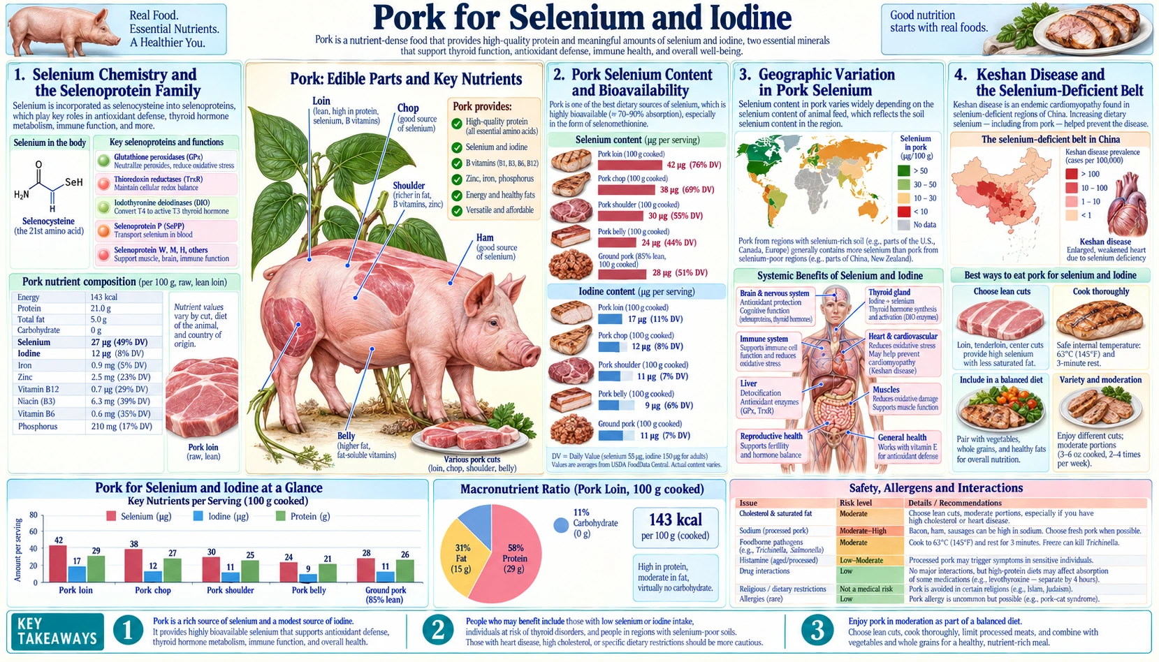
A 3-oz serving of pork delivers 30-50 micrograms of selenium — 60 to 90 percent of the adult Recommended Dietary Allowance (RDA) — incorporated as the highly bioavailable amino acids selenomethionine and selenocysteine. That selenium is the obligate cofactor for the deiodinase enzymes that convert thyroxine (T4) to active triiodothyronine (T3), for glutathione peroxidase antioxidant defense, and for thioredoxin reductase. Pork iodine content is more variable and depends on soil iodine and feed iodine supplementation. Together, selenium and iodine in pork support thyroid function in a way that no single supplement can match. This page covers Keshan disease (the selenium-deficiency cardiomyopathy of central China), Hashimoto's thyroiditis (where selenium repletion reduces antibody titers), the geographic variation in pork selenium content driven by soil chemistry, and the practical question of whether selenium-rich pork from the US Great Plains can substitute for supplements.
Table of Contents
- Selenium Chemistry and the Selenoprotein Family
- Pork Selenium Content and Bioavailability
- Geographic Variation in Pork Selenium
- Keshan Disease and the Selenium-Deficient Belt
- Selenium and Thyroid Function (Deiodinases)
- Hashimoto's Thyroiditis and Selenium Therapy
- Iodine in Pork (Feed, Salt, Iodophor Disinfectants)
- Combined Selenium and Iodine for Thyroid Health
- Cautions (Selenosis, Selenium-Iodine Imbalance)
- Key Research Papers
- Connections
- Featured Videos
Selenium Chemistry and the Selenoprotein Family
Selenium is a trace mineral chemically similar to sulfur, occupying the position directly below sulfur on the periodic table. In biology it is incorporated as selenocysteine (Sec) — the 21st amino acid, encoded by a UGA codon that is reinterpreted in the presence of a SECIS element in the mRNA. About 25 selenoproteins exist in the human genome, each requiring selenium at the active site for catalytic function.
The major selenoprotein families are:
- Glutathione peroxidases (GPx1-8) — the primary intracellular antioxidant defense against hydrogen peroxide and lipid peroxides. GPx1 is cytosolic and ubiquitous; GPx3 circulates in plasma; GPx4 protects membrane lipids from peroxidation and is essential for sperm function and the prevention of ferroptosis.
- Iodothyronine deiodinases (DIO1, DIO2, DIO3) — convert the prohormone T4 to the active hormone T3, or inactivate both to reverse T3 (rT3). Without selenium, the thyroid axis cannot produce active hormone regardless of how much iodine is available.
- Thioredoxin reductases (TrxR1-3) — maintain the reduced state of thioredoxin, a master regulator of cellular redox state and apoptosis signaling.
- Selenoprotein P (SELENOP) — the principal selenium transport protein in plasma; brain and testes have dedicated SELENOP receptors (LRP8 / ApoER2) to ensure preferential selenium delivery to these organs even in deficiency.
- Methionine sulfoxide reductase B1 (MSRB1) — repairs oxidized methionine residues in proteins.
The hierarchy of selenoprotein synthesis in deficiency is well characterized. When dietary selenium drops, the body prioritizes brain and testicular selenoproteins, then thyroid deiodinases, then glutathione peroxidases. Hepatic GPx1 is the first to drop; brain and testicular MSRB1 are the last. This explains why mild selenium deficiency presents as antioxidant defense weakness and thyroid dysfunction before any neurological symptoms emerge.
Pork Selenium Content and Bioavailability
U.S. Department of Agriculture (USDA) values for selenium content in pork, per 3-oz (85 g) cooked serving:
- Pork loin (roasted) — ~37 mcg selenium (67% Recommended Dietary Allowance)
- Pork tenderloin — ~33 mcg selenium (60% Recommended Dietary Allowance)
- Pork shoulder — ~40 mcg selenium (73% Recommended Dietary Allowance)
- Pork chop — ~35 mcg selenium (64% Recommended Dietary Allowance)
- Ham (cured, lean) — ~22 mcg selenium (40% Recommended Dietary Allowance)
- Bacon (3 slices) — ~10 mcg selenium (18% Recommended Dietary Allowance)
- Pork liver — ~50 mcg selenium (91% Recommended Dietary Allowance per 3-oz)
Pork selenium is incorporated principally as selenomethionine (Se-Met), the protein-bound form where selenium replaces sulfur in the methionine amino acid. Selenomethionine is non-specifically incorporated into body proteins in place of methionine and provides a slow-release pool of selenium that can be drawn upon over weeks. Selenocysteine (Sec) is the more functionally active form, directly incorporated into selenoproteins; about 30-40% of pork selenium is in this form.
The bioavailability of pork selenium is high — absorption exceeds 90% in repletion studies, comparable to or better than yeast-derived selenium supplements (selenized yeast) and substantially better than inorganic forms (sodium selenite, sodium selenate, ~50-60% absorption).
Comparing pork to other meaningful animal-source selenium foods (per 3-oz cooked):
- Brazil nuts — ~544 mcg per 1-oz serving (the single most concentrated food source, but variable)
- Pork loin — ~37 mcg
- Beef sirloin — ~31 mcg
- Chicken breast — ~25 mcg
- Tuna — ~92 mcg (highest among fish)
- Cod — ~32 mcg
- Egg — ~15 mcg per large egg
A single pork meal can supply two-thirds of the daily selenium requirement, with high bioavailability, and without the inter-nut variability that makes Brazil nuts an unreliable single-source approach.
Geographic Variation in Pork Selenium
Soil selenium content varies dramatically across the world, and pig selenium content tracks the selenium concentration in the grain that feeds it. The geographic map is well established:
- High-selenium regions — US Great Plains (Dakotas, Wyoming, Nebraska), parts of Canada, Venezuela's alluvial plains, parts of Ireland. Pigs raised on grain grown in these regions deliver the upper end of the selenium content range (40-50+ mcg per 3-oz).
- Moderate-selenium regions — most of Europe, much of North America, Argentina, Australia. Typical pork delivers 30-40 mcg per serving.
- Low-selenium regions — the Keshan disease belt across central China (from northeastern Heilongjiang to southwestern Yunnan), parts of New Zealand, Finland (pre-fertilization), parts of the United Kingdom, parts of Scandinavia.
The most striking case study is Finland, which had documented low population selenium status driven by low-selenium soils. In 1984 Finland implemented a national policy of adding sodium selenate to all agricultural fertilizers. Within two years, mean selenium intake rose three-fold and serum selenium concentrations rose by 70% (Aro 1995). In 1990 the added amount was reduced to 6 mg per kg of fertilizer, which lowered mean intake by 30% and serum selenium by 25% from the highest levels, seen in 1989. The policy has been maintained for over 40 years with no documented harms.
The opposite case is the central China “Keshan belt,” where soil selenium is exceptionally low and population selenium intake from local food can be as low as 10 mcg/day — a fraction of the Recommended Dietary Allowance. Until oral selenium supplementation programs were rolled out in the 1970s, this region had endemic Keshan disease (covered next section).
The practical implication for North American consumers: pork raised on US-grown corn and soy will reliably deliver near-RDA selenium per serving. Pork imported from low-selenium regions or raised on imported feed of unknown origin may deliver less.
Keshan Disease and the Selenium-Deficient Belt
Keshan disease is an endemic dilated cardiomyopathy first described in 1935 in Keshan County, Heilongjiang Province, China. It primarily affects children and women of childbearing age in low-selenium-soil regions of central China. Untreated, the acute form has 80% mortality from cardiogenic shock and arrhythmia.
The clinical features include:
- Dilated cardiomyopathy with biventricular failure
- Cardiogenic shock and severe arrhythmias in acute presentation
- Multifocal myocardial necrosis on histology
- Subacute and chronic forms with progressive heart failure
- Geographic distribution exactly matching low-selenium soil bands
The selenium connection was established in the 1970s through a series of large Chinese trials that randomized children in affected villages to oral sodium selenite supplementation or placebo. Selenium supplementation dramatically reduced Keshan incidence and mortality. Chen's 2012 review of this 50-year story describes several large population-based trials of oral sodium selenite tablets that significantly reduced Keshan disease incidence, and concludes that selenium deficiency is the major cause of the disease, although “other etiological factors could not be ruled out.” The same review notes that these findings became part of the scientific basis for setting the minimum selenium requirement and the Recommended Dietary Allowance. By the 1980s, selenium supplementation programs had nearly eliminated Keshan disease as a public health problem in the affected regions.
The pathogenesis is now understood to involve two factors interacting: (1) selenium deficiency that lowers glutathione peroxidase activity in the heart, leaving cardiomyocytes vulnerable to oxidative damage, and (2) co-infection with normally benign Coxsackie B viruses that become more cardiotoxic when they replicate in selenium-deficient hosts. In a mouse study, a harmless strain of coxsackievirus B3 became virulent and caused heart inflammation (myocarditis) in selenium- and vitamin E-deficient mice, through mutations in the virus's genes; once these mutations had occurred, even well-nourished mice developed disease from the mutated virus (Beck and Levander 2000).
The implications extend beyond Keshan disease to other settings where chronic selenium deficiency and viral infection coincide. For more on dilated cardiomyopathy in general, see our Cardiomyopathy page.
Selenium and Thyroid Function (Deiodinases)
The thyroid gland concentrates more selenium per gram of tissue than any other organ in the body. This is not coincidence — the iodothyronine deiodinases (DIO1, DIO2, DIO3) that activate and inactivate thyroid hormone are selenoproteins, and the gland depends on them for normal hormone metabolism.
The thyroid produces predominantly the prohormone thyroxine (T4), with much smaller amounts of the active hormone triiodothyronine (T3). Most active T3 is generated peripherally by deiodinase enzymes that remove one iodine atom from T4, converting it to T3.
- DIO1 — expressed in liver, kidney, thyroid; produces circulating T3
- DIO2 — expressed in brain, pituitary, brown adipose; produces intracellular T3 for local action
- DIO3 — expressed in placenta, brain, skin; degrades T3 and T4 to inactive forms (reverse T3)
In selenium deficiency, deiodinase activity drops. The clinical signature is a pattern of high T4, low T3, and normal or elevated reverse T3 (rT3) — the “low T3 syndrome.” The thyroid-stimulating hormone (TSH) may be normal, low, or mildly elevated. This pattern can be mistaken for nonthyroidal illness syndrome or for thyroid hormone resistance.
Repletion with adequate dietary selenium — or in deficient patients, supplemental selenium 100-200 mcg/day — restores deiodinase activity and the normal T4:T3 ratio within weeks. For more on the thyroid axis broadly, see our Hypothyroidism page and Thyroid Panel page.
Hashimoto's Thyroiditis and Selenium Therapy
Hashimoto's thyroiditis (chronic lymphocytic thyroiditis) is the most common cause of hypothyroidism in iodine-replete populations and is characterized by autoantibodies against thyroid peroxidase (anti-TPO) and thyroglobulin (anti-Tg). The autoimmune destruction of the thyroid gland leads to progressive hypothyroidism over years to decades.
Selenium has been studied extensively as adjunctive therapy in Hashimoto's. Multiple randomized controlled trials have shown that selenium supplementation (typically 200 mcg/day of selenomethionine for 3-12 months) produces a measurable reduction in anti-TPO antibody titers. The Gärtner 2002 trial in the Journal of Clinical Endocrinology and Metabolism was the first major demonstration: 70 women received 200 mcg/day of sodium selenite or placebo for 3 months, and mean anti-TPO fell to 63.6% of its starting level in the selenium group versus 88% in the placebo group.
Subsequent meta-analyses (Toulis 2010, Wichman 2016) have consistently found anti-TPO reduction with selenium therapy, though the effect size varies across studies and the impact on actual disease progression (need for levothyroxine, progression to overt hypothyroidism) is less clear. The current consensus is that selenium supplementation may be a useful adjunct in selected Hashimoto's patients, particularly those with high baseline anti-TPO titers and confirmed low or low-normal selenium status.
The Wichman 2016 meta-analysis pooled 16 controlled trials. In people already taking levothyroxine (thyroid hormone tablets), anti-TPO levels were lower with selenium after 3 months, and this held at 6 and 12 months; in people not taking levothyroxine, anti-TPO was lower after 3 months but not after 6 or 12 months. The authors rated the quality of the evidence as generally low, found that people taking selenium had a significantly higher risk of reporting side effects (p = 0.036), and wrote that whether these antibody changes matter for clinically relevant measures “remains to be demonstrated.”
A larger 2024 meta-analysis of randomized trials (Huwiler and colleagues, 35 studies) found that in people with Hashimoto's not taking thyroid hormone, selenium lowered TSH slightly (7 cohorts, 869 participants), and that anti-TPO fell in people both with and without thyroid hormone treatment (29 cohorts, 2,358 participants). Side effects were comparable between the selenium and control groups (odds ratio 0.89), free T4, free T3 and thyroid volume did not change significantly, and the authors rated the overall certainty of the evidence as moderate.
An Italian randomized, placebo-controlled trial (Negro 2007) extended this work to pregnant women with thyroid peroxidase antibodies. Of 151 antibody-positive women, 77 took 200 mcg/day of selenomethionine during pregnancy and after birth and 74 took a placebo. Thyroid problems after birth (postpartum thyroid dysfunction) occurred in 28.6% of the selenium group versus 48.6% of the placebo group, and lasting (permanent) hypothyroidism in 11.7% versus 20.3%.
For comparison, a 3-oz serving of pork supplies roughly 30-50 mcg of selenium (toward the upper end when the pigs are raised on grain from high-selenium regions such as the US Great Plains), while the Hashimoto's trials above gave 200 mcg/day as a supplement; none of these trials tested pork in place of supplements. Brazil nuts are another food source, with substantial inter-nut variability — 1-2 Brazil nuts per day can supply Recommended Dietary Allowance selenium but a single nut might contain anywhere from 50 to 500+ mcg. For more, see our Hashimoto's Thyroiditis page.
Iodine in Pork (Feed, Salt, Iodophor Disinfectants)
Iodine content in pork is variable and depends on three sources: (1) iodine in the soil and feed of the production region, (2) iodine added to pig feed as a supplement (typical commercial feed includes 0.3-0.5 mg iodine per kg of feed), and (3) iodine residues from iodophor disinfectants used in dairy and meat processing.
Typical pork iodine content per 3-oz serving:
- US commercial pork (corn-soy fed, supplemented) — 5-15 mcg iodine (3-10% Recommended Dietary Allowance)
- European pork (lower feed supplementation) — 3-10 mcg iodine
- Heritage / pasture-raised pork — highly variable, dependent on soil iodine and forage
- Imported / Asian pork — typically lower unless feed is supplemented
Pork is not a primary iodine source for most populations. Dairy, seaweed, eggs, and iodized salt deliver substantially more iodine per serving. However, in iodine-sufficient regions, regular pork consumption contributes meaningfully to total intake while delivering high selenium that pairs synergistically with iodine for thyroid function.
For populations relying heavily on pork without iodized salt or seaweed access (some inland European populations historically), pork alone is insufficient to prevent goiter or hypothyroidism from iodine deficiency. The Swiss goiter epidemic of the 19th century and the introduction of iodized salt as a public health intervention is the relevant precedent.
Combined Selenium and Iodine for Thyroid Health
Iodine and selenium operate at sequential steps in thyroid hormone biology:
- Iodine is required as raw material for thyroid hormone synthesis — thyroglobulin in the thyroid follicle is iodinated by thyroid peroxidase (TPO) to form mono- and diiodotyrosine, which couple to form T4 and T3.
- Selenium is required for the deiodinase enzymes that convert T4 to T3 peripherally, and for glutathione peroxidase that protects the thyroid follicle from oxidative damage during the iodination reaction (which generates substantial hydrogen peroxide).
Iodine without adequate selenium can produce thyroid damage. Forcing iodine supplementation in selenium-deficient populations has been associated with increased autoimmune thyroid disease — the iodination reaction generates oxidative stress that an under-resourced selenoprotein defense cannot manage, potentially exposing thyroglobulin epitopes that trigger autoantibody formation. This is part of the explanation for why some Eastern European and Chinese regions saw increased Hashimoto's after iodization programs.
Selenium without adequate iodine cannot produce normal thyroid hormone levels — the substrate is missing.
The clinical implication is that thyroid health requires both minerals in adequate supply. Pork from selenium-replete regions supplies selenium, while iodized salt, dairy and seafood supply most dietary iodine. Serum selenium and urinary iodine are the laboratory measures of each mineral's status, and because the two minerals work at different steps, a shortfall or an imbalance in either one affects thyroid hormone production.
Cautions (Selenosis, Selenium-Iodine Imbalance)
- Selenium upper limit (Tolerable Upper Intake Level, UL) — 400 mcg/day for adults. Chronic intake above this can produce selenosis, with symptoms of brittle hair and nails, garlic breath, gastrointestinal upset, peripheral neuropathy, and in extreme cases hepatic and pancreatic toxicity. Regular pork consumption does not approach this limit; the risk is concentrated in inappropriate supplementation or daily Brazil nut consumption.
- Brazil nut variability — a single Brazil nut can contain 50-500 mcg selenium. Recommendations to “eat 1-2 Brazil nuts per day” for selenium can produce intakes ranging from 50 mcg (well below Recommended Dietary Allowance) to 1000+ mcg (toxic). Pork is more predictable.
- SELECT trial cautions — the Selenium and Vitamin E Cancer Prevention Trial (200 mcg/day selenium L-selenomethionine plus vitamin E 400 international units (IU)/day for prostate cancer prevention) was stopped early without benefit and with a small signal of increased type 2 diabetes risk in the selenium arm. The 2009 trial report found that selenium at this dose did not prevent prostate cancer in relatively healthy men, and that the rise in type 2 diabetes in the selenium group was not statistically significant (relative risk 1.07; 99% confidence interval 0.94-1.22; P = .16).
- Selenium and human immunodeficiency virus (HIV) — human immunodeficiency virus-positive patients have lower plasma selenium and selenium supplementation has shown some benefit in reducing disease progression. Standard care includes selenium-adequate nutrition.
- Selenium and statin myopathy — isolated case reports suggest selenium deficiency may worsen statin-induced myopathy; the mechanism would relate to GPx-driven antioxidant defense of muscle. Not established as routine care.
- Iodine excess in salt-restricted populations — modern processed-food eaters can exceed iodine recommendations from iodized salt; this is not typically a problem for selenium-paired intake.
- Pregnancy — both selenium (60 mcg/day Recommended Dietary Allowance) and iodine (220 mcg/day Recommended Dietary Allowance) requirements rise; pork, iodized salt and prenatal vitamins are all sources of these minerals.
Key Research Papers
- Rayman MP (2012). Selenium and human health. The Lancet. — PubMed PMID: 22381456
- Combs GF Jr (2001). Selenium in global food systems. British Journal of Nutrition. 85(5):517-547. — PubMed PMID: 11348568
- Gärtner R et al. (2002). Selenium supplementation in patients with autoimmune thyroiditis decreases thyroid peroxidase antibodies concentrations. Journal of Clinical Endocrinology and Metabolism. 87(4):1687-1691. — PubMed PMID: 11932302
- Wichman J, Winther KH, Bonnema SJ, Hegedüs L (2016). Selenium supplementation significantly reduces thyroid autoantibody levels in patients with chronic autoimmune thyroiditis: a systematic review and meta-analysis. Thyroid. 26(12):1681-1692. — PubMed PMID: 27702392
- Huwiler VV et al. (2024). Selenium supplementation in patients with Hashimoto thyroiditis: a systematic review and meta-analysis of randomized clinical trials. Thyroid. 34(3):295-313. — PubMed PMID: 38243784
- Toulis KA et al. (2010). Selenium supplementation in the treatment of Hashimoto's thyroiditis: a systematic review and a meta-analysis. Thyroid. — PubMed PMID: 20883174
- Negro R et al. (2007). The influence of selenium supplementation on postpartum thyroid status in pregnant women with thyroid peroxidase autoantibodies. Journal of Clinical Endocrinology and Metabolism. 92(4):1263-1268. — PubMed PMID: 17284630
- Lippman SM et al. (2009). Effect of selenium and vitamin E on risk of prostate cancer (SELECT trial). JAMA. — PubMed PMID: 19066370
- Beck MA, Levander OA (2000). Host nutritional status and its effect on a viral pathogen. Journal of Infectious Diseases. 182(Suppl 1):S93-S96. (Keshan / Coxsackie B) — PubMed PMID: 10944489
- Chen J (2012). An original discovery: selenium deficiency and Keshan disease (an endemic heart disease). Asia Pacific Journal of Clinical Nutrition. 21(3):320-326. — PubMed PMID: 22705420
- Schomburg L (2011). Selenium, selenoproteins and the thyroid gland: interactions in health and disease. Nature Reviews Endocrinology. — PubMed PMID: 22009156
- Aro A et al. (1995). Effects of supplementation of fertilizers on human selenium status in Finland. Analyst. — PubMed PMID: 7741238
- Zimmermann MB (2009). Iodine deficiency. Endocrine Reviews. — PubMed PMID: 19460960
- Köhrle J (2013). Selenium and the thyroid. Current Opinion in Endocrinology, Diabetes & Obesity. 20(5):441-448. — PubMed PMID: 23974773