⚠ Evidence warning: Clinical trials or other evidence do not support the grading scheme called the LAM Functional Classification. This page keeps it as a report of what people, websites or other media say.
Lymphangioleiomyomatosis (LAM)
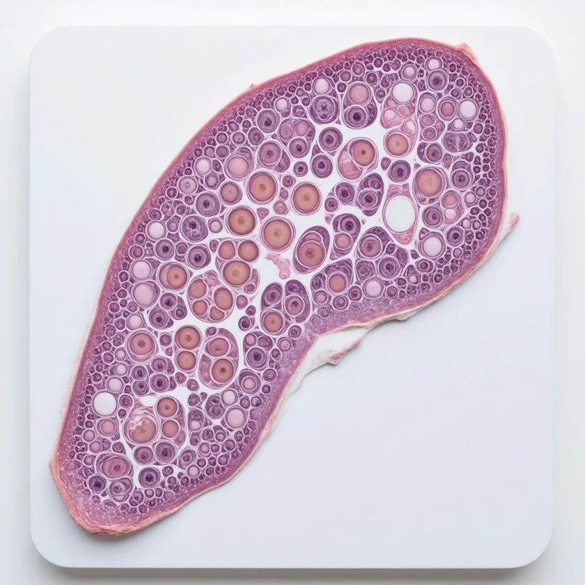
LAM is a rare, progressive, cystic lung disease caused by mutations in the TSC1 or TSC2 genes, leading to mTORC1 hyperactivation and abnormal proliferation of smooth muscle-like LAM cells throughout the lungs and lymphatics. It almost exclusively affects premenopausal women and is characterized by bilateral pulmonary cysts, spontaneous pneumothorax, chylothorax, and renal angiomyolipomas.
Table of Contents
- Disease Overview and Classification
- Pathogenesis: TSC Mutations and mTORC1
- Pulmonary Manifestations
- Extrapulmonary Manifestations
- Symptoms and Clinical Presentation
- Diagnosis: High-Resolution Computed Tomography, VEGF-D, and Biopsy
- Pulmonary Function and Disease Progression
- Treatment: Mechanistic Target of Rapamycin Inhibitors and Supportive Care
- Prognosis and Transplantation
- Key Research Papers
- Featured Videos
Disease Overview and Classification
Lymphangioleiomyomatosis (LAM) is a multisystem disorder characterized by the uncontrolled proliferation of abnormal smooth muscle-like cells — LAM cells — throughout the lung parenchyma, lymphatics, and kidneys. The disease was first described in 1937 and remained a medical curiosity until the 1990s, when the discovery of TSC gene mutations provided its molecular basis and eventually led to targeted mechanistic target of rapamycin (mTOR) inhibitor therapy.
Sporadic LAM (sLAM)
Sporadic LAM accounts for approximately 80–85% of all cases. It arises from a somatic (acquired) mutation in the TSC2 gene occurring in a single progenitor cell. The LAM cells bearing this mutation then proliferate, spread hematogenously and lymphatically, and seed distant organs. sLAM is almost exclusively a disease of women and is most often found during the reproductive years. In the US National Heart, Lung, and Blood Institute registry of 230 women with LAM (sporadic and tuberous sclerosis forms together), ages at enrollment ran from 18 to 76, symptoms began at an average age of 38.9 years and diagnosis came at an average of 41.0 years; the authors called the age range broader than previously appreciated. The strong estrogen dependence of sLAM is evidenced by its rarity before menarche, acceleration during pregnancy, and occasional stabilization after menopause. No germline mutations are present and sLAM does not carry an increased risk for family members.
Tuberous Sclerosis Complex-Associated LAM (TSC-LAM)
TSC-LAM affects individuals with tuberous sclerosis complex (TSC), an autosomal dominant neurocutaneous syndrome caused by germline mutations in TSC1 (hamartin) or TSC2 (tuberin). In a specialist clinic study of 101 women with TSC who had chest computed tomography scans (a referral population), 47.5% met criteria for LAM; prevalence was 27% under age 21 and 81% over age 40, rising about 8% per year, and the authors concluded that most women with TSC ultimately develop cystic changes consistent with LAM. TSC-LAM can affect males, although the disease is far less severe in men — a difference attributed to the lack of estrogen-driven LAM cell proliferation. TSC patients also carry the full burden of extrapulmonary TSC manifestations: cortical tubers causing epilepsy, subependymal giant cell astrocytomas, cardiac rhabdomyomas (often regressing spontaneously in childhood), and renal angiomyolipomas.
Epidemiology
LAM is genuinely rare, with prevalence estimates of 3–7 per million women. Because of this rarity and the nonspecific early presentation, diagnosis is frequently delayed by 3–5 years. The LAM Foundation patient registry has been critical in characterizing the natural history of this disease in an otherwise understudied population.
Pathogenesis: TSC Mutations and mTORC1
The molecular basis of LAM is hyperactivation of mTORC1 (mechanistic target of rapamycin complex 1), a master regulator of cell growth, protein synthesis, and autophagy. Understanding this pathway is essential to understanding both the disease and the rationale for its treatment.
The TSC1/TSC2 Tumor Suppressor Complex
TSC1 (hamartin) and TSC2 (tuberin) form an obligate heterodimer that functions as a GTPase-activating protein (GAP) for the small GTPase Rheb. When active, the TSC1/TSC2 complex converts Rheb-GTP to inactive Rheb-GDP, preventing Rheb from activating mTORC1. Loss-of-function mutations in either TSC1 or TSC2 eliminate this brake: Rheb remains bound to GTP, constitutively activating mTORC1. mTORC1 activation then drives phosphorylation of S6K1 and 4E-BP1, leading to increased ribosomal biogenesis, protein synthesis, cell growth, and inhibition of autophagy — the cellular machinery underlying the hyperproliferative LAM cell phenotype.
LAM Cell Identity and Spread
LAM cells are smooth muscle-like cells that express neural crest markers (HMB-45, a melanoma antigen), smooth muscle markers (alpha-smooth muscle actin, SMA), and sex hormone receptors (estrogen receptor, ER; progesterone receptor, PR). Their precise tissue of origin remains debated, but the leading hypothesis is that they derive from a uterine or pelvic source and spread to the lungs via lymphatic and hematogenous routes. Evidence for this metastatic spread model comes from the observation that LAM recurs in transplanted lungs — new LAM cells from the recipient colonize the donor lung. Recurrence after transplant is rare (4 of 61 recipients in a European series), but it excludes the possibility that LAM is simply a locally generated pulmonary disease.
Estrogen's Role
Estrogen promotes LAM cell survival and proliferation through ER-mediated transcriptional effects and possibly through mTORC1-independent pathways. Clinical observations supporting estrogen dependence include: the near-exclusive premenopausal female predominance, documented disease progression during pregnancy (when estrogen levels are maximally elevated), anecdotal evidence of disease acceleration with exogenous estrogen administration, and case reports of disease stabilization after oophorectomy. However, anti-estrogen therapies (progesterone, tamoxifen, oophorectomy) did not show clear benefit in the pre-mTOR inhibitor era, suggesting estrogen modulation alone is insufficient to halt LAM progression. For progesterone the evidence is retrospective rather than from controlled trials: a study of 275 patients concluded, “within the limitations of a retrospective study,” that progesterone does not slow the decline in lung function, and in a US registry of 410 patients hormonal therapy was linked to a higher risk of death or transplant (hazard ratio 2.93), which the authors said may reflect confounding by indication (sicker patients being the ones treated). The 2016 American Thoracic Society/Japanese Respiratory Society guideline recommends against hormonal therapy.
Pulmonary Manifestations
The lungs bear the brunt of LAM pathology. LAM cells infiltrate the lung parenchyma along lymphatics and airway walls, causing progressive destruction of normal lung architecture and replacement with thin-walled cysts.
Pulmonary Cysts: Formation and Distribution
The hallmark of LAM on imaging is bilateral, diffuse, thin-walled, round-to-oval pulmonary cysts distributed throughout both lungs. The cysts range from a few millimeters to several centimeters in diameter and characteristically have a normal-appearing parenchyma between them — a feature that distinguishes LAM from emphysema (where alveolar destruction eliminates the normal intervening lung). The mechanism of cyst formation involves LAM cell infiltration and partial obstruction of small airways, creating a check-valve effect that traps air distally; protease release by LAM cells then destroys adjacent alveolar walls.
Obstructive Physiology: A Paradox
Despite being classified as an interstitial lung disease, LAM produces an obstructive ventilatory defect on pulmonary function testing — airflow limitation with reduced ratio of forced expiratory volume in 1 second to forced vital capacity and air trapping (elevated residual volume and total lung capacity). This is paradoxical because most interstitial lung diseases produce restriction. The explanation lies in the mechanism: LAM cells obstruct small airways directly, and the loss of elastic recoil from cyst formation reduces the driving pressure for expiratory airflow, mimicking emphysema physiology. The diffusing capacity for carbon monoxide (DLCO) is reduced early and tracks disease severity. Decline in forced expiratory volume in 1 second in untreated LAM is reported at roughly 70–120 mL/year (a retrospective study of 275 patients measured an average of 75 mL/year) — faster than in chronic obstructive pulmonary disease (COPD) — but is highly variable between patients. Not every patient shows the obstructive pattern: in the registry of 230 women, 57.3% had it and 33.9% had normal spirometry at enrollment.
Spontaneous Pneumothorax
In the LAM Foundation registry, 260 of 395 patients (66%) reported at least one spontaneous pneumothorax in their lifetime, and a Korean cohort of 54 patients reported 66.7%. Of the 193 registry patients who answered a detailed questionnaire, 82% had their first pneumothorax before LAM was diagnosed. It is the most dramatic acute presentation of LAM and results from rupture of subpleural cysts. Unlike primary spontaneous pneumothorax (which recurs in about 25–30% of cases), LAM-associated pneumothorax has a recurrence rate exceeding 70% after the first episode — a rate that profoundly affects management. In that registry questionnaire, recurrence was 66% after conservative care, 27% after chemical pleurodesis and 32% after surgery. The 2017 American Thoracic Society/Japanese Respiratory Society guideline makes a conditional recommendation for offering pleurodesis (chemical or surgical) after a first LAM-associated pneumothorax rather than postponing it until the first recurrence, unlike the "watchful waiting" approach used in primary spontaneous pneumothorax.
Chylothorax and Chylous Ascites
Chylothorax — accumulation of chylous (lymph-rich) fluid in the pleural space — is a hallmark complication occurring in approximately 20–30% of LAM patients. It results from LAM cell infiltration and obstruction of thoracic lymphatic vessels, causing lymph to leak into the pleural space. Chylothorax is milky-white in appearance, with triglyceride levels typically exceeding 110 mg/dL and chylomicrons on lipoprotein electrophoresis. Chylous ascites reflects the same process in the peritoneal lymphatics. Both respond to mechanistic target of rapamycin inhibitor therapy — sirolimus reduces lymphatic complications in addition to its pulmonary effects.
Extrapulmonary Manifestations
LAM is a multisystem disease. Extrapulmonary manifestations reflect LAM cell infiltration of abdominal and pelvic lymphatics and parenchymal organs, and in TSC-LAM, the full systemic burden of tuberous sclerosis complex.
Renal Angiomyolipomas (AML)
Angiomyolipomas are benign hamartomas composed of abnormal blood vessels, immature smooth muscle, and fat. They occur in 50–60% of women with sLAM and in over 80% of TSC-LAM patients. AMLs can be identified on computed tomography (CT) by their fat content (Hounsfield units less than -10 in the fat component) or on magnetic resonance imaging (MRI) using fat-suppression sequences. The clinical significance of AMLs relates to their propensity to bleed spontaneously — the abnormal vasculature within AMLs lacks the normal elastic lamina and is prone to aneurysm formation and rupture. AMLs exceeding 4 cm in diameter are at highest risk for spontaneous hemorrhage (Wunderlich syndrome), which can be life-threatening. Management of large or symptomatic AMLs includes mechanistic target of rapamycin inhibitor therapy (first line; in an open-label, nonrandomized trial of 25 patients, sirolimus shrank angiomyolipomas to about 53% of their starting volume at 12 months, and they regrew to about 86% of it one year after the drug was stopped) or selective embolization; surgical resection is avoided to preserve renal parenchyma.
Lymphangioleiomyomas
Lymphangioleiomyomas are LAM cell-lined cystic structures that form within the lymphatic system, most commonly in the retroperitoneum, pelvis, and mediastinum. They appear on computed tomography as thin-walled fluid-filled structures and can grow to considerable size. Symptoms depend on location: retroperitoneal lymphangioleiomyomas may cause abdominal pain, early satiety, or bowel compression. They are distinct from simple lymphoceles and respond to mechanistic target of rapamycin inhibitor therapy.
TSC-Specific Manifestations (TSC-LAM Only)
In TSC-LAM, the full spectrum of tuberous sclerosis manifestations may be present. Cortical tubers are epileptogenic lesions that cause seizures, often beginning in infancy, and vary widely in severity — from pharmacologically controlled epilepsy to refractory status epilepticus. Subependymal giant cell astrocytomas (SEGAs) can obstruct cerebrospinal fluid (CSF) flow causing hydrocephalus and require treatment with mechanistic target of rapamycin inhibitors (everolimus is U.S. Food and Drug Administration-approved for SEGA) or surgery. Cutaneous features include facial angiofibromas (adenoma sebaceum, the classic butterfly facial rash), ash leaf macules (hypomelanotic macules, visible under Wood's lamp), shagreen patches (connective tissue hamartomas on the lower back), and periungual fibromas. Cardiac rhabdomyomas, present in fetal life, frequently involute spontaneously. Retinal hamartomas are incidental findings.
Symptoms and Clinical Presentation
LAM presents insidiously in most patients, with symptoms often attributed to more common conditions such as asthma or anxiety. The average delay from first symptoms to diagnosis is 3–5 years, partly because many physicians have never encountered the disease. Awareness of LAM's characteristic presentations can dramatically shorten this diagnostic odyssey.
Exertional Dyspnea
Progressive exertional dyspnea is the most common presenting symptom, reported by over 80% of patients at diagnosis. It follows an insidious course — initially noticed only during strenuous exercise, then during activities of daily living. The mechanism is multifactorial: obstructive airflow limitation, reduced DLCO from cyst destruction of the alveolar-capillary membrane, and impaired gas exchange efficiency during exercise. Unlike asthma, the dyspnea does not have the classic episodic bronchospastic character, though bronchodilator responsiveness is present in some patients.
Spontaneous Pneumothorax as First Presentation
A spontaneous pneumothorax in a young woman — particularly if recurrent, or occurring without obvious precipitant — is a recognized clue to LAM. In the LAM Foundation registry questionnaire, 82% of respondents had their first pneumothorax before LAM was diagnosed, and patients are often evaluated for "primary spontaneous pneumothorax" for months before the underlying LAM is recognized. The clue is recurrence: primary spontaneous pneumothorax recurs in 25–30% of cases; LAM-associated pneumothorax recurs in over 70%.
Chylous Effusion Presentation
Chylothorax presenting as a unilateral or bilateral pleural effusion — particularly in a young woman — is a highly specific presentation for LAM (or other lymphatic abnormalities). The effusion is often massive, causing dyspnea and reduced exercise tolerance. Thoracentesis revealing milky fluid with high triglycerides confirms chylous origin and is usually followed by high-resolution computed tomography and VEGF-D testing.
Incidental Diagnosis
An increasing proportion of LAM is diagnosed incidentally — cysts are found on computed tomography performed for another reason, or renal AMLs are discovered on abdominal ultrasound. This shift toward incidental detection is expected to improve prognosis as the diagnosed population includes earlier-stage disease.
Diagnosis: High-Resolution Computed Tomography, VEGF-D, and Biopsy
The diagnosis of LAM can often be established without surgical lung biopsy using a combination of high-resolution computed tomography findings, serum VEGF-D levels, and clinical context. Current European Respiratory Society and American Thoracic Society guidelines provide a diagnostic algorithm that spares many patients an invasive procedure.
High-Resolution Computed Tomography
High-resolution computed tomography of the chest is the cornerstone of LAM diagnosis. The characteristic appearance is bilateral, diffusely distributed, thin-walled (wall thickness typically <2 mm), round-to-oval cysts ranging from 2 mm to 2 cm in diameter, with completely normal intervening lung parenchyma. The cysts are uniformly distributed throughout both lungs — upper and lower zones equally — without the upper-lobe predominance of emphysema or the lower-lobe predominance of usual interstitial pneumonia. In early disease, cysts may be few and small; in advanced disease they may replace most of the lung parenchyma. Ground-glass opacities and nodules are not prominent features of LAM (their presence points toward an alternative diagnosis). A characteristic high-resolution computed tomography in the right clinical context (young woman with dyspnea, pneumothorax, or chylothorax) is sufficient to suspect LAM strongly.
Serum VEGF-D: A Diagnostic Biomarker
Vascular endothelial growth factor D (VEGF-D) is produced by LAM cells and is measurable in serum. In a 2010 study of 195 women, a serum VEGF-D above 600 pg/mL was highly associated with LAM (specificity 97.6%) and values above 800 pg/mL were called diagnostically specific in a woman with typical cysts on high-resolution computed tomography. A level above 800 pg/mL is included in the European Respiratory Society and American Thoracic Society diagnostic criteria as a non-invasive confirmatory test. Combined with characteristic high-resolution computed tomography findings, a VEGF-D >800 pg/mL is sufficient to diagnose LAM without tissue biopsy in most cases. VEGF-D sensitivity is reported at approximately 69% in sLAM. In the same 2010 study, 12 of 15 women later diagnosed with LAM by biopsy exceeded 800 pg/mL, median levels were 1,175 pg/mL in sLAM and 3,465 pg/mL in TSC-LAM (separate groups), and the authors state that a negative VEGF-D result does not exclude LAM. VEGF-D also tracks disease activity and may serve as a pharmacodynamic marker for mechanistic target of rapamycin inhibitor therapy.
Diagnostic Criteria (European Respiratory Society and American Thoracic Society)
According to current guidelines, a definitive LAM diagnosis can be made without biopsy if: characteristic or compatible high-resolution computed tomography findings are present plus any one of the following — (1) serum VEGF-D ≥800 pg/mL; (2) renal angiomyolipoma; (3) chylous pleural or peritoneal effusion; (4) lymphangioleiomyoma; (5) confirmed TSC diagnosis. In the absence of these features, tissue is the remaining route: the 2017 American Thoracic Society/Japanese Respiratory Society guideline makes conditional recommendations against a clinical diagnosis on high-resolution computed tomography alone and for considering transbronchial lung biopsy, and surgical lung biopsy (video-assisted thoracoscopic surgery, VATS) is the other option. Biopsy specimens show LAM cell nests — spindle-shaped smooth muscle-like cells expressing HMB-45, SMA, ER, and PR — surrounding the characteristic cysts.
Additional Diagnostic Workup
Abdominal computed tomography or magnetic resonance imaging is used in LAM patients to evaluate for renal AMLs and retroperitoneal lymphangioleiomyomas. Genetic testing for TSC1/TSC2 germline mutations is described for LAM patients regardless of apparent sporadic nature — TSC mutations found on germline testing redefine the diagnosis as TSC-LAM and have implications for family screening. Pulmonary function tests at diagnosis provide a functional baseline. Echocardiography is the test used when pulmonary hypertension is suspected (LAM can cause Group 5 pulmonary hypertension through lymphatic and vascular involvement).
Pulmonary Function and Disease Progression
Monitoring lung function over time is central to LAM management because the disease is progressive and the rate of progression varies substantially between patients. Some women experience rapid decline in forced expiratory volume in 1 second requiring early treatment; others remain stable for years.
Spirometry and DLCO
Pulmonary function in LAM shows an obstructive pattern: reduced ratio of forced expiratory volume in 1 second to forced vital capacity, increased residual volume, air trapping, and disproportionately reduced DLCO relative to forced vital capacity. Bronchodilator responsiveness (improvement in forced expiratory volume in 1 second ≥12% and 200 mL after albuterol) was found in 39 of 130 patients (30%) in a prospective study. Responders had lower forced expiratory volume in 1 second and DLCO and a faster decline in both (p = 0.044 and p = 0.039), and the authors concluded that a positive bronchodilator response is associated with an accelerated decline in lung function. In the MILES trial, baseline VEGF-D was also higher in responders. DLCO correlates with cyst burden on high-resolution computed tomography and with exercise capacity, making it a valuable complement to forced expiratory volume in 1 second for disease staging.
Rate of Decline in Forced Expiratory Volume in 1 Second
In untreated LAM, the mean rate of decline in forced expiratory volume in 1 second has been reported as 70–120 mL/year in most cohort studies — substantially faster than the 30–40 mL/year expected with normal aging or the 40–60 mL/year in chronic obstructive pulmonary disease. There is, however, enormous interindividual variability: some patients lose over 200 mL/year, others show minimal decline over years of follow-up. The LAM Functional Classification (LFC) system uses baseline forced expiratory volume in 1 second (as %predicted) to grade severity: LFC I (>70% predicted), LFC II (51–70%), LFC III (≤50%).
Exercise Testing
Six-minute walk test (6MWT) and cardiopulmonary exercise testing (CPET) provide complementary information about functional impairment. Many LAM patients demonstrate exercise-induced oxygen desaturation disproportionate to their resting lung function — early supplemental oxygen supplementation guidance relies on exercise oximetry. CPET characteristically shows a ventilatory limitation pattern with reduced peak VO2 and VE/VCO2 slope consistent with the obstructive physiology and impaired gas exchange efficiency of LAM.
Treatment: Mechanistic Target of Rapamycin Inhibitors and Supportive Care
The 2011 publication of the MILES trial (Multicenter International LAM Efficacy of Sirolimus trial) fundamentally transformed LAM management by demonstrating that sirolimus stabilizes lung function — ending the era of watchful waiting as the only available strategy for most patients.
Mechanistic Target of Rapamycin Inhibitors: Sirolimus and Everolimus
Sirolimus (rapamycin) inhibits mTORC1 by binding FKBP-12 and allosterically blocking mTORC1 kinase activity, directly countering the TSC2 loss-of-function that drives LAM cell proliferation. In the MILES trial (N Engl J Med, 2011; PubMed PMID: 21410393), sirolimus stabilized forced expiratory volume in 1 second over 12 months (change +1 mL in sirolimus vs −12 mL in placebo per month), and improved forced vital capacity, functional residual capacity, serum VEGF-D, quality of life and functional performance compared to placebo; there was no significant difference in 6-minute walk distance or DLCO. Crucially, however, lung function declined again after sirolimus discontinuation — demonstrating that mechanistic target of rapamycin inhibitors suppress but do not cure the disease, and ongoing treatment is required for sustained benefit. Sirolimus also reduces serum VEGF-D, shrank renal AMLs to about 53% of their starting volume at 12 months in an open-label trial of 25 patients (with regrowth after stopping), and reduces chylous complications. Target trough serum levels are 5–15 ng/mL. Everolimus (a sirolimus analog) has demonstrated similar efficacy for renal AMLs in TSC and is used interchangeably in many patients. Common side effects include mouth ulcers (stomatitis), infections (particularly opportunistic), hyperlipidemia, proteinuria, cytopenias, impaired wound healing, and rarely pneumonitis.
What the Sirolimus Label Says
Sirolimus is approved by the U.S. Food and Drug Administration for treating LAM. The label gives a starting dose of 2 mg a day by mouth, with blood trough levels checked in 10–20 days and the dose adjusted to hold them at 5–15 ng/mL. Its boxed warning states that immunosuppression may increase the chance of infection and the possible development of lymphoma and other cancers, and that use is not recommended in liver or lung transplant patients. The label states that sirolimus can harm a fetus and recommends highly effective contraception during treatment and for 12 weeks after the final dose. It also lists live vaccines (such as measles, mumps and rubella) as ones not to be used during treatment.
Long-Term Results on Sirolimus
In a cohort of 38 patients, yearly change in forced expiratory volume in 1 second went from −2.3% predicted before sirolimus to +1.0% predicted on it. In a national cohort of 47 people treated for a mean of 35.8 months, forced expiratory volume in 1 second changed by a mean of +11 mL a year, ranging from +254 to −148 mL a year, and poor response was tied to lower starting lung function and longer disease. In a 2026 retrospective cohort of 71 patients from Poland, chylous effusions (15 patients) resolved in all by 12 months without recurrence, and about 6% stopped the drug for good.
Resveratrol Added to Sirolimus
Resveratrol is a plant compound. A 2023 open-label phase 2 study added it (250 mg rising to 1,000 mg a day over 24 weeks) to sirolimus in 25 patients. It was well tolerated despite some gut side effects. The main goal, a fall in VEGF-D of at least 42%, was not met, though VEGF-D did fall significantly and quality of life improved modestly. The authors state that larger controlled trials might be warranted.
Indications for Mechanistic Target of Rapamycin Inhibitor Therapy
Not all patients with LAM require immediate mechanistic target of rapamycin inhibitor therapy. Current American Thoracic Society guidelines recommend initiating treatment when any of the following are present: (1) moderate or severe lung function impairment (forced expiratory volume in 1 second <70% predicted); (2) rapid decline in lung function over observation period; (3) symptomatic or large renal AMLs (≥3 cm, or any AML with symptoms); (4) chylous complications (chylothorax, chylous ascites); (5) functional impairment limiting daily activities. Patients with preserved lung function and no complications may be observed with lung function monitoring every 6–12 months before initiating treatment. The 2016 American Thoracic Society/Japanese Respiratory Society guideline recommends sirolimus treatment and VEGF-D testing, and recommends against doxycycline and hormonal therapy as treatments.
Bronchodilators
Inhaled bronchodilators — short-acting beta-2 agonists and long-acting beta-2 agonists — are used for symptom management in patients with bronchodilator-responsive airflow obstruction. Approximately 30% of LAM patients demonstrate clinically meaningful bronchodilator responsiveness. Inhaled corticosteroids may be considered for patients with significant bronchospasm but are not standard therapy for LAM itself.
Pneumothorax Management
The 2017 American Thoracic Society/Japanese Respiratory Society guideline makes a conditional recommendation for offering pleurodesis after an initial pneumothorax rather than simple drainage alone, given the high recurrence rate exceeding 70%. Chemical pleurodesis (talc or doxycycline via thoracoscopy) or surgical pleurodesis (mechanical abrasion or pleurectomy via VATS) are both effective: registry recurrence was 66% after conservative care, 27% after chemical pleurodesis and 32% after surgery. The trade-off is the potential impact on future lung transplantation — prior pleurodesis increases the technical difficulty and risk of lung transplant surgery (in the registry, 14 of 80 transplant patients reported pleural bleeding after surgery, 13 of them with prior pleurodesis), so thoracic surgical input is part of the decision. The same guideline makes a conditional recommendation against pleurodesis being a reason to exclude someone from lung transplantation. Contralateral prophylactic pleurodesis is sometimes considered given the high bilateral risk.
Estrogen Exposure
A 2014 management review states that patients “should be advised against using estrogen-containing contraceptives and foods” because of the potential risks of estrogens in LAM. Combined oral contraceptive pills (estrogen-progestin) are described as contraindicated. Progestin-only contraceptives (progestin-only pills, levonorgestrel IUD, depot medroxyprogesterone) are the alternatives described. Pregnancy in the context of LAM carries reported risks, including documented rapid disease progression during and after pregnancy. A 2024 systematic review of 31 studies (25 of them case reports) lists pneumothorax, preterm delivery, chylothorax and lung-function decline, with worse outcomes when LAM was diagnosed during pregnancy, calls most of the evidence low quality and proposes a strategy for preconception counseling. The US registry of 410 patients found no link between pregnancy and survival or transplant. Assisted reproductive technologies that involve ovarian hyperstimulation are described as calling for caution. Hormone replacement therapy at menopause is generally avoided.
Lung Transplantation
Lung transplantation is an option for end-stage LAM with forced expiratory volume in 1 second <30% predicted, hypoxemia at rest, or rapidly declining function despite optimal medical therapy. LAM-specific considerations include: (1) LAM can recur in the transplanted lung — rarely, in 4 of 61 recipients in a European series — due to recipient LAM cells colonizing the donor lung; (2) prior pleurodesis increases surgical complexity; (3) young patients may outlive first transplant allografts and require retransplantation. Overall post-transplant survival in LAM is comparable to or better than other transplant indications, with 5-year survival approximately 65–70% in experienced centers. A Japanese national survey of 57 recipients reported survival of 86.7% at 1 year and 73.7% at 5 and 10 years. Among 32,337 US lung transplant recipients (2005–2021), 156 had LAM, and LAM was associated with 45% lower mortality than other diagnoses after adjustment; sirolimus use before transplant was associated with higher mortality, which the authors say may reflect baseline differences.
Prognosis and Transplantation
The natural history of LAM has been substantially altered by mechanistic target of rapamycin inhibitor therapy, but the disease remains progressive. Understanding prognostic factors helps guide counseling and treatment intensity decisions.
Prognostic Factors
Worse prognosis is associated with: severe baseline lung function impairment (forced expiratory volume in 1 second <50% predicted at diagnosis), rapid decline in forced expiratory volume in 1 second in the pre-treatment period, diffuse cyst score on high-resolution computed tomography, symptomatic renal AMLs, and a positive bronchodilator response (tied to faster lung-function decline in a prospective study of 130 patients). Conversely, incidental diagnosis (by definition, early disease), preserved DLCO, and good functional capacity at diagnosis are favorable features. Postmenopausal women, while rare, generally have more indolent disease, consistent with the estrogen dependence of LAM cell proliferation.
Life Expectancy in the Modern Era
Historical data from the pre-sirolimus era suggested median survival from symptom onset of approximately 10 years, but this dramatically underestimates the survival of patients diagnosed today — many of whom are diagnosed earlier, receive mechanistic target of rapamycin inhibitors, and may live near-normal lifespans with preserved lung function for decades. Registry data suggest that LAM-attributable 10-year mortality is now below 10% in patients with mild-to-moderate disease who receive appropriate therapy. In a US registry of 410 patients, estimated median transplant-free survival was 29 years from symptom onset and 23 years from diagnosis, with 86% transplant-free survival at 10 years. Death from LAM is most commonly due to respiratory failure from progressive cystic destruction.
Monitoring and Follow-Up
Monitoring described for LAM includes pulmonary function tests and 6MWT every 6–12 months (more frequently in patients on mechanistic target of rapamycin inhibitors or with rapid decline), annual abdominal imaging for AML surveillance, high-resolution computed tomography every 2–3 years or with clinical change, and serum VEGF-D levels as a pharmacodynamic marker during mechanistic target of rapamycin inhibitor therapy. The LAM Foundation runs a patient registry, and clinical trials enroll eligible patients — research into LAM remains active despite the small patient population.
Key Research Papers
- McCormack FX, Inoue Y, Moss J, et al. Efficacy and safety of sirolimus in lymphangioleiomyomatosis. N Engl J Med. 2011;364(17):1595–1606. — PubMed PMID: 21410393
- Johnson SR, Cordier JF, Lazor R, et al. European Respiratory Society guidelines for the diagnosis and management of lymphangioleiomyomatosis. Eur Respir J. 2010;35(1):14–26. — PubMed PMID: 20044458
- Gupta N, Finlay GA, Kotloff RM, et al. Lymphangioleiomyomatosis diagnosis and management: high-resolution chest computed tomography, transbronchial lung biopsy, and pleural disease management. An official American Thoracic Society/Japanese Respiratory Society clinical practice guideline. Am J Respir Crit Care Med. 2017;196(10):1337–1348. — PubMed PMID: 29140122
- Young LR, Almoosa KF, Pollock-Barziv S, Coutinho M, McCormack FX, Sahn SA. Patient perspectives on management of pneumothorax in lymphangioleiomyomatosis. Chest. 2006;129(5):1267–1273. — PubMed PMID: 16685018
- Taveira-DaSilva AM, Steagall WK, Moss J. Lymphangioleiomyomatosis. Cancer Control. 2006;13(4):276–285. — PubMed PMID: 17075565
- Bissler JJ, McCormack FX, Young LR, et al. Sirolimus for angiomyolipoma in tuberous sclerosis complex or lymphangioleiomyomatosis. N Engl J Med. 2008;358(2):140–151. — PubMed PMID: 18184959
- Henske EP, McCormack FX. Lymphangioleiomyomatosis — a wolf in sheep's clothing. J Clin Invest. 2012;122(11):3807–3816. — PubMed PMID: 23114603
- Baldi BG, Freitas CS, Araujo MS, et al. Clinical course and characterisation of lymphangioleiomyomatosis in a Brazilian reference centre. Sarcoidosis Vasc Diffuse Lung Dis. 2014;31(2):129–135. — PubMed PMID: 25078640
- Ando K, Okada Y, Akiba M, et al. Lung transplantation for lymphangioleiomyomatosis in Japan. PLoS One. 2016;11(1):e0146749. — PubMed PMID: 26771878
- Young L, Lee HS, Inoue Y, et al. Serum VEGF-D a concentration as a biomarker of lymphangioleiomyomatosis severity and treatment response: a prospective analysis of the Multicenter International Lymphangioleiomyomatosis Efficacy of Sirolimus (MILES) trial. Lancet Respir Med. 2013;1(6):445–452. — PubMed PMID: 24159565
- Oprescu N, McCormack FX, Byrnes S, Kinder BW. Clinical predictors of mortality and cause of death in lymphangioleiomyomatosis: a population-based registry. Lung. 2013;191(1):35–42. — PubMed PMID: 23007140
- Taveira-DaSilva AM, Moss J. Management of lymphangioleiomyomatosis. F1000Prime Rep. 2014;6:116. — PubMed PMID: 25580270
- McCormack FX, Gupta N, Finlay GR, et al. Official American Thoracic Society/Japanese Respiratory Society clinical practice guidelines: lymphangioleiomyomatosis diagnosis and management. Am J Respir Crit Care Med. 2016;194(6):748–761. — PubMed PMID: 27628078
- Almoosa KF, Ryu JH, Mendez J, et al. Management of pneumothorax in lymphangioleiomyomatosis: effects on recurrence and lung transplantation complications. Chest. 2006;129(5):1274–1281. — PubMed PMID: 16685019
- Chang S, Choi JS, Leem AY, et al. Long-term clinical course and progression of lymphangioleiomyomatosis in a single lung transplant referral centre in Korea. Sci Rep. 2022;12(1):8260. — PubMed PMID: 35585116
- Taveira-DaSilva AM, Steagall WK, Rabel A, et al. Reversible airflow obstruction in lymphangioleiomyomatosis. Chest. 2009;136(6):1596–1603. — PubMed PMID: 19447921
- Cudzilo CJ, Szczesniak RD, Brody AS, et al. Lymphangioleiomyomatosis screening in women with tuberous sclerosis. Chest. 2013;144(2):578–585. — PubMed PMID: 23539171
- Benden C, Rea F, Behr J, et al. Lung transplantation for lymphangioleiomyomatosis: the European experience. J Heart Lung Transplant. 2009;28(1):1–7. — PubMed PMID: 19134523
- Young LR, Vandyke R, Gulleman PM, et al. Serum vascular endothelial growth factor-D prospectively distinguishes lymphangioleiomyomatosis from other diseases. Chest. 2010;138(3):674–681. — PubMed PMID: 20382711
- Taveira-DaSilva AM, Stylianou MP, Hedin CJ, Hathaway O, Moss J. Decline in lung function in patients with lymphangioleiomyomatosis treated with or without progesterone. Chest. 2004;126(6):1867–1874. — PubMed PMID: 15596686
- Ryu JH, Moss J, Beck GJ, et al. The NHLBI lymphangioleiomyomatosis registry: characteristics of 230 patients at enrollment. Am J Respir Crit Care Med. 2006;173(1):105–111. — PubMed PMID: 16210669
- Larson EL, Jenkins RT, Ruck JM, et al. Lung transplantation outcomes and peritransplant sirolimus use in lymphangioleiomyomatosis. Ann Thorac Surg Short Rep. 2024;3(1):133–137. — PubMed PMID: 40098835
- Gupta N, Zhang B, Zhou Y, et al. Safety and efficacy of combined resveratrol and sirolimus in lymphangioleiomyomatosis. Chest. 2023;163(5):1144–1155. — PubMed PMID: 36642366
- Yao J, Taveira-DaSilva AM, Jones AM, et al. Sustained effects of sirolimus on lung function and cystic lung lesions in lymphangioleiomyomatosis. Am J Respir Crit Care Med. 2014;190(11):1273–1282. — PubMed PMID: 25329516
- Bee J, Fuller S, Miller S, Johnson SR. Lung function response and side effects to rapamycin for lymphangioleiomyomatosis: a prospective national cohort study. Thorax. 2018;73(4):369–375. — PubMed PMID: 28993539
- Nowacka-Ejsmont J, Bączek K, Bestry I, et al. Long-term outcomes of sirolimus therapy in sporadic and tuberous sclerosis complex-associated lymphangioleiomyomatosis: a retrospective cohort study. Pol Arch Intern Med. 2026;136(7–8):17320. — PubMed PMID: 42284501
- Champion ML, Maier JG, Bushman ET, et al. Systematic review of lymphangioleiomyomatosis outcomes in pregnancy and a proposed management guideline. Am J Perinatol. 2024;41(S 01):e1508–e1520. — PubMed PMID: 36898409
- Sirolimus prescribing information. DailyMed, U.S. National Library of Medicine. — DailyMed label