Mastocytosis
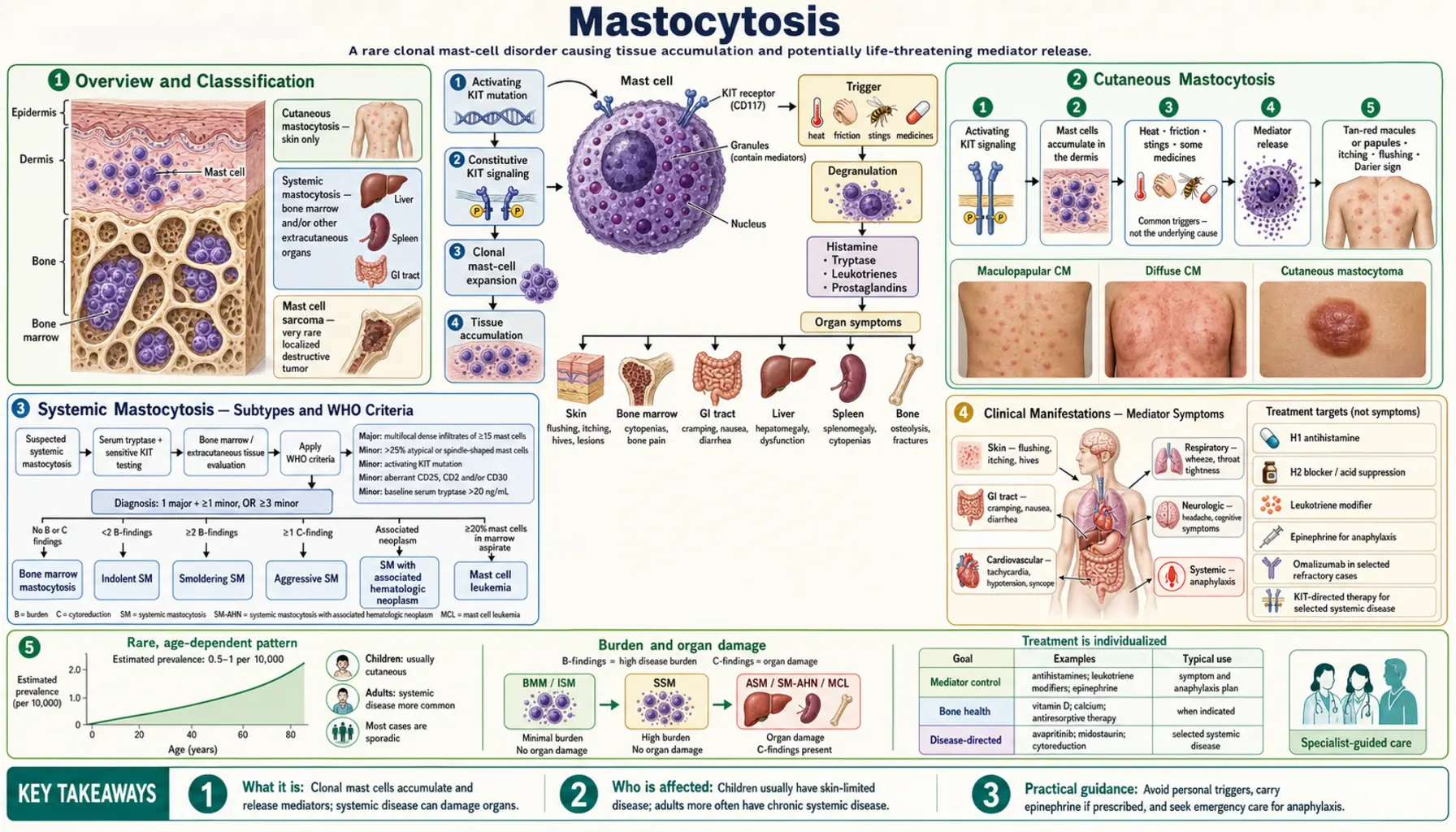
Mastocytosis is a clonal mast cell disorder caused by an activating mutation in the KIT gene (most commonly KIT D816V), which encodes the stem cell factor receptor CD117. This mutation drives ligand-independent signaling, resulting in uncontrolled mast cell proliferation and accumulation in one or more organ systems. The condition spans a wide spectrum from skin-limited disease in children to aggressive systemic forms in adults requiring targeted kinase inhibitor therapy.
Interactive Visualization Histamine, Mast Cells & Allergy — trigger a mast cell yourself Arm a mast cell with immunoglobulin E (IgE), let the allergen cross-link it, and watch histamine burst out to raise a wheal — then block the H1 receptor and see symptoms fall while histamine still pours out. Launch →Table of Contents
- Overview and Classification
- Cutaneous Mastocytosis
- Systemic Mastocytosis — Subtypes and WHO Criteria
- Clinical Manifestations — Mediator Symptoms
- Bone Manifestations and Osteoporosis
- Diagnosis — Laboratory and Pathology
- Treatment — Symptomatic Management
- Treatment — Advanced Systemic Mastocytosis
- Hereditary Alpha-Tryptasemia
- Key Research Papers
- Connections
- Featured Videos
Overview and Classification
Mastocytosis arises from a clonal expansion of mast cells carrying an activating mutation, most commonly KIT D816V — a single amino acid substitution (aspartate to valine at position 816) in the activation loop of the KIT tyrosine kinase receptor. This substitution renders the receptor constitutively active without requiring stem cell factor (SCF) ligand binding, driving autonomous mast cell proliferation.
The KIT gene encodes CD117, the stem cell factor receptor expressed on mast cells, hematopoietic progenitors, and melanocytes. In normal physiology, SCF binding induces receptor dimerization and downstream signaling through RAS/MAPK and PI3K/AKT pathways. The D816V mutation bypasses this requirement. This mutation is found in over 90% of adult systemic mastocytosis cases and in a significant proportion of pediatric cases.
The two major categories of mastocytosis are:
- Cutaneous Mastocytosis (CM): mast cell accumulation confined to the skin; predominantly affects children; most cases resolve by puberty
- Systemic Mastocytosis (SM): mast cell infiltration of extracutaneous organs, most critically the bone marrow; predominantly affects adults; requires specific WHO diagnostic criteria for classification and subtyping
Cutaneous Mastocytosis
Cutaneous mastocytosis is the most common form of mastocytosis in children and is characterized by mast cell infiltration of the dermis without systemic organ involvement.
Urticaria Pigmentosa (Maculopapular Cutaneous Mastocytosis)
Urticaria pigmentosa is the most frequent form of CM, also termed maculopapular cutaneous mastocytosis (MPCM). It presents as tan or reddish-brown macules and papules distributed across the trunk, extremities, and occasionally the face. These lesions represent focal dermal accumulations of mast cells. In children, lesions tend to be fewer and larger; in adults (who can develop CM as a variant of systemic disease), lesions are typically more numerous and smaller.
Darier's Sign
Darier's sign is the pathognomonic physical finding in mastocytosis. When a skin lesion is firmly stroked, the mechanical stimulation triggers mast cell degranulation, releasing histamine and other mediators locally. This produces a localized wheal-and-flare reaction (urticaria) overlying the stroked lesion within minutes. A positive Darier's sign strongly supports the diagnosis of cutaneous mastocytosis and distinguishes mast cell infiltrates from other pigmented skin lesions such as lentigines or melanocytic nevi.
Pediatric Course and Prognosis
Childhood cutaneous mastocytosis carries an excellent prognosis. The majority of children with CM — particularly those with onset in infancy — experience spontaneous regression of skin lesions by puberty as mast cell burden naturally decreases. Systemic involvement is minimal in pure CM. Known mast cell triggers in children include heat, friction and certain medications, which can provoke mediator release and symptomatic episodes, including flushing and, rarely, anaphylaxis. In a German series of 120 consecutive patients, 9% of the children had ever had anaphylaxis, and all of them had extensive skin involvement (Brockow 2008).
Other CM Subtypes
- Diffuse cutaneous mastocytosis (DCM): rare, occurs in infants; widespread mast cell infiltration of the entire skin surface; can cause extensive blistering (bullous formation) due to massive mast cell degranulation; more clinically severe than MPCM
- Solitary mastocytoma: a single mast cell tumor nodule, usually appearing in the first months of life; most resolve spontaneously; generally excellent prognosis; biopsy confirms the diagnosis if needed
Systemic Mastocytosis — Subtypes and WHO Criteria
Systemic mastocytosis is defined by clonal mast cell infiltration of extracutaneous organs. The bone marrow is the primary site of involvement and the key diagnostic organ. SM is predominantly a disease of adults; the KIT D816V mutation is present in the vast majority of cases.
WHO 2022 Diagnostic Criteria
The diagnosis of SM requires fulfillment of either 1 major criterion plus 1 minor criterion, or at least 3 minor criteria. The list below follows the updated criteria of the 2021 international consensus proposal (Valent 2021):
Major criterion:
- Multifocal dense mast cell aggregates (clusters of 15 or more mast cells) in bone marrow biopsy sections and/or in sections of other extracutaneous organs
Minor criteria:
- 25% or more of the mast cells are spindle-shaped or have atypical morphology
- Detection of an activating KIT point mutation at codon 816 (typically D816V) or in other critical regions of KIT, in bone marrow or another extracutaneous organ
- Mast cells in bone marrow, blood, or another extracutaneous organ express one or more of CD2, CD25 and CD30, in addition to normal mast cell markers (CD117, tryptase); CD30 was added as a marker in the 2021 update
- Serum total tryptase persistently exceeds 20 ng/mL (does not count if there is an unrelated myeloid neoplasm; in a person with known hereditary alpha-tryptasemia the tryptase level is adjusted for the extra alpha-tryptase gene copies)
The 2021 update also defines bone marrow mastocytosis as SM without skin lesions and without B- or C-findings.
SM Subtypes (in Order of Increasing Severity)
- Indolent SM (ISM): fulfills SM criteria; no C-findings (organ damage); no evidence of associated hematologic neoplasm; normal peripheral blood counts; best prognosis among SM subtypes; near-normal life expectancy; symptoms driven primarily by mediator release rather than organ infiltration
- Smoldering SM (SSM): higher mast cell burden than ISM (often serum tryptase >200 ng/mL, or >30% mast cells on BM biopsy, or splenomegaly without C-findings); no frank organ damage yet; intermediate risk of progression
- SM with Associated Hematologic Neoplasm (SM-AHN): formerly called SM-AHNMD; SM occurs alongside a separate clonal myeloid or lymphoid neoplasm (most commonly chronic myelomonocytic leukemia, myelodysplastic syndrome, or acute myeloid leukemia); prognosis driven largely by the associated neoplasm
- Aggressive SM (ASM): presence of one or more C-findings indicating organ damage attributable to mast cell infiltration (see below); requires cytoreductive therapy
- Mast Cell Leukemia (MCL): the rarest and most severe form; defined by circulating mast cells comprising 20% or more of the white cell differential on peripheral blood smear; alternatively, >20% mast cells on bone marrow aspirate smear regardless of peripheral blood; median survival historically measured in months; currently treated with avapritinib
C-Findings (Organ Damage Markers)
C-findings represent end-organ damage attributable to mast cell infiltration and define the threshold between smoldering/indolent and aggressive disease:
- Absolute neutrophil count below 1.0 × 10&sup9;/L
- Hemoglobin below 10 g/dL
- Platelet count below 100 × 10&sup9;/L
- Alanine aminotransferase (ALT) or aspartate aminotransferase (AST) elevated more than 3-fold above the upper limit of normal with or without ascites
- Serum albumin below 3.5 g/dL
- Pathologic fractures due to mast cell-induced osteolysis
- Large liver or spleen with portal hypertension and impaired liver function
- Malabsorption with weight loss due to gastrointestinal (GI) mast cell infiltration
Clinical Manifestations — Mediator Symptoms
A defining feature of mastocytosis — across all subtypes — is the release of preformed and newly synthesized mast cell mediators. These episodes can occur spontaneously or be provoked by identifiable triggers. The main mediators involved include histamine, tryptase, prostaglandin D2, heparin, leukotrienes (LTC4, LTD4), and cytokines (interleukin-6 (IL-6), tumor necrosis factor-alpha).
Flushing
Episodic flushing — sudden reddening and warmth of the face, neck, and upper chest — is one of the most common presenting symptoms. Flushing in mastocytosis is driven primarily by histamine and prostaglandin D2 rather than the catecholamine-mediated flushing of pheochromocytoma. Common triggers include:
- Heat (hot baths, hot weather, exercise)
- Alcohol — even small quantities
- Emotional stress
- Nonsteroidal anti-inflammatory drugs (especially aspirin, which can trigger massive prostaglandin-related flushing)
- Opioid analgesics (particularly codeine and morphine, which directly stimulate mast cell degranulation)
- Iodinated contrast media (intravenous dye)
- Hymenoptera venom (bee and wasp stings)
- Physical friction to skin lesions
Urticaria and Pruritus
Generalized or localized itching is nearly universal in cutaneous disease and common in SM. Mechanical stimulation (Darier's sign) can provoke hive formation at lesion sites. Aquagenic urticaria (itching triggered by water contact) also occurs in some patients.
Gastrointestinal Symptoms
Gastrointestinal mast cell accumulation produces multiple effects. Histamine stimulates gastric acid hypersecretion via H2 receptors on parietal cells, leading to peptic ulcer disease, epigastric pain, and reflux. Mast cell infiltration of the intestinal wall causes cramping, bloating, diarrhea, and in severe cases malabsorption with weight loss. Abdominal pain is among the most debilitating symptoms for many patients with SM and frequently requires multi-drug management.
Neurological and Cognitive Symptoms
Headache, difficulty concentrating, and the constellation of symptoms collectively termed "brain fog" are reported frequently. These symptoms may be histamine-mediated (histamine crosses the blood-brain barrier and modulates neurological function via H1 and H3 receptors) or related to prostaglandin and cytokine release. Depression and anxiety are also increased in mastocytosis populations.
Anaphylaxis
Anaphylaxis — systemic, potentially life-threatening mast cell degranulation — is a major concern in all SM patients. Several features are distinctive:
- SM patients, including those with indolent disease, have a paradoxically high rate of severe anaphylaxis relative to their overall disease burden
- Hymenoptera venom (bee, wasp, yellow jacket) is the classic and most dangerous trigger; venom-triggered anaphylaxis in mastocytosis can be fatal without rapid epinephrine administration
- Anaphylaxis may also occur without an identifiable trigger (idiopathic)
- Standard anaphylaxis symptoms apply: urticaria, angioedema, bronchospasm, hypotension, loss of consciousness
- In a German study of 120 consecutive patients, anaphylaxis had occurred in 49% of adults but only 9% of children, and in 56% of adults with systemic disease versus 13% with skin-only disease; 48% of the adults' reactions were severe and 38% caused unconsciousness. The authors recommend an emergency set of medication that includes epinephrine (adrenaline).
- In the same study, the triggers adults named most often were insect stings (19%), foods (16%) and medicines (9%), and in 26% of reactions only a combination of different triggers came before the attack. No trigger was identified in 67% of reactions in children versus 13% in adults. People who had had anaphylaxis had higher baseline tryptase (average 60.2 ng/mL) than those who had not (21.2 ng/mL).
Importantly, serum tryptase measured 1-3 hours after an anaphylactic episode is markedly elevated in virtually all SM patients (due to the large mast cell burden); a persistently elevated tryptase weeks after the acute event suggests SM as the underlying diagnosis.
Bone Manifestations and Osteoporosis
Skeletal disease is a major and underrecognized complication of systemic mastocytosis, even in the indolent subtype. Mast cells release multiple mediators that directly perturb bone remodeling:
- Histamine stimulates osteoclast activity via H1 and H2 receptors
- Heparin promotes osteoclast differentiation and inhibits osteoblast function
- Tryptase activates protease-activated receptors on bone cells
- Interleukin-6 and tumor necrosis factor-alpha increase RANKL expression, further driving osteoclast-mediated bone resorption
Osteoporosis
Osteoporosis occurs at significantly higher rates in SM patients compared to age-matched controls. Fractures — including vertebral compression fractures — are common and can be the presenting manifestation of previously undiagnosed SM. The lumbar spine is most frequently affected. Osteoporosis in SM can be severe even in young patients and in the absence of other C-findings, which is why bone density is commonly checked in all SM patients regardless of subtype. A 2016 review describes osteoporosis as one of the most frequent manifestations of SM, particularly in adults, often complicated by repeated fragility fractures, mainly of the vertebrae (Rossini 2016).
A DEXA (dual-energy X-ray absorptiometry) scan is commonly done at the time of diagnosis in SM. Bisphosphonate therapy (alendronate orally, or zoledronate intravenously for severe cases) is used for patients with low bone mineral density (T-score below -2.5) or with prevalent fragility fractures, and DEXA is commonly repeated each year during treatment. Calcium and vitamin D supplements are commonly added.
Osteosclerosis
A paradoxical increase in bone density — osteosclerosis — occurs in a subset of SM patients, particularly affecting the axial skeleton (spine, pelvis). This results in abnormally dense, thickened trabeculae on imaging. Patients may simultaneously have osteosclerotic regions alongside osteoporotic regions, creating a mixed lytic/sclerotic pattern on radiographs or computed tomography (CT) scans. Osteosclerosis is more common in patients with higher mast cell burden and advanced SM subtypes. Despite the radiographic density, structural bone quality is abnormal and fracture risk may still be elevated.
Radiologic Assessment
Plain radiographs of the spine and pelvis can identify mixed lytic/sclerotic changes. Computed tomography provides higher resolution detail of bone architecture. A nuclear medicine bone scan (technetium-99m) can help identify sites of active bone turnover. Magnetic resonance imaging (MRI) of the spine is sensitive for detecting mast cell infiltration of vertebral marrow (hypointense on T1, variable on T2) and associated marrow edema.
Diagnosis — Laboratory and Pathology
Diagnosis of mastocytosis integrates clinical assessment, laboratory studies, imaging, and tissue biopsy. A systematic approach is essential given the heterogeneity of presentations.
Serum Tryptase
Serum total tryptase is the most clinically useful biomarker for mastocytosis. Tryptase is a serine protease stored in mast cell secretory granules. In healthy individuals, baseline serum tryptase is typically below 11.4 ng/mL. Key interpretive points:
- A persistently elevated baseline tryptase above 20 ng/mL (measured at least 24 hours after any acute event) is a minor diagnostic criterion for SM
- Tryptase levels are proportional to total body mast cell burden and correlate with SM subtype severity
- Transient elevation during anaphylaxis (peak at 1-3 hours, returning to baseline within 24 hours) contrasts with the persistent elevation seen in SM
- Hereditary alpha-tryptasemia (HαT) can cause elevated baseline tryptase without mastocytosis and is told apart through genetic testing
KIT D816V Mutation Testing
Detection of KIT D816V is a minor diagnostic criterion and critical for treatment selection. Testing options include:
- Peripheral blood allele-specific polymerase chain reaction (PCR): highly sensitive modern assays can detect the mutation in blood in the majority of SM patients, enabling a less invasive initial screen
- Bone marrow polymerase chain reaction: more sensitive than peripheral blood; particularly important when blood-based testing is negative but clinical suspicion remains high
- KIT D816V positivity also determines eligibility for avapritinib therapy in advanced SM
Bone Marrow Biopsy
Bone marrow trephine biopsy is required to fulfill the major diagnostic criterion for SM and to assess disease burden. A comprehensive biopsy evaluation includes:
- Hematoxylin and eosin (H&E) staining for overall morphology and detection of mast cell aggregates
- Giemsa staining for mast cell granule visualization
- Immunohistochemistry: CD117 (marks mast cells), tryptase (confirms mast cell identity), CD25 (aberrant expression in clonal mast cells — not present on normal mast cells), CD2 (variable aberrant expression)
- The finding of multifocal aggregates of 15 or more mast cells is the major diagnostic criterion
Skin Biopsy
If urticaria pigmentosa lesions are present, punch biopsy of a representative lesion confirms dermal mast cell infiltration. Staining with tryptase or CD117 highlights the accumulated mast cells. A positive skin biopsy in the appropriate clinical context (with Darier's sign) strongly supports the diagnosis and may spare some patients the need for immediate bone marrow evaluation.
Additional Laboratory Studies
- Complete blood count with differential: cytopenias (low neutrophils, anemia, thrombocytopenia) indicate C-findings; eosinophilia may accompany SM-AHN
- Comprehensive metabolic panel: liver function tests for hepatic involvement; albumin level
- 24-hour urine N-methylhistamine: elevated in active mast cell disease; useful for monitoring mediator burden
- Urinary prostaglandin D2 metabolites (11-beta-PGF2-alpha): elevation indicates active prostaglandin D2 release
- Computed tomography scan of chest/abdomen/pelvis: assesses for organomegaly (liver, spleen, lymph nodes), lymphadenopathy, ascites
- DEXA scan: bone mineral density baseline at diagnosis
Treatment — Symptomatic Management
Symptom control targeting mast cell mediator effects is the foundation of treatment for all mastocytosis patients, including those with CM and indolent SM who do not require cytoreductive therapy.
Antihistamines
Antihistamines are the usual first-line treatment for patients with symptoms:
- H1 antihistamines (cetirizine, loratadine, fexofenadine): control flushing, urticaria, pruritus, and reduce anaphylaxis risk; non-sedating agents preferred for daily use; higher doses than standard allergy dosing are often required
- H2 antihistamines (famotidine): reduce gastric acid hypersecretion, control gastrointestinal symptoms, and provide additional systemic H1-type symptom coverage; commonly taken 30 minutes before meals. Ranitidine, once used here too, is no longer available in the United States: on 1 April 2020 the U.S. Food and Drug Administration asked manufacturers to withdraw all prescription and over-the-counter ranitidine because of an impurity, N-nitrosodimethylamine (NDMA), that can increase over time.
Epinephrine Auto-Injector
The 120-patient anaphylaxis study described above concluded that adults, and children with extensive skin disease, have an increased risk of severe anaphylaxis, and its authors recommend an emergency set of medication including epinephrine (Brockow 2008). In practice, people with systemic mastocytosis — and people with skin-only disease who have had anaphylaxis or significant mediator symptoms — are commonly prescribed two epinephrine auto-injectors (0.3 mg each). Two devices are prescribed because a single dose may be insufficient for severe anaphylaxis in SM patients given their high mast cell burden. Training patients, family members and caregivers to recognize anaphylaxis and use the injector is part of this practice. The reasoning given is that using epinephrine when it turns out not to be needed carries less risk than delaying it during anaphylaxis.
Cromolyn Sodium
Oral cromolyn sodium (disodium cromoglycate) is a mast cell stabilizer taken four times daily before meals and at bedtime. It is poorly absorbed from the gastrointestinal tract, meaning its primary action is local stabilization of intestinal mast cells. It is effective for abdominal cramping, diarrhea, bloating, and abdominal pain in SM. It has minimal systemic side effects given its poor absorption.
Bisphosphonates and Bone Protection
- Oral bisphosphonates (alendronate 70 mg weekly): used for SM patients with osteoporosis (T-score < -2.5) or osteopenia with fragility fracture. A 2016 review calls bisphosphonates the most rational treatment for bone involvement in SM, while noting that only a few small studies, using many different drugs and schedules, support it (Rossini 2016)
- Intravenous zoledronate (5 mg annually): used for severe osteoporosis, poor tolerance of oral tablets, or kidney considerations within safe limits; in the same review, zoledronate had the best evidence for gains in bone density and for suppressing bone turnover
- Calcium and vitamin D3 supplements are commonly given alongside (doses often quoted are 1,000-1,200 mg calcium and 1,000-2,000 international units (IU) of vitamin D3 daily)
- DEXA scans are commonly repeated each year during treatment
Trigger Avoidance
Known triggers that patients commonly learn to recognize and keep away from include:
- Nonsteroidal anti-inflammatory drugs (NSAIDs): aspirin and other nonsteroidal anti-inflammatory drugs can trigger massive prostaglandin D2-mediated flushing and anaphylaxis in susceptible SM patients; people who have taken them before without a reaction often continue to use them
- Opioids: codeine and morphine directly stimulate mast cell degranulation through a non-IgE mechanism; fentanyl has substantially less mast cell degranulating activity and is relatively safer for procedural analgesia in mastocytosis patients
- Iodinated contrast media: premedication with an H1 antihistamine, an H2 antihistamine and a corticosteroid before computed tomography or angiographic contrast is common practice, with the radiology team told in advance
- Anesthesia: in a review of 29 anesthetic procedures in 22 children with mastocytosis, routine anesthetic techniques were used and no serious adverse events occurred; the authors concluded that departing from routine anesthesia is not necessarily needed, but that careful preparation to treat possible reactions is advised (Carter 2008)
- Temperature extremes: very hot or cold environments, sudden temperature changes
- Alcohol: even small amounts can trigger degranulation in sensitive patients
Venom Immunotherapy
Venom immunotherapy (VIT) — subcutaneous desensitization with Hymenoptera venom extract — is widely used for SM patients who have experienced venom-triggered anaphylaxis or who have venom sensitization (positive skin test or specific immunoglobulin E). Key points for SM-specific VIT:
- VIT dramatically reduces the risk of future severe anaphylaxis from Hymenoptera stings, potentially from >50% risk per sting to less than 5%
- In the general population, VIT is typically administered for 3-5 years; for SM patients, VIT is often continued lifelong because the risk of anaphylaxis recurs upon discontinuation in the setting of persistent elevated mast cell burden
- Epinephrine auto-injectors are usually still carried during VIT
Treatment — Advanced Systemic Mastocytosis
Advanced SM — including aggressive SM (ASM), SM with associated hematologic neoplasm (SM-AHN), and mast cell leukemia (MCL) — requires cytoreductive therapy to reduce mast cell burden and reverse organ damage.
Avapritinib
Avapritinib represents the most significant therapeutic advance in mastocytosis treatment. It is a highly selective type I kinase inhibitor designed specifically to inhibit the D816V mutant form of KIT. This selectivity is critical: the KIT D816V mutation is in the activation loop of the kinase and confers resistance to imatinib and other earlier-generation tyrosine kinase inhibitors that target the inactive (type II) kinase conformation.
- U.S. Food and Drug Administration (FDA) approval for advanced SM (2021): avapritinib 200 mg once daily for ASM, SM-AHN, and MCL; in the phase 1 EXPLORER trial the overall response rate was 75% in 53 response-evaluable patients (36% complete remission), and in the interim analysis of the phase 2 PATHFINDER trial it was 75% in 32 response-evaluable patients (19% complete remission) at a starting dose of 200 mg daily; in PATHFINDER, serum tryptase fell by half or more in 93% of patients and bone marrow mast cells in 88% (DeAngelo 2021; Gotlib 2021)
- U.S. Food and Drug Administration approval for indolent SM (2023): avapritinib 25 mg once daily for ISM patients with severe, refractory mediator symptoms; lower dose achieves meaningful symptom control while reducing the intracranial bleeding risk observed at higher doses
- PIONEER trial (indolent SM): 212 people with moderate-to-severe indolent SM were randomized two to one to avapritinib 25 mg once daily or placebo, both with usual symptom care. Over 24 weeks the total symptom score fell by 15.6 points on avapritinib versus 9.2 points on placebo, and 54% of people on the drug (none on placebo) had their serum tryptase cut by half or more; swelling (edema) and raised alkaline phosphatase were more common on avapritinib (Gotlib 2023)
- Common side effects: periorbital edema, dizziness, cognitive effects ("avapritinib brain fog"), nausea; intracranial bleeding is a serious concern — in EXPLORER it occurred in 13% of patients overall but in only 1% of those without severely low platelets (below 50 × 10&sup9;/L) — particularly in patients on anticoagulation
Midostaurin
Midostaurin was the first U.S. Food and Drug Administration-approved drug for advanced SM (2017). It is a multi-kinase inhibitor targeting KIT D816V along with FLT3, PKC-alpha, PKC-beta, and VEGFR. It is less KIT D816V-selective than avapritinib. In the pivotal open-label trial (89 evaluable patients with mastocytosis-related organ damage, 100 mg twice daily), the overall response rate was 60%, and 45% had a major response — complete resolution of at least one type of mastocytosis-related organ damage. Median overall survival was 28.7 months, and 9.4 months in the 16 patients with mast cell leukemia. New or worsening severe low neutrophil counts, anemia and low platelet counts occurred in 24%, 41% and 29% of patients, mostly in those whose counts were already low (Gotlib 2016). Midostaurin 100 mg twice daily (with food) is the approved regimen. Common side effects include nausea, vomiting, and fatigue. It is also approved for FLT3-mutated acute myeloid leukemia, which is relevant for SM-AHN patients with concurrent AML.
Cladribine (2-CdA)
Cladribine, a purine nucleoside analog, was a principal cytoreductive agent for ASM before the KIT inhibitor era. It induces mast cell apoptosis through incorporation into DNA during replication and inhibition of DNA repair. Cladribine reduces mast cell burden and improves C-findings in a subset of patients but produces significant immunosuppression (particularly CD4+ T cell depletion). It remains an option in resource-limited settings or in patients not eligible for KIT inhibitors.
Interferon-Alpha
Interferon-alpha (with or without corticosteroids) was used in earlier decades as cytoreductive therapy for ASM. Response rates were moderate and treatment was associated with substantial toxicity (flu-like symptoms, depression, cytopenias). With the availability of avapritinib and midostaurin, interferon-alpha has largely been displaced from frontline advanced SM treatment but may still be used in certain situations.
Allogeneic Stem Cell Transplantation
Allogeneic hematopoietic stem cell transplantation (allo-HSCT) is considered for patients with MCL or refractory ASM who are medically fit for the procedure and have a suitable donor. Allo-HSCT can achieve long-term remission but carries substantial treatment-related mortality (20-30% in most series for advanced SM patients given their typically older age and organ dysfunction). The availability of effective KIT inhibitors has altered the treatment sequencing, and allo-HSCT is generally reserved for patients who fail or are intolerant to avapritinib-based therapy.
Hereditary Alpha-Tryptasemia
Hereditary alpha-tryptasemia (HαT) is a recently characterized germline condition that needs to be told apart from mastocytosis in patients with elevated baseline serum tryptase and systemic symptoms.
Genetics and Mechanism
HαT is caused by increased copy numbers of the TPSAB1 gene, which encodes alpha-tryptase. Normally, individuals carry two copies of TPSAB1 (one per chromosome). In HαT, one or both chromosomes carry additional TPSAB1 copies (duplex, triplex, or tetraplex), resulting in constitutively increased tryptase production even in the absence of mast cell clonal expansion. HαT follows an autosomal dominant inheritance pattern and is present in approximately 5% of the general population (4.4% of the control group in a 2021 study, Greiner 2021).
Clinical Presentation
HαT produces a multisystem symptom complex that substantially overlaps with mastocytosis and mast cell activation syndrome:
- Elevated baseline serum tryptase (often 8-40 ng/mL), sometimes exceeding the 20 ng/mL minor SM criterion
- Flushing, urticaria, pruritus
- Gastrointestinal symptoms including abdominal pain and diarrhea
- Connective tissue abnormalities (joint hypermobility, which may satisfy criteria for hypermobile Ehlers-Danlos syndrome)
- Autonomic dysfunction (orthostatic intolerance, postural orthostatic tachycardia syndrome-like features)
- Atopy and allergic sensitivities
Distinguishing HαT from Mastocytosis
The distinction is critical because the prognosis and management differ substantially:
- In HαT: KIT D816V mutation is absent; bone marrow biopsy does not show mast cell aggregates; WHO SM criteria are not fulfilled; no clonal mast cell disease
- In SM: KIT D816V typically present; BM biopsy shows aggregates; WHO criteria are met
- TPSAB1 copy number genotyping (available as a clinical laboratory test) confirms HαT
- HαT and SM can coexist in the same patient, and HαT may amplify the tryptase elevation of SM, making the serum tryptase threshold less specific in this context. In a 2021 study of 180 mastocytosis patients and 180 sex-matched controls, HαT was found in 17.2% of the people with mastocytosis versus 4.4% of controls; mastocytosis patients with HαT had higher tryptase (median 49.6 versus 34.5 ng/mL) and had insect-venom reactions and severe heart-and-circulation reactions or anaphylaxis far more often, and the authors propose HαT as a marker of a person's risk of severe anaphylaxis (Greiner 2021)
Management of HαT focuses on symptom control (antihistamines, avoiding triggers) rather than cytoreductive therapy; no mast cell cytoreduction is indicated because there is no clonal expansion.
Key Research Papers
- Valent P, Horny HP, Escribano L, et al. "Diagnostic criteria and classification of mastocytosis: a consensus proposal." Leuk Res. 2001;25(7):603-625. — PubMed PMID: 11377686
- Valent P, Akin C, Hartmann K, et al. "Updated Diagnostic Criteria and Classification of Mast Cell Disorders: A Consensus Proposal." HemaSphere. 2021;5(11):e646. — PubMed PMID: 34901755
- Pardanani A. "Systemic mastocytosis in adults: 2019 update on diagnosis, risk stratification and management." Am J Hematol. 2019;94(3):363-377. — PubMed PMID: 30536695
- Gotlib J, Reiter A, Radia DH, et al. "Efficacy and safety of avapritinib in advanced systemic mastocytosis: interim analysis of the phase 2 PATHFINDER trial." Nat Med. 2021;27(12):2192-2199. — PubMed PMID: 34873345
- DeAngelo DJ, Radia DH, George TI, et al. "Safety and efficacy of avapritinib in advanced systemic mastocytosis: the phase 1 EXPLORER trial." Nat Med. 2021;27(12):2183-2191. — PubMed PMID: 34873347
- Gotlib J, Kluin-Nelemans HC, George TI, et al. "Efficacy and Safety of Midostaurin in Advanced Systemic Mastocytosis." N Engl J Med. 2016;374(26):2530-2541. — PubMed PMID: 27355533
- Greiner G, Sprinzl B, Górska A, et al. "Hereditary α tryptasemia is a valid genetic biomarker for severe mediator-related symptoms in mastocytosis." Blood. 2021;137(2):238-247. — PubMed PMID: 32777817
- Gotlib J, Castells M, Elberink HO, et al. "Avapritinib versus Placebo in Indolent Systemic Mastocytosis." NEJM Evid. 2023;2(6):EVIDoa2200339. — PubMed PMID: 38320129
- Brockow K, Jofer C, Behrendt H, Ring J. "Anaphylaxis in patients with mastocytosis: a study on history, clinical features and risk factors in 120 patients." Allergy. 2008;63(2):226-232. — PubMed PMID: 18186813
- Rossini M, Zanotti R, Orsolini G, et al. "Prevalence, pathogenesis, and treatment options for mastocytosis-related osteoporosis." Osteoporos Int. 2016;27(8):2411-2421. — PubMed PMID: 26892042
- Carter MC, Uzzaman A, Scott LM, Metcalfe DD, Quezado Z. "Pediatric mastocytosis: routine anesthetic management for a complex disease." Anesth Analg. 2008;107(2):422-427. — PubMed PMID: 18633019
- Akin C. "Mast cell activation syndromes." J Allergy Clin Immunol. 2017;140(2):349-355. — PubMed PMID: 28780942
PubMed Topic Searches
Connections
- Pain & Allergy
- Histamine, Mast Cells & Allergy — interactive animation
- Mast Cell Activation Syndrome (MCAS)
- Allergies
- Anaphylaxis
- Hereditary Angioedema
- Common Variable Immunodeficiency
- Mast Cell Activation Syndrome: History and Discovery
- Chronic Granulomatous Disease
- Tryptase — serum total tryptase is the primary biomarker used to screen for and monitor mastocytosis.