Osteomyelitis
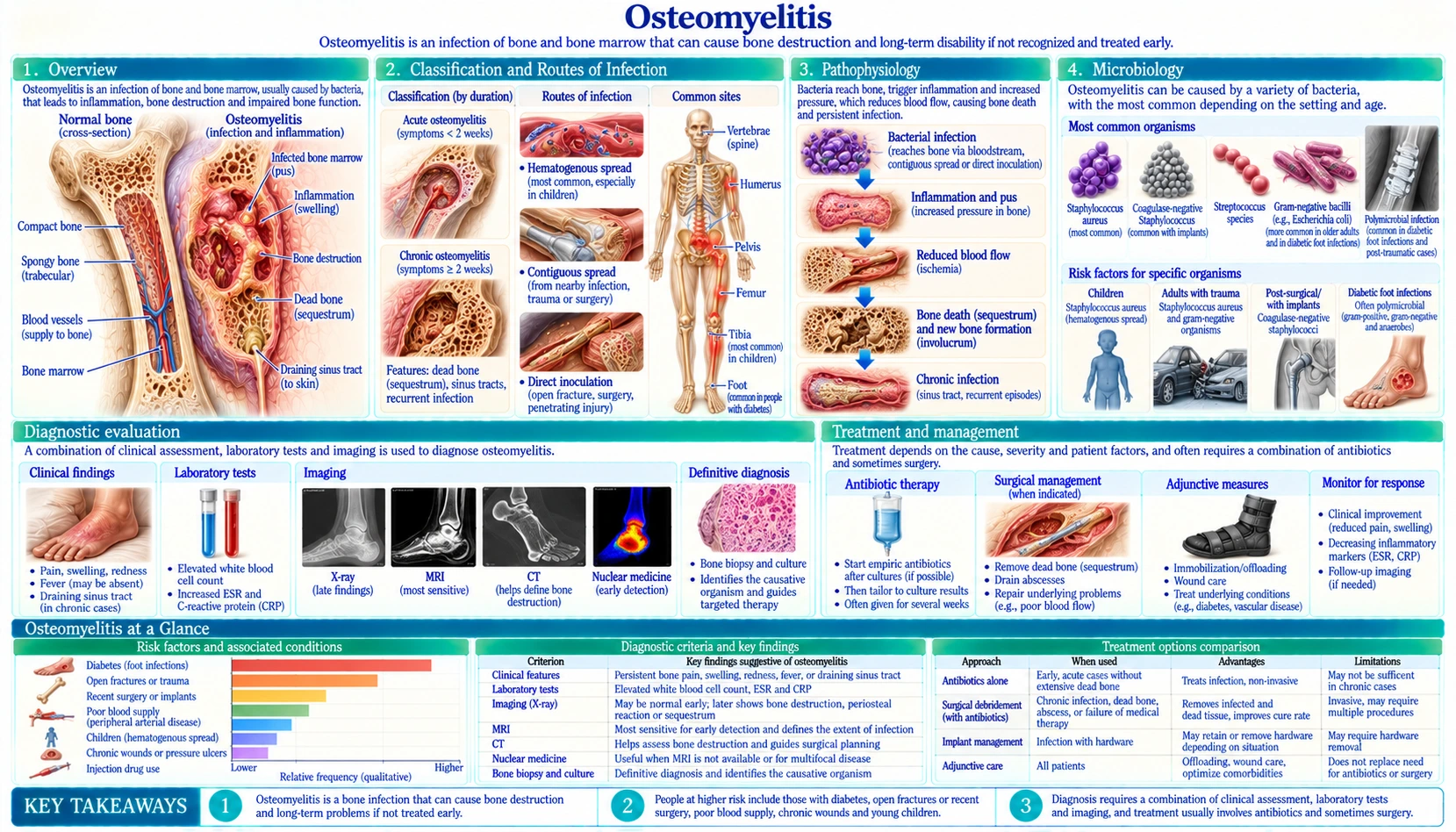
Table of Contents
1. Overview
Osteomyelitis is an infection of bone. A 2004 review in The Lancet (Lew and Waldvogel) notes that the high success rates antimicrobial therapy achieves in most infectious diseases have not yet been achieved in bone and joint infections, owing to the physiological and anatomical characteristics of bone. Infected bone can die (form a sequestrum), shelter bacteria inside biofilm, and become the site of an inflammatory response that adds to local damage.
The condition spans a wide spectrum, from an acute, bloodstream-seeded infection in a healthy child to a long-standing chronic infection with draining sinus tracts and dead bone. Lew and Waldvogel describe chronic osteomyelitis as associated with avascular necrosis of bone and sequestrum formation, with surgical debridement necessary for cure in addition to antibiotics, whereas acute osteomyelitis can respond to antibiotics alone. The type of osteomyelitis shapes the treatment strategy: medical or surgical, and how long antibiotics are given.
In a population-based study of 760 cases in Olmsted County, Minnesota, from 1969 to 2009 (Kremers 2015), the age- and sex-adjusted annual incidence of osteomyelitis was 21.8 per 100,000 person-years. It rose from 11.4 per 100,000 in 1969–1979 to 24.4 per 100,000 in 2000–2009; the rise was concentrated in people older than sixty and was partly driven by diabetes-related osteomyelitis (2.3 to 7.6 per 100,000). The authors called the reasons unclear, possibly including changes in diagnosing patterns or a higher prevalence of risk factors such as diabetes. For vertebral osteomyelitis alone, French national hospital data for 2002–2003 gave an incidence of 2.4 per 100,000 (Grammatico 2008).
2. Classification and Routes of Infection
A common way to classify osteomyelitis is by route of bacterial entry. Lew and Waldvogel list the types in order of decreasing frequency: secondary to a contiguous focus of infection (after trauma, surgery, or insertion of a joint prosthesis), secondary to vascular insufficiency (in diabetic foot infections), and of hematogenous (bloodstream) origin.
Hematogenous osteomyelitis
Bacteria reach bone via the bloodstream from a distant site — a urinary tract infection, intravenous catheter, skin furuncle, endocarditis, or occult bacteremia. This is the classic route in children, where the anatomy of the metaphyseal microvasculature creates a uniquely susceptible niche:
- The terminal capillaries of the metaphysis make sharp turns back toward the growth plate, creating sluggish flow and functional sinusoids.
- These sinusoids lack phagocytic lining cells, so circulating bacteria can adhere and multiply without immediate immune clearance.
- The long bones are therefore the usual site in children; a 2012 systematic review of children's hematogenous osteomyelitis (Dartnell) found the tibia and femur the most commonly affected long bones and Staphylococcus aureus the most common organism detected.
In adults, hematogenous seeding most commonly targets the vertebral bodies (vertebral osteomyelitis / spondylodiscitis), reflecting the rich arterial supply of the vertebral end-plates. The infection typically begins at the end-plate, spreads across the disc space to the adjacent vertebral body, and can extend into the epidural space causing an abscess that threatens the spinal cord.
Contiguous-focus osteomyelitis
Bacteria reach bone from an adjacent soft-tissue infection. The classic example is the diabetic foot: a plantar ulcer overlying a bony prominence becomes chronically infected; bacteria erode through soft tissue to invade the underlying metatarsal or phalanx. Vascular disease and peripheral neuropathy (reducing pain sensation and impairing wound healing) contribute in people with diabetes. In the probe-to-bone test, an ulcer is probed with a sterile blunt metal probe to feel for bone. In a 1995 study of 76 infected foot ulcers in 75 hospitalized diabetic patients (Grayson), osteomyelitis was present in 66% of ulcers, and palpating bone had a sensitivity of 66%, a specificity of 85% and a positive predictive value of 89%. A 2016 systematic review of seven studies (Lam) pooled a sensitivity of 0.87 and a specificity of 0.83, and concluded that the test can rule in osteomyelitis in high-risk patients and rule it out in low-risk patients.
Direct inoculation osteomyelitis
Open fractures, penetrating trauma (lawn-mower injuries, gun-shot wounds), bite wounds, and orthopedic procedures (joint replacement, internal fixation of fractures, spinal surgery) introduce bacteria directly into bone. Biofilm formation on hardware is a central challenge in implant-associated infection.
Acute and chronic osteomyelitis
Osteomyelitis is also described as acute or chronic (Lew and Waldvogel 2004):
- Acute osteomyelitis can respond to antibiotics alone.
- Chronic osteomyelitis is associated with avascular necrosis of bone and formation of sequestrum (dead bone); surgical debridement is necessary for cure in addition to antibiotic therapy.
3. Pathophysiology
Once bacteria establish a foothold in bone, a destructive cycle begins:
- Bacterial seeding and initial multiplication. S. aureus is particularly adept at bone invasion — it expresses surface adhesins (fibronectin-binding proteins, bone sialoprotein receptors) that allow it to attach to bone matrix proteins. The bacteria initially proliferate in a planktonic (free-floating) state before transitioning to biofilm communities embedded in polysaccharide matrix.
- Inflammatory response and ischemia. Bacterial products activate the innate immune response, triggering a massive neutrophil influx and cytokine release (IL-1, IL-6, TNF-α). The resulting edema and purulent exudate raise intraosseous pressure, compressing the already tenuous cortical blood supply. Subperiosteal pus strips the periosteum from the cortex, further disrupting the outer cortical blood supply.
- Cortical necrosis and sequestrum formation. Segments of cortical bone that lose their blood supply die, becoming a sequestrum — a fragment of avascular dead bone that cannot be resorbed by the body and acts as a protected reservoir for bacteria.
- Involucrum. The periosteum attempts to wall off the infection by forming new reactive bone around the infected area, creating a shell of new bone called the involucrum. Openings in the involucrum (cloacae) allow pus and bone fragments to drain to the skin surface through sinus tracts, the hallmark of chronic osteomyelitis.
- Biofilm persistence. Bacteria within the sequestrum exist in slow-metabolizing biofilm communities, with reduced susceptibility to antibiotics. Lew and Waldvogel describe surgical debridement as necessary, in addition to antibiotics, to cure chronic osteomyelitis with sequestrum.
4. Microbiology
The causative organism varies considerably with patient age, risk factors, and route of infection:
- Staphylococcus aureus was the most commonly isolated organism in a 2009 systematic review of pyogenic vertebral osteomyelitis (Mylona) and the most common organism detected in a 2012 systematic review of children's hematogenous osteomyelitis (Dartnell). In the Olmsted County population study, 44% of all osteomyelitis cases involved S. aureus (Kremers 2015).
- Source of infection in vertebral osteomyelitis: in the Mylona review, the urinary tract was the commonest source of infection and diabetes mellitus the most common underlying illness.
- Salmonella and sickle cell disease: a 2023 systematic review of osteomyelitis in sickle cell anemia (Hegeman) found nontyphoid Salmonella the most common pathogen cultured (117 of 192 cases, 60.9%), followed by S. aureus (41 of 192, 21.8%). Salmonella was most often identified in children under 12 presenting with acute osteomyelitis; the S. aureus cases presented at an older average age.
- Mycobacterium tuberculosis causes tuberculous spondylitis (Pott's disease). In French national hospital data for 2002–2003, M. tuberculosis was reported in 31% of vertebral osteomyelitis cases and Staphylococcus species in 38% (Grammatico 2008).
- Kingella kingae is being isolated increasingly in children (Dartnell 2012). A 2011 review (Yagupsky) reports that in previously healthy children more than 95% of K. kingae infections are diagnosed between 6 and 48 months of age, that the organism colonizes the posterior pharynx, that the presentation is often subtle and may come with normal acute-phase reactants, and that it is usually susceptible to beta-lactam antibiotics, with infections typically responding well to medical treatment.
5. Clinical Presentation
Hematogenous osteomyelitis in children
The typical picture is a child with acute focal bone pain, tenderness, and unwillingness to use the affected limb, often with swelling and warmth over the bone. Fever is not universal: in the Dartnell 2012 systematic review, 40% of children were afebrile on admission. In neonates, the presentation can be subtle — pseudoparalysis (not moving the limb) and irritability without striking fever.
Vertebral osteomyelitis in adults
A 2009 systematic review of 14 studies and 1,008 patients with pyogenic vertebral osteomyelitis (Mylona) summarizes the presentation:
- Back pain and fever were the most common presenting symptoms; 62% of patients were men, and the lumbar spine was the most commonly affected area.
- Delayed diagnosis is common because of the insidious nature and rarity of the disease (Pupaibool 2015).
- Neurologic deficits and relapses were each described in 32% of cases; attributable mortality was 6%.
- Associated conditions: diabetes mellitus was the most common underlying illness in the Mylona review. In French hospital data, the median age was 59, incidence increased with age, and the most frequent comorbidities were septicaemia (27%) and endocarditis (9%) (Grammatico 2008).
Diabetic foot osteomyelitis
The challenge here is that osteomyelitis often coexists with Charcot neuroarthropathy and neuropathic ulceration in a limb that lacks pain sensation. Swelling, redness, warmth, and purulent drainage from a foot ulcer are the presenting features. The diagnostic question is whether the underlying bone is infected, inflamed from Charcot changes, or both.
6. Diagnosis
Laboratory studies
Inflammatory markers (ESR and CRP), white blood cell count and blood cultures are part of the work-up. In children, the Dartnell 2012 systematic review found that clinical examination, blood tests and radiological tests are only reliable for diagnosis in combination.
Blood cultures can identify the organism without a bone sample. In the Mylona review of vertebral osteomyelitis, CT-guided or open biopsy yielded the causative organism more often than blood cultures (77% versus 58%).
Imaging
- Plain radiographs: periosteal reaction, lytic lesions and sequestrum are radiographic findings of osteomyelitis. In the Mylona review of vertebral osteomyelitis, plain radiography showed abnormalities in 89% of cases, while bone scanning and CT or MRI were positive in 94%.
- MRI shows marrow and soft-tissue changes; in vertebral osteomyelitis the typical pattern involves the adjacent vertebral end-plates and the disc, and contrast enhancement shows abscesses and epidural extension. In a 2005 meta-analysis of imaging for chronic osteomyelitis (Termaat), MRI had a pooled sensitivity of 84% and specificity of 60%.
- CT shows cortical destruction and sequestrum, and is used to guide needle biopsy. In a 2015 meta-analysis of image-guided spinal biopsy for spontaneous vertebral osteomyelitis (Pupaibool), biopsy had a sensitivity of 52.2% and a specificity of 99.9%.
- Nuclear medicine: in the Termaat meta-analysis of chronic osteomyelitis, bone scintigraphy had a sensitivity of 82% but the lowest specificity (25%); leukocyte scintigraphy had a sensitivity of 61% overall (84% in the peripheral skeleton, 21% in the axial skeleton) and a specificity of 77%; and FDG-PET was the most sensitive technique (96%) with a specificity of 91%. The authors concluded that FDG-PET has the highest diagnostic accuracy for chronic osteomyelitis and is superior to leukocyte scintigraphy in the axial skeleton.
Bone biopsy and culture
Lew and Waldvogel describe early diagnosis, including bone sampling for microbiological and pathological examination, as the key to targeted, long-lasting antimicrobial therapy. For vertebral osteomyelitis, the microbiological yield of image-guided needle biopsy reported across studies varies between 36% and 91% (Pupaibool 2015); its pooled sensitivity of 52.2% and specificity of 99.9% mean it performs well in confirming the diagnosis but only moderately in ruling it out.
7. Antibiotic Treatment
The cornerstone of osteomyelitis treatment is prolonged antibiotic therapy tailored to the causative organism and its susceptibility profile. Key principles:
Duration
In a French open-label non-inferiority trial of 359 adults with microbiologically confirmed pyogenic vertebral osteomyelitis (Bernard 2015, PubMed PMID: 25468170), 6 weeks of antibiotics was non-inferior to 12 weeks: 90.9% of patients in each group were classified as cured at 1 year. A 2012 review of chronic osteomyelitis in adults (Spellberg and Lipsky) found no evidence that antibiotic therapy for more than 4–6 weeks improves outcomes compared with shorter regimens, and called the optimal duration uncertain. For children, the Dartnell 2012 systematic review reports that antibiotic treatment is usually sufficient to eradicate the infection, with a short intravenous course and early conversion to oral treatment.
The OVIVA trial (Li 2019, PubMed PMID: 30699315) randomly assigned 1,054 adults treated for bone or joint infection at 26 UK centres to oral or intravenous antibiotics for the first 6 weeks of therapy. Definitive treatment failure within 1 year occurred in 14.6% of the intravenous group and 13.2% of the oral group, meeting the trial's non-inferiority criterion; serious adverse events did not differ significantly, and catheter complications were more common with intravenous therapy (9.4% vs 1.0%). The Spellberg and Lipsky review likewise found that oral and parenteral therapies achieve similar cure rates in chronic osteomyelitis.
Choice of antibiotic
Antibiotic choice depends on the organism identified and its susceptibility profile. The Spellberg and Lipsky review notes that oral antibiotics are available that achieve adequate levels in bone, that oral therapy is a reasonable choice for osteomyelitis caused by susceptible organisms, and that adding rifampin to other antibiotics may improve cure rates. Kingella kingae is usually susceptible to beta-lactam antibiotics (Yagupsky 2011). The 2015 Infectious Diseases Society of America (IDSA) guideline on native vertebral osteomyelitis in adults (Berbari 2015) contains evidence- and opinion-based recommendations on diagnosis and on management with antimicrobial therapy, with or without surgery. Decisions about specific antibiotics and doses belong with a clinician.
8. Surgical Management
Not all osteomyelitis is treated surgically. Situations in which surgery is used include:
- Chronic osteomyelitis with sequestrum: Lew and Waldvogel describe surgical debridement (removal of the dead bone) as necessary for cure in addition to antibiotics.
- Failure of antibiotic therapy for acute osteomyelitis.
- Subperiosteal or soft-tissue abscess requiring drainage.
- Vertebral osteomyelitis with epidural abscess causing neurologic deficit: surgical decompression.
- Vertebral instability from extensive bone destruction: instrumented spinal fusion.
- Diabetic foot osteomyelitis: a 2017 review (Senneville and Robineau) notes that surgical removal of all infected bone was once considered standard, but that a medical approach has now proven efficacy in selected situations; the two approaches are described as complementary, and the review expects antibiotic-resistant gram-negative bacteria to narrow the place of the medical approach. Amputation is used in some cases.
- Implant-associated osteomyelitis: DAIR (Debridement, Antibiotics, and Implant Retention) is an option used for early infections with a stable implant; other cases are managed with implant removal, dead-space management (antibiotic-loaded cement spacer) and later re-implantation.
Dead-space management after radical debridement is a key surgical challenge. Options include antibiotic-impregnated calcium sulfate or hydroxyapatite beads (release high local antibiotic concentrations), muscle flap coverage (imports vascularized tissue to a poorly perfused area), and bone transport via the Ilizarov method for large segmental defects.
9. Complications and Prognosis
Reported complications of osteomyelitis include:
- Septicemia and septic shock: Hematogenous osteomyelitis, particularly in neonates and immunocompromised patients, can progress rapidly to life-threatening bacteremia.
- Pathologic fracture: Extensive cortical destruction weakens bone enough to fracture with minimal trauma.
- Growth disturbance in children: Infection involving the physis (growth plate) can cause premature closure, resulting in limb-length discrepancy or angular deformity.
- Squamous cell carcinoma arising in chronic draining sinus tracts (Marjolin ulcer): a rare complication of long-standing chronic osteomyelitis.
- Amyloidosis: Chronic systemic inflammation from long-standing osteomyelitis can cause secondary (AA) amyloid deposition in kidneys and other organs.
- Relapse: in the Mylona review of pyogenic vertebral osteomyelitis, relapses were described in 32% of cases.
For children, the Dartnell review reports that antibiotic treatment is usually sufficient to eradicate the infection. In the Bernard 2015 trial of adults with pyogenic vertebral osteomyelitis, 90.9% of patients in each arm were classified as cured at 1 year, and 14 (8%) and 12 (7%) patients died in the 6-week and 12-week groups. Attributable mortality was 6% in the Mylona review, and 3% of patients died in the French hospital data (Grammatico 2008). In the OVIVA trial of adults with bone and joint infection, definitive treatment failure within 1 year occurred in 13–15% of patients.
Key Research Papers
- Li HK, Rombach I, Zambellas R, et al. (2019). Oral versus Intravenous Antibiotics for Bone and Joint Infection. N Engl J Med 380(5):425-436 — OVIVA: 1,054 adults; treatment failure at 1 year 13.2% oral vs 14.6% intravenous, meeting non-inferiority. — PubMed PMID: 30699315
- Lew DP, Waldvogel FA (2004). Osteomyelitis. Lancet 364(9431):369-79 — Review of osteomyelitis types, the role of bone sampling, and why chronic osteomyelitis with sequestrum needs debridement. — PubMed PMID: 15276398
- Berbari EF, Kanj SS, Kowalski TJ, et al. (2015). 2015 Infectious Diseases Society of America (IDSA) Clinical Practice Guidelines for the Diagnosis and Treatment of Native Vertebral Osteomyelitis in Adults. Clin Infect Dis 61(6):e26-46 — IDSA guideline: evidence- and opinion-based recommendations for diagnosing and managing native vertebral osteomyelitis in adults. — PubMed PMID: 26229122
- Termaat MF, Raijmakers PG, Scholten HJ, et al. (2005). The accuracy of diagnostic imaging for the assessment of chronic osteomyelitis: a systematic review and meta-analysis. J Bone Joint Surg Am 87(11):2464-71 — Meta-analysis of imaging in chronic osteomyelitis: FDG-PET pooled sensitivity 96%, specificity 91%; bone scintigraphy specificity 25%. — PubMed PMID: 16264122
- Lam K, van Asten SA, Nguyen T, et al. (2016). Diagnostic Accuracy of Probe to Bone to Detect Osteomyelitis in the Diabetic Foot: A Systematic Review. Clin Infect Dis 63(7):944-8 — Systematic review of seven studies: probe-to-bone pooled sensitivity 0.87, specificity 0.83. — PubMed PMID: 27369321
- Zimmerli W (2010). Clinical practice. Vertebral osteomyelitis. N Engl J Med 362(11):1022-9 — Clinical-practice review of vertebral osteomyelitis (no abstract in PubMed). — PubMed PMID: 20237348
- Hotchen AJ, Dudareva M, Ferguson JY, et al. (2019). The BACH classification of long bone osteomyelitis. Bone Joint Res 8(10):459-468 — Applies and refines the BACH classification of long-bone osteomyelitis (bone involvement, antimicrobial resistance, soft-tissue coverage, host status). — PubMed PMID: 31728184
- Grammatico L, Baron S, Rusch E, et al. (2008). Epidemiology of vertebral osteomyelitis (VO) in France: analysis of hospital-discharge data 2002-2003. Epidemiol Infect 136(5):653-60 — French hospital-discharge data 2002–2003: vertebral osteomyelitis incidence 2.4 per 100,000; 3% of patients died. — PubMed PMID: 17568478
- Senneville E, Robineau O (2017). Treatment options for diabetic foot osteomyelitis. Expert Opin Pharmacother 18(8):759-765 — Review: medical treatment of diabetic foot osteomyelitis has proven efficacy in selected situations; surgery and medical treatment are complementary. — PubMed PMID: 28406331
- Mylona E, Samarkos M, Kakalou E, et al. (2009). Pyogenic vertebral osteomyelitis: a systematic review of clinical characteristics. Semin Arthritis Rheum 39(1):10-7 — Systematic review of 14 studies (1,008 patients): neurologic deficits and relapses each in 32%; attributable mortality 6%. — PubMed PMID: 18550153
- Spellberg B, Lipsky BA (2012). Systemic antibiotic therapy for chronic osteomyelitis in adults. Clin Infect Dis 54(3):393-407 — Review: oral and parenteral therapy achieve similar cure rates; no evidence that more than 4–6 weeks of antibiotics improves outcomes. — PubMed PMID: 22157324
- Peltola H, Pääkkönen M (2014). Acute osteomyelitis in children. N Engl J Med 370(4):352-60 — Review of acute osteomyelitis in children (no abstract in PubMed). — PubMed PMID: 24450893
- Bernard L, Dinh A, Ghout I, et al. (2015). Antibiotic treatment for 6 weeks versus 12 weeks in patients with pyogenic vertebral osteomyelitis: an open-label, non-inferiority, randomised, controlled trial. Lancet 385(9971):875-82 — Randomized trial of 359 adults with pyogenic vertebral osteomyelitis: 6 weeks non-inferior to 12 weeks, 90.9% cured at 1 year in each arm. — PubMed PMID: 25468170
- Grayson ML, Gibbons GW, Balogh K, et al. (1995). Probing to bone in infected pedal ulcers. A clinical sign of underlying osteomyelitis in diabetic patients. JAMA 273(9):721-3 — 76 infected diabetic foot ulcers: probing to bone had sensitivity 66%, specificity 85%, positive predictive value 89%. — PubMed PMID: 7853630
- Pupaibool J, Vasoo S, Erwin PJ, et al. (2015). The utility of image-guided percutaneous needle aspiration biopsy for the diagnosis of spontaneous vertebral osteomyelitis: a systematic review and meta-analysis. Spine J 15(1):122-31 — Meta-analysis of image-guided spinal biopsy: sensitivity 52.2%, specificity 99.9%. — PubMed PMID: 25058561
- Kremers HM, Nwojo ME, Ransom JE, et al. (2015). Trends in the epidemiology of osteomyelitis: a population-based study, 1969 to 2009. J Bone Joint Surg Am 97(10):837-45 — Olmsted County, 1969–2009: osteomyelitis incidence 21.8 per 100,000 person-years, rising over time. — PubMed PMID: 25995495
- Dartnell J, Ramachandran M, Katchburian M (2012). Haematogenous acute and subacute paediatric osteomyelitis: a systematic review of the literature. J Bone Joint Surg Br 94(5):584-95 — Systematic review of children's hematogenous osteomyelitis: 40% afebrile on admission; S. aureus most common; short IV course with early oral conversion. — PubMed PMID: 22529075
- Yagupsky P, Porsch E, St Geme JW 3rd (2011). Kingella kingae: an emerging pathogen in young children. Pediatrics 127(3):557-65 — Review of Kingella kingae infection in young children. — PubMed PMID: 21321033
- Hegeman EM, Bates T, Lynch T, et al. (2023). Osteomyelitis in Sickle Cell Anemia: Does Age Predict Risk of Salmonella Infection?. Pediatr Infect Dis J 42(8):e262-e267 — Systematic review: nontyphoid Salmonella in 60.9% and S. aureus in 21.8% of osteomyelitis cases in sickle cell anemia. — PubMed PMID: 37079601
PubMed Topic Searches
The links below run live searches on PubMed, the U.S. National Library of Medicine's database of biomedical literature.
- PubMed: Osteomyelitis antibiotic treatment outcomes
- PubMed: Vertebral osteomyelitis MRI diagnosis
- PubMed: Diabetic foot osteomyelitis
- PubMed: Pediatric hematogenous osteomyelitis
- PubMed: MRSA osteomyelitis treatment
- PubMed: Chronic osteomyelitis surgical debridement
- PubMed: Oral antibiotics bone joint infection OVIVA
- PubMed: Implant-associated osteomyelitis biofilm
- PubMed: Tuberculous spondylitis Pott disease
- PubMed: Sickle cell disease osteomyelitis Salmonella
- PubMed: FDG-PET CT osteomyelitis diagnosis
- PubMed: Osteomyelitis bone biopsy culture yield
Connections
- Orthopedics
- Septic Arthritis — bone and joint infections often coexist; hematogenous osteomyelitis frequently seeds adjacent joint spaces, especially the hip in children.
- Stress Fracture — bone pain with imaging changes that can mimic osteomyelitis; differentiation may require MRI and inflammatory markers.
- Hip Fracture — orthopedic hardware placed for fracture fixation is a major risk factor for direct-inoculation and implant-associated osteomyelitis.
- Osteoarthritis — joint arthroplasty (hip, knee) places hardware that can become infected.
- Diabetes — osteomyelitis can develop beneath diabetic foot ulcers; diabetes-related osteomyelitis rose between 1969 and 2009 in a Minnesota population study.
- Spinal Stenosis — vertebral osteomyelitis and epidural abscess can cause acute spinal cord compression, requiring distinction from degenerative stenosis.
- All Conditions — complete disease index.