Femoroacetabular Impingement (FAI)
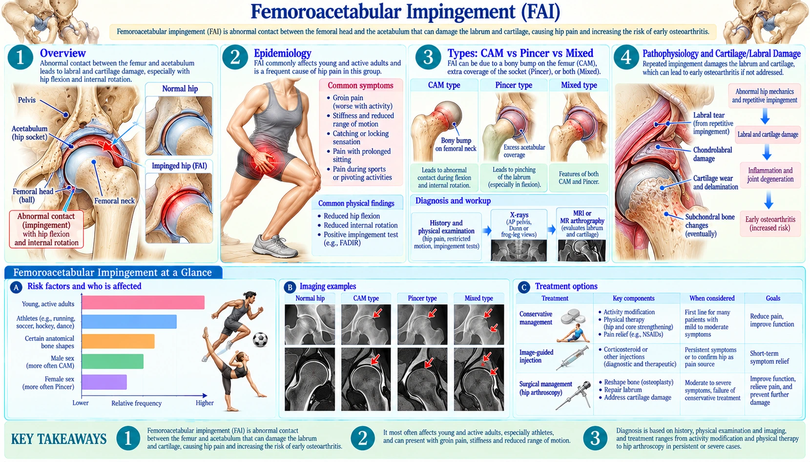
Table of Contents
- Overview
- Epidemiology
- Types: CAM vs Pincer vs Mixed
- Pathophysiology and Cartilage/Labral Damage
- Clinical Presentation and Examination Tests
- Alpha Angle and Imaging (X-ray, MRI Arthrogram, CT)
- Conservative Treatment
- Hip Arthroscopy: Surgery and Techniques
- Rehabilitation and Return to Sport
- Prognosis and Conversion to Total Hip Arthroplasty
- Key Research Papers
- Connections
- Featured Videos
1. Overview
Femoroacetabular impingement, usually shortened to FAI, is a hip condition described mainly in young, active adults: the 2018 UK FASHIoN trial report calls FAI syndrome "an important cause of hip pain in young adults" (Griffin 2018). People with it often report a deep ache in the groin that is worse with long sitting or squatting.
FAI occurs when there is abnormal contact between the femoral head-neck junction and the rim of the acetabulum — the socket of the hip joint — during hip movement. The hip is a ball-and-socket joint: the "ball" is the rounded head of the femur (thigh bone), and the "socket" is the acetabulum, a cup-shaped hollow in the pelvis. In FAI, either the ball is not round where it meets the neck (cam morphology), the socket covers too much of the ball (pincer morphology), or both. In surgical observations, cam impingement sheared the acetabular cartilage off the bone during flexion, and pincer impingement crushed the labrum (the ring of fibrocartilage around the socket rim) between the rim and the femoral neck (Beck 2005).
The usual symptom is deep groin pain. Some patients show where it hurts by cupping a hand in a C-shape around the front and side of the hip, a gesture clinicians call the "C-sign". The pain tends to be activity-related, worse with prolonged sitting, getting up from a low chair, squatting, pivoting, and sports that demand deep hip flexion and rotation.
The bone shapes themselves are common in people with no symptoms: a systematic review of 26 imaging studies (2,114 asymptomatic hips) found cam morphology in 37% and pincer morphology in 67% (Frank 2015). For that reason the 2016 Warwick Agreement introduced the term "FAI syndrome" and stated that a diagnosis requires appropriate symptoms, positive clinical signs and imaging findings (Griffin 2016).
The Warwick Agreement lists conservative care, rehabilitation, and arthroscopic or open surgery as treatments (Griffin 2016). Randomised trials comparing surgery with physiotherapist-led care are summarised in the surgery section below.
2. Epidemiology
Prevalence of the bony shapes in people without symptoms: In the 2015 systematic review of asymptomatic volunteers (mean age 25.3 years), the prevalence of cam morphology was 37%, with a range of 7% to 100% between studies — 54.8% in athletes versus 23.1% in the general population. Pincer morphology was found in 67% (range 61–76%), but the authors noted that pincer deformity was poorly defined across studies. Labral injury was seen on MRI in 68.1% of asymptomatic hips in the 7 studies that reported it (Frank 2015). Having the bone shape, in other words, is not the same as having FAI syndrome.
Who develops symptoms: The trials of FAI syndrome enrolled young and middle-aged adults — aged 16 and over in UK FASHIoN (Griffin 2018), 18 to 60 in FAIT (Palmer 2019) and 18 to 50 in FIRST (FIRST Investigators 2021). In a systematic review of 28 studies of athletes with radiographic cam deformity, cam lesions were most commonly reported in soccer, followed by hockey and American football (Knapik 2019).
Sex differences: The same review identified 1,160 male and 53 female athletes with cam deformity, and male athletes had higher mean alpha angles (59.9° versus 48.3°) (Knapik 2019). In Beck's 2005 surgical series of 302 hips, only 26 had an isolated cam and 16 an isolated pincer impingement (Beck 2005).
Cam morphology develops during growth: Prospective studies of young athletes found that cam morphology is acquired during skeletal maturation. In 63 pre-professional soccer players aged 12–19 followed for about 2.4 years, the prevalence of a head-neck prominence in hips with an open growth plate rose from 2.1% to 17.7%, with no significant increase after the proximal femoral growth plate closed (Agricola 2014). An MRI study of 25 adolescent ice hockey players found that cam morphologies developed early in the final growth spurt, predominantly between 13 and 16 years of age (Hanke 2021). Agricola and colleagues suggested that the formation of a cam deformity might be prevented by adjusting athletic activities during a short period of skeletal growth; that idea has not been tested in a trial.
3. Types: CAM vs Pincer vs Mixed
FAI is categorized into three types based on where the abnormal contact originates. The type (or combination) shapes how imaging is read and what a surgeon would reshape.
CAM Impingement
In CAM impingement, the problem is with the femoral head-neck junction. Instead of a smooth, waisted concavity where the spherical head transitions into the femoral neck, a bony prominence or "cam lesion" sits at the anterosuperior head-neck junction — the non-spherical femoral head described by Beck and colleagues (Beck 2005). This is often described as a loss of the normal "head-neck offset."
As the hip flexes, the non-spherical part of the head is forced into the socket. In Beck's surgical series, cam impingement caused damage to the anterosuperior acetabular cartilage with separation between the labrum and the cartilage: during flexion the cartilage was sheared off the bone by the non-spherical femoral head while the labrum itself remained untouched (Beck 2005).
The usual measurement for cam morphology is the alpha angle, a geometric measure of the shape of the femoral head-neck junction on imaging. Cut-off values differ between studies; the prospective athlete studies cited on this page counted an alpha angle above 60° as cam morphology (Agricola 2014; Hanke 2021). In Nötzli's original 2002 MRI study, the alpha angle averaged 74.0° in patients with impingement and 42.0° in asymptomatic controls (Nötzli 2002).
Pincer Impingement
In pincer impingement, the problem is excessive acetabular cover of the femoral head (Beck 2005). Forms described include a socket that faces backward (acetabular retroversion), an unusually deep socket (coxa profunda) and protrusio acetabuli. The result is that the femoral neck bumps into the edge of the rim during hip flexion.
The damage pattern differs from CAM. In Beck's series, the labrum was crushed between the acetabular rim and the femoral neck during movement, causing degeneration and ossification (bone forming within the labrum), and the cartilage damage ran circumferentially in only a narrow strip (Beck 2005).
A radiographic sign of acetabular retroversion is the crossover sign on a standard AP pelvis X-ray: the anterior acetabular wall projects lateral to the posterior wall at some point, so the anterior rim "crosses over" the posterior rim.
Mixed Impingement
Many hips show elements of both. In Beck's 2005 series of 302 analysed hips, only 26 had an isolated cam and 16 an isolated pincer impingement, and the authors noted that labral damage indicates ongoing impingement and rarely occurs alone (Beck 2005). In UK FASHIoN, randomisation was stratified by impingement type (Griffin 2018).
4. Pathophysiology and Cartilage/Labral Damage
This section covers the normal anatomy of the hip socket and what the research describes going wrong when impingement repeats over time.
The Labrum: The Hip's Sealing Ring
The acetabular labrum is a ring of fibrocartilage around the rim of the socket. In a study of 55 cadaver hips, it added to both the articulating surface area of the acetabulum (28.8 cm² without the labrum, 36.8 cm² with it) and its volume (31.5 cm³ without, 41.1 cm³ with); the labrum was wider anteriorly and superiorly than posteriorly (Tan 2001).
The labrum also acts as a seal. In a laboratory study of 8 cadaver hips, the labrum directly controlled the escape of fluid from the central compartment of the joint during loading; pivoting positions improved the seal, while positions combining flexion with internal rotation (stooping) let fluid escape more easily. The authors suggested that altering the relationship between the labrum and the femoral head may disrupt this seal and leave the joint at risk of pathological changes over time (Dwyer 2014).
Labral tears can produce clicking, catching, and sharp pain with certain hip positions, in addition to the deeper background ache of impingement.
Labral Tear Patterns
Labral damage in FAI follows the impingement type (Beck 2005):
- Labral-chondral separation: In cam impingement, the labrum and the adjacent cartilage separate as the cartilage is sheared off the bone. Torn but healthy labral tissue can be reattached surgically.
- Degeneration and ossification: In pincer impingement, repeated crushing degenerates the labrum and can turn it partly to bone. Badly degenerated tissue may not be repairable; debridement or reconstruction are the alternatives described.
Labral tears are also common in people without symptoms: the 2015 systematic review found labral injury on non-contrast MRI in 68.1% of asymptomatic hips in the studies that reported it (Frank 2015).
Cartilage Damage Progression
In cam FAI, the typical finding is damage to the anterosuperior acetabular cartilage, which separates from the bone at the cartilage-labrum junction (Beck 2005). This kind of damage can be subtle on standard MRI.
Ganz and colleagues, drawing on more than 600 surgical dislocations of the hip, proposed in 2003 that femoroacetabular impingement is a mechanism of early osteoarthritis in most non-dysplastic hips, and that early surgery might slow the degenerative process (Ganz 2003). Beck's 2005 study concluded that both cam and pincer impingement lead to osteoarthritis of the hip (Beck 2005). Whether surgery protects the joint is less settled: in the Australian FASHIoN trial (99 participants), the primary outcome — a 12-month MRI measure of cartilage metabolism (dGEMRIC) — showed no statistically significant difference between arthroscopy and physiotherapist-led care, and the authors concluded that the greater symptom benefit after surgery was not explained by better cartilage metabolism (Hunter 2021).
5. Clinical Presentation and Examination Tests
The Warwick Agreement states that a diagnosis of FAI syndrome requires appropriate symptoms, positive clinical signs and imaging findings — no single element is enough on its own (Griffin 2016).
Typical Symptoms
- Deep groin pain is the main symptom, felt in the front of the groin and sometimes spreading into the thigh. Patients often describe it as being "inside the hip."
- The "C-sign": When asked to point to where it hurts, some patients cup the hand in a C-shape around the groin and the side of the hip.
- Activity-related pain that is typically worse with:
- Prolonged sitting (especially in low chairs or car seats)
- Rising from a seated position
- Squatting and deep hip flexion
- Pivoting, cutting, and rotational sport movements
- Stair or hill climbing
- Clicking or catching sensations in the hip, which may come from a labral tear.
- Gradual onset: FAI usually develops over months or years rather than after a single injury, and it can be mistaken for a groin strain or hip-flexor problem.
- Reduced hip range of motion, particularly internal rotation with the hip flexed. In the adolescent ice hockey study, pain on the impingement test and/or internal rotation below 20° became more frequent as cam morphology developed (Hanke 2021).
Key Physical Examination Tests
Several provocative tests are used to look for FAI and labral pathology:
- FADIR test (Flexion-ADduction-Internal Rotation), also called the anterior impingement test. The examiner flexes the hip, brings it toward the midline and rotates it inward; a positive test reproduces the patient's groin pain. A 2015 meta-analysis found pooled sensitivity of 0.94 to 0.99 for FADIR, but with wide uncertainty in its diagnostic odds ratio, and concluded that FADIR and the flexion-internal rotation test possess only screening accuracy: a negative test makes FAI less likely, a positive test does not confirm it (Reiman 2015).
- FABER test (Flexion-ABduction-External Rotation), also called the Patrick test. The examiner places the heel on the opposite knee (figure-4 position) and gently presses the knee toward the table; groin pain or a knee that sits higher than on the other side points to the hip.
- Log roll test: With the patient lying on the back and the leg straight, the examiner rolls the leg inward and outward. Pain or restricted internal rotation is suspicious for a problem inside the joint.
- Thomas test: Checks for tightness of the hip flexors.
The same 2015 review found that few hip examination tests have been studied in enough high-quality studies to direct clinical decisions (Reiman 2015), which is one reason the Warwick Agreement requires imaging findings as well as symptoms and signs (Griffin 2016).
6. Alpha Angle and Imaging (X-ray, MRI Arthrogram, CT)
Imaging in FAI is used to confirm the bony shape, measure how severe it is, assess the labrum and cartilage, and plan any surgery.
Plain X-Rays: The Starting Point
X-ray evaluation of FAI usually starts with two views:
- AP (anteroposterior) pelvis X-ray, taken with the pelvis in a standardized position. Findings include:
- Lateral center-edge angle (LCEA): how much of the femoral head is covered by the acetabulum. In the review of asymptomatic volunteers, the mean lateral center-edge angle was 31.2° (Frank 2015); the FAIT trial excluded hip dysplasia, defined as a center-edge angle below 20° (Palmer 2019).
- Crossover sign: the anterior acetabular wall projects lateral to the posterior wall at some point, indicating acetabular retroversion (a form of pincer morphology).
- Coxa profunda (a deep socket) and protrusio acetabuli, the more extreme forms of over-coverage.
- Cam deformity: loss of the normal head-neck offset; a "pistol grip deformity" describes the shape seen in severe cam morphology.
- Lateral views (for example a Dunn view or frog-leg lateral) profile the anterosuperior head-neck junction, where cam lesions most commonly occur, and are used to measure the alpha angle. The soccer study cited above used standardized AP and frog-leg lateral radiographs (Agricola 2014).
The Alpha Angle: Key Measurement for CAM Morphology
The alpha angle is the most widely used measure of cam deformity. It was described by Nötzli and colleagues in 2002, who measured the anterior margin of the waist of the femoral neck on tilted axial MRI scans passing through the center of the femoral head, with good reproducibility among four observers (Nötzli 2002).
Conceptually, the alpha angle measures where the femoral head stops being a sphere at its front. A circle is fitted to the femoral head; one line runs along the axis of the femoral neck through the center of that circle, and a second line runs from the center to the point where the bone first leaves the circle. The angle between the two lines is the alpha angle.
In Nötzli's study, the angle averaged 74.0° in symptomatic patients and 42.0° in controls (Nötzli 2002). In asymptomatic volunteers across 17 studies the mean was 54.1° (Frank 2015). Studies use different thresholds for "cam morphology"; the prospective athlete studies on this page used an angle above 60° (Agricola 2014; Hanke 2021). Values can differ between X-ray, MRI and CT.
MRI and MR Arthrography for Labral Tears
MRI is used to look at the labrum and cartilage. In MR arthrography (MRA), contrast is injected into the hip joint before the scan so that it outlines tears. A 2011 meta-analysis of 19 papers (881 hips) found that conventional MRI detected labral tears with a sensitivity of 66% and specificity of 79%, and MRA with a sensitivity of 87% and specificity of 64%; MRA appeared superior on ROC analysis, and the authors called both "useful adjuncts" (Smith 2011).
A 2017 meta-analysis of 20 labral-tear studies found that CT arthrography had the strongest overall diagnostic accuracy (pooled sensitivity 0.91, specificity 0.89). A positive MRI/MRA or ultrasound result raised the probability of a labral tear only by a minimal to small degree, and the authors judged that the results have limited generalizability given the very high pre-test probability in the studies (Reiman 2017).
MRI and MRA can show:
- Labral tears: location, shape and extent
- Labral degeneration and paralabral cysts (fluid-filled cysts next to tears)
- Articular cartilage defects and delamination
- Cam lesion shape and the alpha angle
- Associated problems such as synovitis or ligamentum teres tears
CT Scan: Surgical Planning for Complex Cases
CT scanning with 3D reconstruction gives a detailed picture of bone shape and is used for surgical planning in complex cases, including:
- Alpha angle measurement at several positions around the head-neck junction (a cam lesion is often not uniform)
- Acetabular version (how far the socket faces forward or backward)
- Computer simulation of hip motion to show where bone contact occurs
- Planning how much bone to remove
CT involves ionizing radiation and is not a first-line test.
7. Conservative Treatment
Not everyone with FAI has surgery. The Warwick Agreement lists conservative care and rehabilitation alongside arthroscopic or open surgery as suitable treatments (Griffin 2016). In UK FASHIoN, patients given a physiotherapist-led programme also improved: their mean iHOT-33 score rose from 35.6 to 49.7 at 12 months (Griffin 2018). A 2020 meta-analysis of physiotherapist-led treatment for hip-related pain found that it might improve pain and function, but there were no full-scale placebo-controlled trials, and its authors called for high-quality RCTs (Kemp 2020).
Activity Modification
Activity modification is part of conservative care in the trials: the FAIT comparison arm was "physiotherapy and activity modification" (Palmer 2019). Positions described as loading the front rim of the socket include deep hip flexion, hip flexion combined with internal rotation, prolonged sitting in low chairs and hard pivoting in sport. In the cadaver seal study, stooping positions combining flexion with internal rotation also weakened the labral seal (Dwyer 2014). Programmes generally aim to reduce these provoking positions rather than stop all exercise.
Anti-Inflammatory Medication
A 2024 systematic review describes FAI syndrome as initially managed conservatively with oral anti-inflammatory drugs and physiotherapy (Takla 2024). These medicines treat pain and inflammation; they do not change the shape of the bone. Questions about any medicine, including its risks, belong with a clinician.
Physical Therapy
The physiotherapy arms of the randomised trials give the clearest description of what conservative programmes contain:
- UK FASHIoN — "personalised hip therapy": an individualised, supervised and progressive physiotherapist-led programme (Griffin 2018).
- FAIT: a goal-based programme tailored to individual needs, with emphasis on core stability and movement control, delivered in at most eight physiotherapy sessions over five months (Palmer 2019).
Intra-Articular Injection
An image-guided injection of local anaesthetic and/or corticosteroid into the hip joint is used both to relieve pain and as a diagnostic test: relief suggests that the pain comes from inside the joint. Its value in predicting surgical results is mixed. A 2024 systematic review of seven studies (637 patients) found that only two reported a statistically significant link between injection response and outcome after arthroscopy; in the other five, a positive response increased the odds of a good outcome but not significantly. The authors concluded that injections can be a useful prognostic tool but are not a consistently reliable predictor (Takla 2024).
8. Hip Arthroscopy: Surgery and Techniques
Surgery is one of the treatments listed by the Warwick Agreement, by arthroscopy or by open surgery (Griffin 2016). Hip arthroscopy is a minimally invasive procedure: the surgeon works through small incisions using a camera (arthroscope) and specialized instruments.
The Procedure: What Happens in the Operating Room
Hip arthroscopy for FAI is performed under general or spinal anesthesia. Because the hip is a deep joint, the leg is placed in traction so that the ball is pulled slightly away from the socket and the instruments can enter; traction is released for the parts of the procedure on the femoral neck (the peripheral compartment).
The surgeon works through several small portals, moving the camera and instruments between them. In the FIRST trial, cam and pincer lesions were resected using fluoroscopic (real-time X-ray) guidance (FIRST Investigators 2021).
Femoroplasty (CAM Resection)
For cam impingement, the surgeon performs a femoroplasty (osteochondroplasty): the cam lesion is shaved down with a motorized burr to restore the concave head-neck offset. Imaging and moving the hip through its range during surgery are used to check that the impingement has been removed. In FAIT, the surgery was described as excising the bone that impinged during hip movements (Palmer 2019).
Acetabuloplasty (Pincer Resection)
For pincer impingement, the surgeon performs an acetabuloplasty: trimming the over-covering acetabular rim to reduce contact with the femoral neck. The amount removed is balanced against the need to keep enough socket coverage for a stable hip.
Labral Repair Versus Debridement
A torn labrum can be trimmed away (debridement) or reattached to the rim with suture anchors (repair). A 2020 meta-analysis of five studies (four observational, one randomised) found no significant difference between the two in the Non-Arthritic Hip Score, surgical failure rate or complications, while modified Harris Hip Score, pain (VAS) and satisfaction favoured repair. The authors concluded that both are effective for symptomatic FAI, with a trend toward better results after repair (Wu 2020). In FIRST, surgeons were instructed to repair the labrum when it was mechanically unstable; 88.3% of participants had a labral tear and 60.3% of those were repaired (FIRST Investigators 2021).
Labral Reconstruction
When labral tissue is badly degenerated, calcified or absent — often in revision surgery — there may not be enough healthy tissue to reattach. A labral reconstruction replaces it with a graft, either the patient's own tissue (an autograft, for example from the iliotibial band) or donor tissue (an allograft). It is more technically demanding than a primary repair.
The Randomised Trials: Surgery Versus Physiotherapy and Versus Lavage
UK FASHIoN (Griffin 2018, Lancet): a pragmatic, assessor-blinded trial at 23 NHS hospitals randomised 348 people aged 16 and over with FAI syndrome and no osteoarthritis to hip arthroscopy (171) or personalised hip therapy (177). At 12 months, mean iHOT-33 scores (hip-related quality of life) improved from 39.2 to 58.8 after arthroscopy and from 35.6 to 49.7 after hip therapy; the adjusted difference was 6.8 points (95% CI 1.7–12.0) in favour of arthroscopy, just above the minimum clinically important difference of 6.1 points. Patient-reported adverse events (most commonly muscle soreness) were reported by 72% of the arthroscopy group and 60% of the hip-therapy group; five of six serious adverse events in the arthroscopy group were treatment-related, and one patient developed a hip joint infection after surgery (Griffin 2018). The full report added that 24% of hip-therapy patients chose to have arthroscopy between 1 and 3 years, 12% of arthroscopy patients had revision arthroscopy, and 7% of hip-therapy patients and 2% of arthroscopy patients had a hip replacement within 3 years; the trial did not show that arthroscopy was cost-effective within the first 12 months (Griffin 2022).
FAIT (Palmer 2019, BMJ): 222 participants aged 18 to 60 at seven NHS sites were randomised to arthroscopic surgery (112) or physiotherapy and activity modification (110). At eight months, the hip outcome score for activities of daily living was 10.0 points higher (95% CI 6.4–13.6) after surgery, above the trial's 9-point minimum clinically important difference; no serious adverse events were reported in either group (Palmer 2019).
Australian FASHIoN (Hunter 2021): 99 participants; the cartilage MRI primary outcome did not differ, while hip-related quality of life (iHOT-33) improved by 14 points more after arthroscopy at 12 months (Hunter 2021).
FIRST (2021, American Journal of Sports Medicine): rather than comparing surgery with physiotherapy, this trial at 10 centres in Canada, Finland and Denmark compared arthroscopic osteochondroplasty (reshaping the bone) with arthroscopic lavage (washing out the joint without reshaping it), with labral repair allowed in both arms. Among 214 participants analysed, pain at 12 months did not differ between groups (mean difference 0.11 on a 100-point scale); both groups improved, and the only significant secondary difference favoured lavage. By 24 months, reoperations were fewer after osteochondroplasty (8 of 105) than after lavage (19 of 104) (FIRST Investigators 2021).
Pooled evidence: a 2021 meta-analysis of three randomised trials (574 participants for the main analysis) found high-certainty evidence that arthroscopy gave better hip-specific quality of life than non-operative care at 12 months (mean difference 11.02 points), but low-quality evidence of similar outcomes at 24 months; its authors wrote that placebo trials are needed to establish the efficacy of hip arthroscopy (Ferreira 2021).
9. Rehabilitation and Return to Sport
Post-operative rehabilitation protocols vary widely. A 2023 systematic review of 24 studies (2,231 patients) found seven different terms for weight-bearing instructions, most of them some form of partial weight-bearing, and only 75% of studies reported their rehabilitation protocol; it concluded that there is not enough comparative evidence to support specific weight-bearing restrictions after hip arthroscopy for FAI (Hemstock 2023). The phases below describe the general shape of published programmes, not a fixed schedule.
Phase 1: Protected Weight-Bearing
After surgery, weight-bearing is usually limited for a period, often with crutches, to protect any labral repair. How long depends on the surgeon's protocol and on what was done in the joint, such as the extent of labral repair or cartilage work.
Some protocols include a continuous passive motion (CPM) machine, which moves the hip gently through a range of motion. Early passive range-of-motion exercises are typically limited to pain-free arcs that do not stress the repair.
Phase 2: Range of Motion and Muscle Activation
As weight-bearing progresses and crutches are stopped, therapy shifts to restoring hip range of motion and activating the surrounding muscles. Typical goals:
- Restoring hip flexion, external rotation and internal rotation
- Activating the gluteal muscles
- Restoring a normal walking pattern, including correcting a hip drop with each step (Trendelenburg gait) caused by weak hip abductors
- Low-impact exercise such as pool work and stationary cycling
Hip-flexor weakness can be a problem early after surgery, particularly if the iliopsoas tendon was released, and is addressed directly in rehabilitation.
Phase 3: Strengthening
Progressive loading of the hip in functional movement patterns:
- Closed-chain strengthening: squats (with depth progressed gradually), leg press, step-ups, lunges
- Hip abductor and external rotator strengthening: clamshells, lateral band walks, single-leg stance
- Progressive resistance training for the quadriceps, hamstrings and hip extensors
- Balance and proprioception training
- Light, straight-line jogging once strength and walking pattern have recovered
Phase 4: Return-to-Sport Training
Sport-specific training includes cutting, pivoting, deceleration and skill work, with return guided by strength, hop testing and movement quality rather than time alone. A 2019 meta-analysis of 38 studies (1,773 patients, mean follow-up 28.1 months) found pooled return-to-sport rates of 93% at any level, 82% at the pre-operative level, 89% in competitive athletes and 94% in professional athletes; a shorter duration of symptoms before surgery was associated with a higher rate of return (Memon 2019).
10. Prognosis and Conversion to Total Hip Arthroplasty
This section summarises what the research reports about results after treatment, for conservative care and for surgery.
Surgical Outcomes
Beyond the randomised trials described above, longer-term data come mostly from case series:
- A 2023 systematic review of 12 studies with a minimum 10-year follow-up (1,344 hips) found that 8 of the 10 studies reporting patient-reported outcomes showed significant improvement after primary hip arthroscopy at long-term follow-up; rates of secondary arthroscopy ranged from 4.5% to 24% (Lee 2023).
- Return to sport: 93% at any level and 82% at the pre-operative level in the 2019 meta-analysis (Memon 2019).
- In the randomised trials, arthroscopy improved hip-related quality of life more than physiotherapist-led care at 8–12 months (Griffin 2018; Palmer 2019), but the pooled benefit was not shown at 24 months (Ferreira 2021), and reshaping the bone did not reduce pain at 12 months more than a joint lavage did (FIRST Investigators 2021).
Predictors of Good and Poor Outcome
Factors the research links with outcome:
- Age and cartilage damage: older age and chondral (cartilage) damage were the most commonly cited predictors of conversion to total hip arthroplasty in the 10-year review (Lee 2023).
- Arthritis grade and labral repair: in the same review, patients with a Tönnis grade below 1 who had labral repair had greater than 90% survivorship — that is, no hip replacement (Lee 2023). The Tönnis grade is an X-ray scale of hip osteoarthritis, with higher grades meaning more advanced arthritis.
- Established arthritis: for hips with FAI and Tönnis grade 2 or greater, a 2022 systematic review of 11 studies found the evidence contradictory and insufficient, with conversion to hip replacement of 0–9% in four studies and 35–70% in four others (Andronic 2022). The randomised trials excluded people with osteoarthritis (Griffin 2018; Palmer 2019).
- Symptom duration: a shorter duration of symptoms before surgery was associated with a higher return-to-sport rate (Memon 2019).
- Injection response: relief after a pre-operative joint injection is not a consistently reliable predictor of surgical outcome (Takla 2024).
Conversion to Total Hip Arthroplasty
Whether arthroscopy for FAI delays or prevents hip replacement has not been settled by randomised evidence. Reported rates:
- In UK FASHIoN, 2% of the arthroscopy group and 7% of the personalised-hip-therapy group had a hip replacement within 3 years (Griffin 2022).
- In case series with 10 to 20 years of follow-up, conversion to total hip arthroplasty varied from 0% to 44.1% (Lee 2023).
- In hips with Tönnis grade 2 or greater arthritis, reported conversion rates ranged from 0–9% to as high as 35–70% (Andronic 2022).
Ganz and colleagues proposed in 2003 that early surgery for FAI might slow the degenerative process in young patients (Ganz 2003); the Australian FASHIoN trial did not find better cartilage metabolism after surgery at 12 months (Hunter 2021), and the UK FASHIoN investigators wrote that further follow-up will show whether the benefits of arthroscopy are maintained (Griffin 2022). Decisions about treatment belong with a clinician.
Key Research Papers
- Griffin DR, Dickenson EJ, Wall PDH, et al.; FASHIoN Study Group (2018). Hip arthroscopy versus best conservative care for the treatment of femoroacetabular impingement syndrome (UK FASHIoN): a multicentre randomised controlled trial. Lancet 391(10136):2225–2235 — PubMed PMID: 29893223
- Griffin DR, Dickenson EJ, Achana F, et al. (2022). Arthroscopic hip surgery compared with personalised hip therapy in people over 16 years old with femoroacetabular impingement syndrome: UK FASHIoN RCT. Health Technol Assess 26(16):1–236 — PubMed PMID: 35229713
- Palmer AJR, Ayyar Gupta V, Fernquest S, et al.; FAIT Study Group (2019). Arthroscopic hip surgery compared with physiotherapy and activity modification for the treatment of symptomatic femoroacetabular impingement: multicentre randomised controlled trial. BMJ 364:l185 (correction published 2021) — PubMed PMID: 30733197
- Femoroacetabular Impingement Randomized Controlled Trial (FIRST) Investigators; Ayeni OR, Karlsson J, Heels-Ansdell D, et al. (2021). Osteochondroplasty and labral repair for the treatment of young adults with femoroacetabular impingement: a randomized controlled trial. Am J Sports Med 49(1):25–34 — PubMed PMID: 32970955
- Hunter DJ, Eyles J, Murphy NJ, et al. (2021). Multi-centre randomised controlled trial comparing arthroscopic hip surgery to physiotherapist-led care for femoroacetabular impingement (FAI) syndrome on hip cartilage metabolism: the Australian FASHIoN trial. BMC Musculoskelet Disord 22(1):697 — PubMed PMID: 34399702
- Ferreira GE, O'Keeffe M, Maher CG, et al. (2021). The effectiveness of hip arthroscopic surgery for the treatment of femoroacetabular impingement syndrome: a systematic review and meta-analysis. J Sci Med Sport 24(1):21–29 — PubMed PMID: 32616421
- Griffin DR, Dickenson EJ, O'Donnell J, et al. (2016). The Warwick Agreement on femoroacetabular impingement syndrome (FAI syndrome): an international consensus statement. Br J Sports Med 50(19):1169–1176 — PubMed PMID: 27629403
- Frank JM, Harris JD, Erickson BJ, et al. (2015). Prevalence of femoroacetabular impingement imaging findings in asymptomatic volunteers: a systematic review. Arthroscopy 31(6):1199–1204 — PubMed PMID: 25636988
- Knapik DM, Gaudiani MA, Camilleri BE, et al. (2019). Reported prevalence of radiographic cam deformity based on sport: a systematic review of the current literature. Orthop J Sports Med 7(3):2325967119830873 — PubMed PMID: 30915376
- Agricola R, Heijboer MP, Ginai AZ, et al. (2014). A cam deformity is gradually acquired during skeletal maturation in adolescent and young male soccer players: a prospective study with minimum 2-year follow-up. Am J Sports Med 42(4):798–806 — PubMed PMID: 24585362
- Hanke MS, Schmaranzer F, Steppacher SD, et al. (2021). A cam morphology develops in the early phase of the final growth spurt in adolescent ice hockey players: results of a prospective MRI-based study. Clin Orthop Relat Res 479(5):906–918 — PubMed PMID: 33417423
- Ganz R, Parvizi J, Beck M, et al. (2003). Femoroacetabular impingement: a cause for osteoarthritis of the hip. Clin Orthop Relat Res (417):112–120 — PubMed PMID: 14646708
- Beck M, Kalhor M, Leunig M, Ganz R (2005). Hip morphology influences the pattern of damage to the acetabular cartilage: femoroacetabular impingement as a cause of early osteoarthritis of the hip. J Bone Joint Surg Br 87(7):1012–1018 — PubMed PMID: 15972923
- Nötzli HP, Wyss TF, Stoecklin CH, et al. (2002). The contour of the femoral head-neck junction as a predictor for the risk of anterior impingement. J Bone Joint Surg Br 84(4):556–560 — PubMed PMID: 12043778
- Tan V, Seldes RM, Katz MA, et al. (2001). Contribution of acetabular labrum to articulating surface area and femoral head coverage in adult hip joints: an anatomic study in cadavera. Am J Orthop (Belle Mead NJ) 30(11):809–812 — PubMed PMID: 11757858
- Dwyer MK, Jones HL, Hogan MG, et al. (2014). The acetabular labrum regulates fluid circulation of the hip joint during functional activities. Am J Sports Med 42(4):812–819 — PubMed PMID: 24557859
- Reiman MP, Goode AP, Cook CE, et al. (2015). Diagnostic accuracy of clinical tests for the diagnosis of hip femoroacetabular impingement/labral tear: a systematic review with meta-analysis. Br J Sports Med 49(12):811 — PubMed PMID: 25515771
- Smith TO, Hilton G, Toms AP, et al. (2011). The diagnostic accuracy of acetabular labral tears using magnetic resonance imaging and magnetic resonance arthrography: a meta-analysis. Eur Radiol 21(4):863–874 — PubMed PMID: 20859632
- Reiman MP, Thorborg K, Goode AP, et al. (2017). Diagnostic accuracy of imaging modalities and injection techniques for the diagnosis of femoroacetabular impingement/labral tear: a systematic review with meta-analysis. Am J Sports Med 45(11):2665–2677 — PubMed PMID: 28129509
- Kemp JL, Mosler AB, Hart H, et al. (2020). Improving function in people with hip-related pain: a systematic review and meta-analysis of physiotherapist-led interventions for hip-related pain. Br J Sports Med 54(23):1382–1394 — PubMed PMID: 32376673
- Takla A, Gunatilake K, Ma N, Moaveni A (2024). Can intra-articular hip injections predict arthroscopy outcomes for femoroacetabular impingement syndrome? A systematic review. J Orthop 50:122–129 — PubMed PMID: 38214002
- Wu ZX, Ren WX, Ren YM, Tian MQ (2020). Arthroscopic labral debridement versus labral repair for patients with femoroacetabular impingement: a meta-analysis. Medicine (Baltimore) 99(19):e20141 — PubMed PMID: 32384496
- Hemstock R, Mulhall D, Didyk J, et al. (2023). Postoperative weight-bearing restrictions and rehabilitation protocols after hip arthroscopy for femoroacetabular impingement: a systematic review. J Hip Preserv Surg 10(3–4):220–227 — PubMed PMID: 38162279
- Memon M, Kay J, Hache P, et al. (2019). Athletes experience a high rate of return to sport following hip arthroscopy. Knee Surg Sports Traumatol Arthrosc 27(10):3066–3104 — PubMed PMID: 29627931
- Lee MS, Kim DN, Moran J, et al. (2023). Patients undergoing primary hip arthroscopy report favorable outcomes at minimum 10 year follow-up: a systematic review. Arthroscopy 39(2):459–475 — PubMed PMID: 36334852
- Andronic O, Claydon-Mueller LS, Cubberley R, et al. (2022). Inconclusive and contradictory evidence for outcomes after hip arthroscopy in patients with femoroacetabular impingement and osteoarthritis of Tönnis grade 2 or greater: a systematic review. Arthroscopy 38(7):2307–2318.e1 — PubMed PMID: 35093497
PubMed Topic Searches
- PubMed search: Hip arthroscopy for femoroacetabular impingement systematic review
- PubMed search: Arthroscopic management of FAI 2-year follow-up
- PubMed search: Outcomes of arthroscopic treatment of FAI minimum 5-year follow-up
- PubMed search: Surgical treatment of FAI improves hip kinematics a computer-assisted model
- PubMed search: The concept of femoroacetabular impingement current status and future perspectives
- PubMed search: Outcomes after arthroscopic treatment of FAI are CAM and mixed morphologies associated
- PubMed search: The prevalence of cam deformity in asymptomatic adults a systematic review
- PubMed search: Clinical presentation of FAI demographics hip range of motion and radiographic findings
- PubMed search: Rehabilitation exercise progression for the gluteus medius muscle with consideration for iliopsoas
PubMed: Femoroacetabular Impingement — Live search for the latest FAI research.
The links below run live searches on PubMed, the U.S. National Library of Medicine's database of biomedical literature. Use them to explore the current evidence on femoroacetabular impingement — morphology, surgical techniques, rehabilitation, and long-term outcomes — and to find newer studies as they are published.
- PubMed: FAI outcomes and results
- PubMed: Hip arthroscopy labral repair
- PubMed: CAM impingement femoroplasty
- PubMed: Acetabular labrum tear diagnosis
- PubMed: FAI alpha angle measurement
- PubMed: FAI and hip osteoarthritis progression
- PubMed: Hip arthroscopy return to sport
- PubMed: Pincer impingement acetabular retroversion
- PubMed: FAI physical therapy and rehabilitation
- PubMed: CAM morphology in youth athletes
- PubMed: Labral reconstruction hip allograft
- PubMed: FAI conversion to total hip arthroplasty
13. Connections
- All Conditions — the full directory of health conditions covered on this site.
- Orthopedics — all musculoskeletal and orthopedic conditions.
- Hip Fracture — fracture of the proximal femur; shares the hip anatomy but a very different injury mechanism and treatment pathway.
- ACL Tear — the most studied sport-related ligament injury; shares the theme of young athletes, arthroscopic surgery, and structured rehabilitation.
- Meniscus Tear — fibrocartilage injury in the knee; shares the suction-seal functional parallel with the acetabular labrum, and both structures are prioritized for repair over debridement.
- Bursitis — trochanteric bursitis is a common differential diagnosis for lateral hip pain that can coexist with or mimic FAI; distinguishing the two is important.
- Stress Fracture — repetitive-load bone injury seen in the same athlete populations; femoral neck stress fractures can present with groin pain mimicking FAI.
- Low Back Pain — lumbar pathology commonly refers pain to the hip and groin and must be differentiated from intra-articular hip pain; FAI and lumbar spine pathology can coexist.