⚠ Evidence warning: Clinical trials or other evidence do not support the statement that sorafenib is approved in some countries for relapsed osteosarcoma. This page keeps it as a report of what people, websites or other media say.
Osteosarcoma

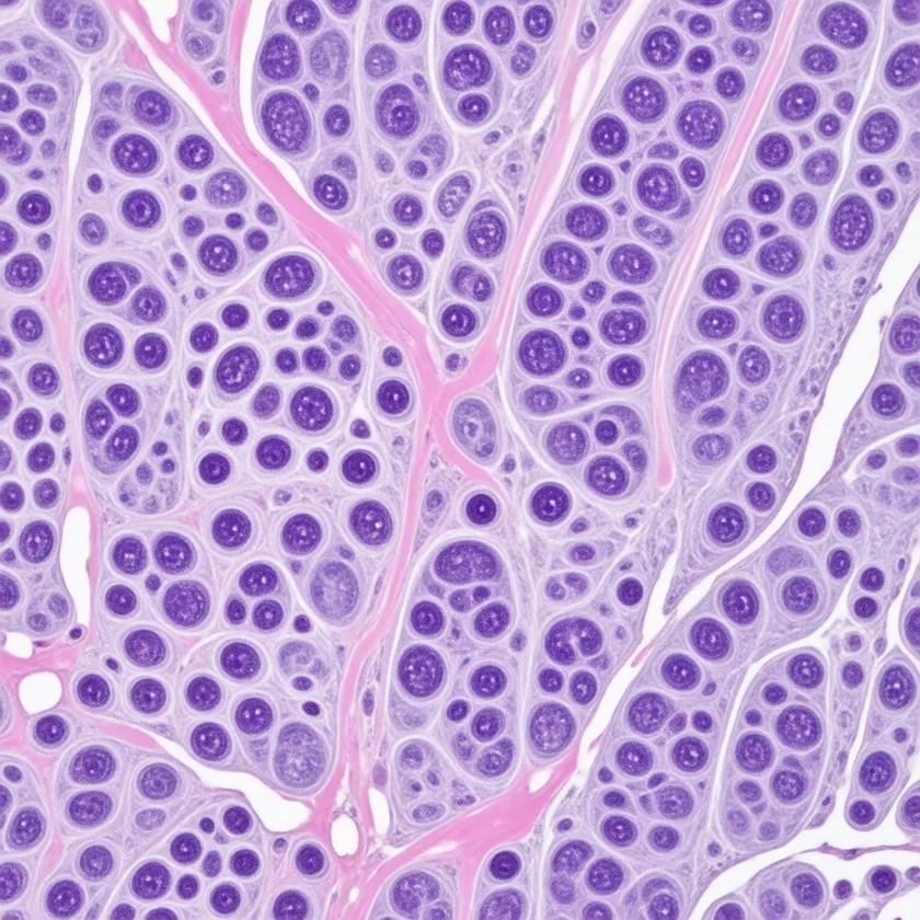
- Overview and Epidemiology
- Pathophysiology and Cell of Origin
- Genetic Risk Factors
- Clinical Presentation
- Imaging and Staging
- Biopsy and Diagnosis
- Chemotherapy (Neoadjuvant and Adjuvant)
- Surgical Treatment and Limb Salvage
- Histologic Response and Prognosis
- Metastatic and Recurrent Disease
- Key Research Papers
- PubMed Topic Searches
- Connections
- Featured Videos
Overview and Epidemiology
In plain words: osteosarcoma is a cancer that starts in bone, most often in teenagers during their growth spurt. The usual first sign is bone pain that keeps getting worse and comes on at night, often with a swelling near the knee, and it is easily mistaken for a sports injury or growing pains. The usual treatment path is an X-ray and magnetic resonance scan of the bone, a chest scan, a needle biopsy, about 10 to 12 weeks of chemotherapy, an operation to take out the tumor (now usually saving the limb), more chemotherapy, and then years of follow-up chest scans.
Osteosarcoma is the most common primary malignant tumor of bone. In the National Cancer Data Base (1985–2003) it made up 11,961 of 26,437 cases of the three main bone sarcomas, ahead of chondrosarcoma (9,606) and Ewing sarcoma (4,870). In the United States, roughly 800 new cases are diagnosed each year, with a global incidence of 3–4 cases per million population annually. Despite its relative rarity, osteosarcoma carries substantial morbidity and, when metastatic, significant mortality — making it one of the most intensively studied bone tumors in pediatric and adolescent oncology.
The age distribution is bimodal. The first and dominant peak occurs in adolescents and young adults between ages 10 and 20, tightly correlated with the pubertal growth spurt. During rapid longitudinal bone growth, the proliferating metaphyseal cells are at highest risk for the replication errors and oncogenic mutations that initiate osteosarcoma. Patients diagnosed during this peak tend to be statistically taller than their unaffected peers — a compelling epidemiologic signal that rapid skeletal growth itself is a permissive factor. A second, smaller peak occurs in adults over age 60, most often in the setting of Paget's disease of bone, prior radiation therapy, or other preexisting bone pathology. The male-to-female ratio is approximately 1.4:1, most pronounced in the adolescent peak and attributable in part to the longer duration of male pubertal bone growth.
Anatomically, osteosarcoma shows a strong predilection for the metaphysis of long bones — the flared end of the shaft adjacent to the growth plate where remodeling activity is highest. The distal femur is the single most common site, accounting for approximately 40% of cases. The proximal tibia accounts for roughly 20%, and the proximal humerus for about 10%. Together these three locations account for 70% of all extremity osteosarcomas. Less commonly, osteosarcoma arises in the pelvis, proximal femur, jaw (craniofacial osteosarcoma), or axial skeleton; these locations carry a worse prognosis due to surgical access limitations. The National Cancer Institute’s physician summary states that approximately 20% to 25% of patients present with clinically detectable metastatic disease; of these metastases, 80–85% involve the lungs, 10–15% involve other bones, and a small fraction involve regional lymph nodes.
A United States registry analysis of 5,016 patients (1975–2017) found that Black patients had the highest incidence across all ages combined, that incidence rose significantly in children aged 0 to 9, and that second osteosarcomas after an earlier cancer (“subsequent” osteosarcoma) in people up to age 24 increased 3-fold since the 2000s.
Pathophysiology and Cell of Origin
Osteosarcoma arises from primitive mesenchymal cells that retain the capacity to produce malignant osteoid — the unmineralized organic matrix of bone. The defining histologic criterion is the production of osteoid or immature woven bone directly by malignant spindle cells, distinguishing osteosarcoma from other bone sarcomas that lack this matrix-producing capacity. This osteoid production occurs even in variants classified as "lytic," "blastic," or "mixed" on plain radiography, reflecting differences in the ratio of matrix synthesis to mineralization rather than a fundamental change in the cell of origin.
The World Health Organization classifies osteosarcoma into several histologic subtypes. Conventional high-grade central osteosarcoma — the most common form — is further divided into osteoblastic (~50%), chondroblastic (~25%), and fibroblastic (~25%) subtypes based on the predominant matrix produced. These subtypes do not strongly influence treatment but affect radiographic appearance and biopsy interpretation. Telangiectatic osteosarcoma is a blood-filled cystic variant that can mimic aneurysmal bone cyst on imaging; it behaves as aggressive high-grade osteosarcoma. Small cell osteosarcoma resembles Ewing sarcoma histologically and may require molecular testing to distinguish. Low-grade central osteosarcoma and parosteal osteosarcoma (arising on the bone surface) are low-grade variants with a markedly better prognosis; they are treated surgically without chemotherapy in most cases. Periosteal osteosarcoma is an intermediate-grade surface variant.
At the molecular level, conventional high-grade osteosarcoma is characterized by genomic instability of extraordinary degree — complex structural rearrangements, chromothripsis events, and whole-genome duplication are common. Mutational burden varies widely. Unlike many carcinomas driven by single oncogenic driver mutations (e.g., KRAS in pancreatic cancer), osteosarcoma is defined by the loss of key tumor suppressors — particularly TP53 and RB1 — rather than by activating oncogene mutations. Elevated alkaline phosphatase (ALP) is the most consistent serum biomarker, reflecting increased osteoblastic activity, and correlates with tumor bulk. Lactate dehydrogenase (LDH) elevation correlates with tumor necrosis and high proliferative rate and independently predicts worse outcome.
Genetic Risk Factors
The vast majority of osteosarcoma cases are sporadic, arising from somatic mutations in dividing metaphyseal cells without a heritable predisposition. However, several germline syndromes confer dramatically elevated risk, providing critical insights into the molecular pathogenesis of the disease.
Later sequencing studies measured how often inherited faults turn up in patients. Among 1,004 patients assessed and not selected for family history, 281 (28.0%) carried a harmful variant in a cancer-susceptibility gene, compared with 12.1% and 9.3% in two comparison groups. In a separate study of 765 patients, 9.5% of those under age 30 carried a Li-Fraumeni-linked mutation (3.8%) or a rare TP53 variant (5.7%), and none aged 30 or older did.
Hereditary retinoblastoma is the strongest known predisposition syndrome for osteosarcoma. Germline loss-of-function mutations in RB1 — the gene encoding the retinoblastoma protein, a master regulator of the G1/S cell cycle checkpoint — confer approximately a 500-fold increase in osteosarcoma risk, particularly in patients who received radiation therapy to the orbit. Even without radiation, the cumulative lifetime risk of osteosarcoma in bilateral retinoblastoma survivors is approximately 13%. In a cohort of 1,604 retinoblastoma survivors, the cumulative incidence of any second cancer 50 years after diagnosis was 51.0% with hereditary retinoblastoma and 5.0% with nonhereditary retinoblastoma. This association established RB1 as a critical osteosarcoma suppressor gene, and somatic loss of RB pathway function is now recognized in ~70% of all osteosarcomas through various mechanisms including deletion, mutation, and promoter methylation.
Li-Fraumeni syndrome, caused by germline TP53 mutations, is the second major predisposition syndrome. TP53 encodes p53, the "guardian of the genome" that orchestrates DNA damage responses, apoptosis, and senescence. Li-Fraumeni families are at elevated risk for osteosarcoma, soft-tissue sarcoma, breast cancer, brain tumors, and adrenocortical carcinoma, often at young ages. Osteosarcoma accounts for approximately 12% of cancers in Li-Fraumeni kindreds. Somatic TP53 mutations or deletions occur in 30–50% of sporadic osteosarcomas, and the two tumor suppressors — p53 and Rb — cooperate to restrain osteoblastic proliferation; loss of both is highly oncogenic in mouse models.
Rothmund-Thomson syndrome (RTS type 2) and Werner syndrome are caused by mutations in RECQL4 and WRN, respectively — members of the RecQ DNA helicase family. These proteins maintain genomic stability during DNA replication and repair. RTS type 2 patients have a 30–40% lifetime osteosarcoma risk. Werner syndrome patients have premature aging and approximately 10% osteosarcoma risk. Both syndromes illustrate how replication stress and DNA repair deficiency drive the chromosomal catastrophe characteristic of osteosarcoma.
Hereditary multiple osteochondromatosis (mutations in EXT1/EXT2) predisposes primarily to chondrosarcoma but carries a small osteosarcoma risk. Paget's disease of bone (predominantly SQSTM1 mutations) causes pathologically disordered bone remodeling; approximately 1% of patients develop secondary osteosarcoma, most commonly in the femur, humerus, or pelvis. Fibrous dysplasia and sites of prior therapeutic radiation (typically appearing 10–20 years after radiation with a latency inversely related to dose) are additional secondary osteosarcoma risk factors in adults.
Genome-wide association studies have identified susceptibility loci on chromosomes 6p21.3 (near GRM4) and 2p25.2 that modestly increase population-level osteosarcoma risk, though these associations explain only a small fraction of sporadic cases.
Clinical Presentation
The most common presenting symptom is progressive, worsening pain at the involved site. Early in the course, pain is often intermittent and activity-related, easily dismissed as sports injury or "growing pains" — a misattribution that is extremely common in the adolescent peak age group. As the tumor enlarges, pain becomes continuous, nocturnal, and eventually severe enough to disrupt sleep. The median delay from symptom onset to diagnosis is 3–4 months, partly because the initial presentation is indistinguishable from much more common benign conditions (patellar tendinopathy, Osgood-Schlatter disease, muscular strain).
On examination, there is typically a palpable soft tissue or bony mass with local warmth and tenderness over the metaphysis. The overlying skin may appear stretched or have prominent superficial veins due to the increased vascularity of the tumor. Range of motion of the adjacent joint may be restricted. Pathological fracture occurs at presentation in approximately 10% of cases, most often in lytic or telangiectatic variants; it significantly complicates surgical planning because fracture hematoma can contaminate tissue planes and potentially convert a limb-salvageable case to amputation.
Constitutional symptoms — fever, night sweats, and unintentional weight loss — are present in a minority of osteosarcoma patients, in contrast to Ewing sarcoma where they are more prominent. Fever occurs more often in advanced or necrotic tumors. Elevated alkaline phosphatase is found in approximately 50% of patients and often correlates with tumor volume and osteoblastic activity; it normalizes with successful treatment and may re-elevate at relapse. Elevated lactate dehydrogenase is found in a smaller subset and carries independent adverse prognostic significance in multivariate analyses.
Symptoms of metastatic disease at diagnosis include dyspnea or cough (pulmonary metastases, which are the dominant site), pain at other skeletal sites (bone metastases), and, rarely, neurological symptoms from cord or nerve involvement. Complete staging before treatment is standard practice, because the presence of lung metastases fundamentally changes the prognosis and alters the surgical strategy.
Imaging and Staging
The initial imaging study for any suspected bone tumor is a plain radiograph of the involved bone in two planes. Osteosarcoma has characteristic but variable radiographic features. The most pathognomonic signs reflect aggressive periosteal reaction and cortical destruction:
The "sunburst" pattern (or "sunray" spiculation) describes radiating spicules of periosteal new bone that form along tumor vasculature as the tumor elevates and breaches the periosteum. The Codman triangle is the triangular shadow of reactive periosteal new bone at the junction between the tumor and normal bone, formed when the periosteum is lifted by the expanding mass. Both signs indicate aggressive periosteal reaction and are not specific to osteosarcoma — they can occur in Ewing sarcoma and other aggressive processes — but their combination with metaphyseal location in an adolescent is highly suggestive. The intramedullary lesion itself may be lytic, blastic (dense sclerosis from osteoid mineralization), or mixed. A soft tissue mass extending beyond the cortex is common in large tumors.
After plain radiography, magnetic resonance imaging (MRI) of the entire involved bone (not just the region of visible tumor) is mandatory. Magnetic resonance imaging characterizes: (1) intramedullary tumor extent and proximity to the growth plate; (2) soft tissue mass extent and relationship to neurovascular structures; (3) skip metastases — separate tumor foci within the same bone proximal to the main tumor, present in 1–3% of cases, identifiable only by whole-bone magnetic resonance imaging and requiring excision as part of surgical treatment. T1-weighted sequences best define marrow involvement; short tau inversion recovery (STIR) or T2 fat-suppressed sequences best display soft tissue extension and edema; gadolinium enhancement with subtraction improves delineation of viable tumor margins — relevant when planning biopsy and re-evaluating response after neoadjuvant chemotherapy.
Computed tomography (CT) of the chest (without contrast) is the most sensitive modality for detecting pulmonary metastases, and the National Cancer Institute’s physician summary states that as many as 20% of patients have radiographically detectable metastases at diagnosis, with the lung the most common site. Nodules as small as 3–4 mm can be detected and are regarded with suspicion in the context of a bone sarcoma, even when below the threshold for biopsy.
Whole-body bone scintigraphy (technetium-99m bone scan) or positron emission tomography with computed tomography (PET-CT) detects distant skeletal metastases and regional lymph node involvement. This combined scan, using the tracer 18F-fluorodeoxyglucose, is increasingly used because it evaluates both bone and soft tissue in one study and provides a baseline standardized uptake value (SUV) that may be followed to assess chemotherapy response. The Enneking surgical staging system (Stage I low-grade, Stage II high-grade, Stage III any grade with metastases; subdivided A intracompartmental / B extracompartmental) guides surgical planning and provides a standardized language for comparing outcomes across institutions.
Biopsy and Diagnosis
Biopsy is the essential step that establishes histologic diagnosis before any treatment is initiated. A critical principle in musculoskeletal oncology is that biopsy is performed at the treating orthopaedic oncology referral center, or at minimum after consultation with the surgeon who will perform the definitive resection. This is not administrative formality — a poorly planned biopsy can irrevocably compromise the resection and convert a limb-salvageable patient to one requiring amputation.
The biopsy tract is contaminated with tumor cells. In extremity sarcomas, the tract is excised en bloc with the tumor at definitive surgery. Surgical teaching therefore describes: (1) longitudinal (not transverse) biopsy incisions on the limbs, which permit en bloc resection of the tract; (2) a path through the minimum number of muscle compartments; (3) meticulous hemostasis, because hematoma spreads tumor cells along fascial planes; (4) a path clear of the neurovascular bundle and joints. A transverse or poorly placed incision that violates the joint capsule or crosses the neurovascular bundle may render the limb unsalvageable.
Core needle biopsy (CNB) under computed tomography or ultrasound guidance is preferred when the soft tissue component is accessible, providing adequate tissue for histology, immunohistochemistry, molecular testing, and cytogenetics with minimal contamination. A coaxial technique using a single entry point minimizes tract contamination. When core needle biopsy is inadequate (e.g., purely intraosseous sclerotic tumors with minimal soft tissue mass, very small lesions, or technically challenging locations), an open incisional biopsy is performed.
The pathologist's report confirms: (1) high-grade malignant spindle cells producing osteoid or woven bone (the sine qua non of osteosarcoma); (2) histologic subtype; (3) grade. Immunohistochemistry for SATB2 (a nuclear osteoblastic marker) can assist in challenging cases, particularly when distinguishing osteosarcoma from Ewing sarcoma or undifferentiated pleomorphic sarcoma. Molecular testing (fluorescence in situ hybridization, FISH, for the EWSR1 gene) is routinely performed to exclude Ewing sarcoma from the differential. Final diagnosis at a center experienced in musculoskeletal pathology is essential, as diagnostic error rates are meaningfully higher at low-volume institutions.
Chemotherapy (Neoadjuvant and Adjuvant)
The introduction of multiagent chemotherapy in the 1970s–1980s transformed osteosarcoma from a disease with 5-year survival of approximately 15–20% (surgery alone) to one with 60–70% survival for localized disease today. The landmark T-10 protocol developed by Gerald Rosen at Memorial Sloan Kettering established that intensive neoadjuvant (pre-operative) chemotherapy could be followed by limb-sparing surgery without compromising survival, and that histologic response to neoadjuvant treatment was a powerful prognostic marker — a paradigm that still governs contemporary treatment.
The current standard regimen for high-grade osteosarcoma is MAP: high-dose Methotrexate + doxorubicin + cisPlatin (the letter A is left over from a trade name of doxorubicin). This triplet, delivered over 10–12 weeks preoperatively and 12–18 weeks postoperatively, remains the international standard after decades of randomized trials failed to improve upon it with additional agents.
High-dose methotrexate is administered at 8–12 g/m² over 4–6 hours (the EURAMOS-1 standard regimen used 12 g/m² over 4 hours) with aggressive hydration and urinary alkalinization. Leucovorin (folinic acid) rescue begins 24 hours after the start of infusion and continues until serum methotrexate levels fall below a defined threshold (typically 0.1–0.05 µmol/L). Methotrexate levels are measured at 24, 48, and 72 hours; delayed clearance — due to third-spacing, renal impairment, or drug interactions — requires prolonged leucovorin rescue. Toxicities include mucositis, renal tubular injury (methotrexate precipitation in acidic urine), hepatotoxicity, and myelosuppression.
Doxorubicin (75 mg/m² per cycle) contributes significantly to tumor necrosis but carries cumulative cardiotoxicity risk. Lifetime doxorubicin dose is carefully tracked; echocardiographic surveillance is mandatory during and after treatment. Cisplatin (120 mg/m² per cycle) causes dose-limiting nephrotoxicity (aggressive intravenous (IV) hydration with mannitol diuresis required), ototoxicity (audiograms at baseline and each cycle), peripheral neuropathy, nausea, and myelosuppression.
The pivotal EURAMOS-1 study (2,260 patients registered; randomized results published in 2015 and 2016, outcomes for the whole group in 2019) tested whether escalating treatment with ifosfamide and etoposide in poor histologic responders, or adding pegylated interferon-α2b in good responders, improved outcomes over MAP alone. Among 618 poor responders, event-free survival did not differ (hazard ratio 0.98), and severe non-blood side effects (grade 4) were more frequent with the added drugs (24% vs 12%); the authors wrote that the results do not support adding them and define the standard of care for this group. Among 716 good responders, 3-year event-free survival was 76% for the whole group, and adding interferon was “not statistically different” from MAP alone (hazard ratio 0.83, P = .214), with a considerable proportion never starting interferon or stopping it early; long-term follow-up for events and survival was continuing. This landmark study closed several major research questions and shifted attention toward novel agents and immunotherapy.
Survival has plateaued. United States registry analyses report that survival “did not improve significantly from 1984 to 2004” and has “remained relatively unchanged over recent decades.” In the full EURAMOS-1 group of 2,260 patients, 5-year event-free survival was 54% and overall survival 71%. In the same study, 50% of patients reached at least 90% necrosis at surgery, and severe (grade 3–4) neutropenia, a drop in infection-fighting white cells, affected 83% of pre-operative cycles, with 59% complicated by infection.
Mifamurtide (MTP-PE) — a synthetic analogue of muramyl dipeptide that activates macrophages — was authorised in the European Union on 6 March 2009 (European Medicines Agency record) for use in combination with MAP based on a Children's Oncology Group trial (Meyers et al., 2008) showing improved overall survival when added to three-drug chemotherapy. It is not U.S. Food and Drug Administration-approved in the United States. Its role remains debated. The trial had a 2 x 2 factorial design and two primary endpoints: 6-year overall survival improved from 70% to 78% (P = .03, hazard ratio 0.71), while event-free survival showed only a trend (P = .08).
Surgical Treatment and Limb Salvage
Surgery remains the only curative modality in osteosarcoma. The goal is en bloc resection with wide surgical margins — removing the tumor with a surrounding cuff of normal uninvolved tissue on all sides, with the biopsy tract excised en bloc. Intralesional resection (debulking) is not considered acceptable; even microscopically positive margins (R1) are associated with markedly increased local recurrence rates. Achieving negative margins while maximizing functional preservation is the central surgical challenge.
Limb salvage is now achieved in 80–90% of patients at high-volume orthopaedic oncology centers, a dramatic improvement from the 1970s when amputation was the default. The shift was enabled by: (1) neoadjuvant chemotherapy reducing tumor volume and creating a reactive pseudocapsule that facilitates dissection; (2) advanced magnetic resonance imaging staging accurately defining resection margins; (3) improvements in endoprosthetic implant design; and (4) refinements in surgical technique and reconstruction options.
The most common reconstruction after resection of distal femur or proximal tibia osteosarcoma in adults is modular endoprosthetic replacement — a custom or modular metallic prosthesis replacing the resected bone and articulating with the remaining joint. Modern modular systems allow intraoperative adjustment of length and can be expanded in growing children using noninvasive electromagnetic lengthening mechanisms (expandable prostheses). These implants are durable and allow early weight-bearing but carry risks of aseptic loosening, periprosthetic infection, structural failure, and need for revision surgery at 10–15 years.
Biological reconstruction using massive osteoarticular allografts — cadaveric bone segments matched for size — preserves the articular surface and allows tendon and ligament reattachment. Allografts have excellent long-term durability when they heal but carry significant early risks of nonunion, fracture, and infection. Allograft-prosthetic composites combine the advantages of both. In young children where bone growth and limb length equality are paramount concerns, rotationplasty (Van Nes procedure) — resecting the mid-femur and rotating the lower leg 180° to use the ankle as a functional knee — provides an excellent functional outcome with high activity levels, despite the dramatic appearance. Psychological acceptance is high among families who choose it. In one small long-term series, 10 survivors followed for a mean of 13.8 years had a mean function score of 82.7% and a mean global health score of 85.8%, and nine reported no or only minor functional restrictions.
Amputation remains necessary when: tumor involves the major neurovascular bundle without reconstruction options; obtaining clear margins around a locally very large or deeply infiltrating tumor is not possible; a pathological fracture has contaminated the surgical field extensively; or infection or failed prior reconstruction makes the limb unsalvageable. Above-knee amputation with a well-fitted prosthesis provides excellent functional outcomes and is curative — survival is equivalent to limb salvage when equal margins are achieved.
Skip lesions — separate intramedullary tumor foci within the same bone — are identified on preoperative magnetic resonance imaging and resected en bloc with the primary tumor; the resection level is extended to include all skip foci with negative margins.
Surgery for pulmonary metastases (metastasectomy, by open chest surgery called thoracotomy or by video-assisted thoracoscopic surgery, VATS) is an important component of treatment for metastatic disease and selected cases of relapse. Complete resection of all macroscopic pulmonary disease, confirmed by manual palpation at open thoracotomy in many protocols, is associated with the best salvage outcomes.
Histologic Response and Prognosis
The most powerful prognostic factor in osteosarcoma after neoadjuvant chemotherapy is the degree of tumor necrosis in the surgical specimen. Pathologists grade the response by examining the entire resected tumor and estimating the percentage of viable versus necrotic tumor cells. Good histologic response is defined as ≥90% necrosis (Huvos grade III or IV); poor response is defined as less than 90% necrosis. This threshold was established empirically from the early Rosen T-10 trial data and has been validated across all subsequent large trials.
Patients who achieve ≥90% necrosis have 5-year event-free survival of approximately 65–70% for localized disease and overall survival of 70–80%. Poor responders have markedly worse outcomes: 5-year event-free survival of 40–50% and overall survival of 55–65%. Despite intensive investigation, no treatment modification in poor responders has improved these outcomes — EURAMOS-1 showed that adding ifosfamide/etoposide to adjuvant MAP did not improve event-free survival in poor responders (101 deaths on MAP vs 92 with the added drugs). This remains one of the most critical unmet needs in osteosarcoma.
Other significant prognostic factors include:
- Metastatic disease at presentation: 5-year overall survival approximately 20–30%, compared to 65–70% for localized disease. Patients with resectable lung-only metastases achieve better outcomes (30–40%) than those with bone or multifocal metastases (<20%).
- Surgical margins: Wide negative margins are associated with substantially lower local recurrence rates than marginal or positive margins. Local recurrence portends poor prognosis for overall survival.
- Tumor size and location: Axial tumors (pelvis, spine) have worse outcomes than extremity tumors, reflecting surgical difficulty in obtaining wide margins. Tumor volume >200 mL is an adverse factor.
- Elevated serum lactate dehydrogenase: Independent adverse prognostic factor in multiple multivariate analyses.
- Age: Older patients (second peak, secondary osteosarcoma) generally have worse outcomes than younger patients, partly reflecting higher rates of axial disease and comorbidities limiting chemotherapy delivery.
Registry figures show how much the outlook differs by age and tumor type. In the National Cancer Data Base (cases from 1985 to 1998), relative 5-year survival for osteosarcoma as a whole was 53.9%: 60% under age 30, 50% at ages 30 to 49 and 30% at age 50 or older. By type it was 85.9% for parosteal, 52.6% for high-grade and 17.8% for tumors arising in Paget disease.
Long-term survivors face substantial late effects from treatment: anthracycline-related cardiomyopathy, cisplatin-related sensorineural hearing loss and peripheral neuropathy, methotrexate-related cognitive effects, infertility, secondary malignancies (particularly leukemia from topoisomerase II inhibitors), and endoprosthetic failure requiring revision surgery. A Rizzoli institute report with at least 10 years of follow-up found that relapses, heart muscle disease and second cancers are possible even 5 years or more after treatment begins, and its authors recommended long-term follow-up.
Metastatic and Recurrent Disease
The National Cancer Institute’s physician summary states that approximately 20% to 25% of patients present with clinically detectable metastatic disease at diagnosis. Historically, lung-only metastases were considered potentially curable; modern treatment incorporates MAP chemotherapy followed by resection of the primary tumor and surgical metastasectomy (thoracotomy with manual palpation of all pulmonary parenchyma to detect nodules not identified on computed tomography). When complete surgical resection of all macroscopic disease is achieved, approximately 30–40% of patients with lung-only metastases survive 5 years — a meaningful proportion, justifying aggressive surgical intent. The National Cancer Institute’s summary gives the event-free figure for the same group: for metastases limited to the lungs with complete surgical remission, 5-year event-free survival is approximately 20% to 25%. Bilateral thoracotomies (staged or simultaneous) are performed when disease is bilateral.
Bone metastases at presentation carry an extremely poor prognosis; 5-year overall survival is typically <10–15% regardless of treatment. Current trials investigate whether high-dose chemotherapy with autologous stem cell rescue improves outcomes in this group.
Recurrent osteosarcoma occurs in 30–40% of patients with initially localized disease, typically within 2 years of completing treatment. Isolated pulmonary relapse — the most common recurrence pattern — remains potentially salvageable with aggressive surgical metastasectomy. Second-line chemotherapy regimens for relapsed osteosarcoma include: ifosfamide with or without etoposide; gemcitabine + docetaxel; sorafenib (multikinase inhibitor, approved in some countries for relapsed osteosarcoma based on phase II data; a nonrandomized phase II trial in 35 patients reported median progression-free survival of 4 months); regorafenib; and cyclophosphamide + topotecan. None of these achieves cure in the majority of patients; 5-year post-relapse survival is approximately 20–25% even with aggressive treatment.
Two randomized, placebo-controlled phase 2 trials tested regorafenib in metastatic osteosarcoma that had progressed. In SARC024 (42 patients), median time without progression was 3.6 months vs 1.7 months on placebo, with no statistically significant difference in overall survival (placebo patients could cross over to the drug), and 64% of those first assigned to regorafenib had severe (grade 3–4) side effects attributed to treatment. In REGOBONE, 17 of 26 patients (65%) on regorafenib were free of progression at 8 weeks, compared with none of 12 on placebo. Cabozantinib was tested in a single-arm trial with no comparison group (CABONE): tumors shrank in 5 of 42 patients (12%) and 14 (33%) were free of progression at 6 months.
Multiple clinical trials are investigating novel strategies: immune checkpoint inhibitors (PD-1/PD-L1 blockade; initial results disappointing, possibly due to low tumor mutational burden), chimeric antigen receptor T cells (CAR-T cells) targeting GD2 (a disialoganglioside expressed on osteosarcoma), antibody-drug conjugates targeting human epidermal growth factor receptor 2 (HER2) (expressed in a subset of osteosarcomas), cyclin-dependent kinase 4/6 (CDK4/6) inhibitors exploiting RB pathway loss, muramyl tripeptide (mifamurtide) in adjuvant settings, and bisphosphonates targeting the bone microenvironment. The Bone Sarcoma Research Group, Children's Oncology Group, and European consortia operate ongoing trials that enroll patients with recurrent disease.
Follow-up after completing treatment consists of chest computed tomography every 3 months for 2 years, then every 4 months in year 3, then every 6 months through year 5, then annually — reflecting the dominant pattern of early pulmonary relapse. Plain radiographs of the primary site assess local control and endoprosthetic status. Alkaline phosphatase and lactate dehydrogenase are measured at each visit as potential biochemical markers of recurrence, though their sensitivity as early relapse indicators is limited.
Key Research Papers
- Meyers PA et al. Osteosarcoma: the addition of muramyl tripeptide to chemotherapy improves overall survival — a report from the Children's Oncology Group. Journal of Clinical Oncology 2008; 26:633–638. — PubMed PMID: 18235123
- Bacci G et al. Long-term outcome for patients with nonmetastatic osteosarcoma of the extremity treated at the Istituto Ortopedico Rizzoli according to the Istituto Ortopedico Rizzoli/osteosarcoma-2 protocol: an updated report. Journal of Clinical Oncology 2000; 18:4016–4027. — PubMed PMID: 11118462
- Rosen G et al. Preoperative chemotherapy for osteogenic sarcoma: selection of postoperative adjuvant chemotherapy based on the response of the primary tumor to preoperative chemotherapy. Cancer 1982; 49:1221–1230. — PubMed PMID: 6174200
- Whelan JS et al. EURAMOS-1, an international randomised study for osteosarcoma: results from pre-randomisation treatment. Annals of Oncology 2015; 26:407–414. — PubMed PMID: 25421877
- Smeland S et al. Survival and prognosis with osteosarcoma: outcomes in more than 2000 patients in the EURAMOS-1 (European and American Osteosarcoma Study) cohort. European Journal of Cancer 2019; 109:36–50. — PubMed PMID: 30685685
- Bielack SS et al. Methotrexate, Doxorubicin, and Cisplatin (MAP) Plus Maintenance Pegylated Interferon Alfa-2b Versus MAP Alone in Patients With Resectable High-Grade Osteosarcoma and Good Histologic Response to Preoperative MAP: First Results of the EURAMOS-1 Good Response Randomized Controlled Trial. Journal of Clinical Oncology 2015; 33:2279–2287. — PubMed PMID: 26033801
- Savage SA et al. Genome-wide association study identifies two susceptibility loci for osteosarcoma. Nature Genetics 2013; 45:799–803. — PubMed PMID: 23727862
- Bielack SS et al. Prognostic factors in high-grade osteosarcoma of the extremities or trunk: an analysis of 1,702 patients treated on neoadjuvant Cooperative Osteosarcoma Study Group protocols. Journal of Clinical Oncology 2002; 20:776–790. — PubMed PMID: 11821461
- Marina NM et al. Comparison of MAPIE versus MAP in patients with a poor response to preoperative chemotherapy for newly diagnosed high-grade osteosarcoma (EURAMOS-1): an open-label, international, randomised controlled trial. Lancet Oncology 2016; 17:1396–1408. — PubMed PMID: 27569442
- Robison LL and Hudson MM. Survivors of childhood and adolescent cancer: life-long risks and responsibilities. Nature Reviews Cancer 2014; 14:61–70. — PubMed PMID: 24304873
- Wong FL et al. Cancer incidence after retinoblastoma. Radiation dose and sarcoma risk. JAMA 1997; 278:1262–1267. — PubMed PMID: 9333268
- Enneking WF et al. A system for the surgical staging of musculoskeletal sarcoma. Clinical Orthopaedics and Related Research 1980; 153:106–120. — PubMed PMID: 7449206
- Damron TA et al. Osteosarcoma, chondrosarcoma, and Ewing's sarcoma: National Cancer Data Base Report. Clinical Orthopaedics and Related Research 2007; 459:40–47. — PubMed PMID: 17414166
- Mirabello L et al. Osteosarcoma incidence and survival rates from 1973 to 2004: data from the Surveillance, Epidemiology, and End Results Program. Cancer 2009; 115:1531–1543. — PubMed PMID: 19197972
- Cole S et al. Osteosarcoma: A Surveillance, Epidemiology, and End Results program-based analysis from 1975 to 2017. Cancer 2022; 128:2107–2118. — PubMed PMID: 35226758
- Mirabello L et al. Frequency of Pathogenic Germline Variants in Cancer-Susceptibility Genes in Patients With Osteosarcoma. JAMA Oncology 2020; 6:724–734. — PubMed PMID: 32191290
- Mirabello L et al. Germline TP53 variants and susceptibility to osteosarcoma. Journal of the National Cancer Institute 2015; 107:djv101. — PubMed PMID: 25896519
- Grignani G et al. A phase II trial of sorafenib in relapsed and unresectable high-grade osteosarcoma after failure of standard multimodal therapy: an Italian Sarcoma Group study. Annals of Oncology 2012; 23:508–516. — PubMed PMID: 21527590
- Davis LE et al. Randomized Double-Blind Phase II Study of Regorafenib in Patients With Metastatic Osteosarcoma. Journal of Clinical Oncology 2019; 37:1424–1431. — PubMed PMID: 31013172
- Duffaud F et al. Efficacy and safety of regorafenib in adult patients with metastatic osteosarcoma: a non-comparative, randomised, double-blind, placebo-controlled, phase 2 study. Lancet Oncology 2019; 20:120–133. — PubMed PMID: 30477937
- Italiano A et al. Cabozantinib in patients with advanced Ewing sarcoma or osteosarcoma (CABONE): a multicentre, single-arm, phase 2 trial. Lancet Oncology 2020; 21:446–455. — PubMed PMID: 32078813
- Wafa H et al. Evaluation of the long-term functional outcome and quality of life after rotationplasty in the management of primary malignant bone tumors about the knee in Children. SICOT-J 2026; 12:27. — PubMed PMID: 42127302