Anal Cancer
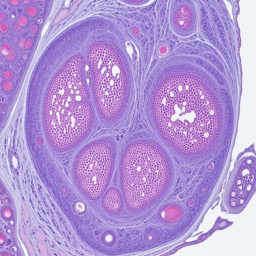
Anal cancer — most commonly squamous cell carcinoma of the anus (anal SCC) — is a rising malignancy strongly driven by human papillomavirus (HPV), particularly human papillomavirus 16. Once treated exclusively with radical surgery, anal cancer is now managed with sphincter-sparing chemoradiation (the Nigro protocol), achieving 70–80% complete response rates and preserving anal function in the majority of patients. Unlike cervical cancer, whose incidence has fallen sharply with Pap screening, anal cancer incidence is increasing — particularly among human immunodeficiency virus-positive men who have sex with men — underscoring the need for expanded human papillomavirus vaccination and high-risk screening programs.
Table of Contents
1. Overview and Epidemiology
Anal cancer comprises squamous cell carcinoma of the anus (anal SCC; approximately 85% of all anal cancers) plus rarer histological subtypes including adenocarcinoma, melanoma, and neuroendocrine tumors. Anal SCC is fundamentally a human papillomavirus-driven malignancy sharing its molecular pathogenesis with cervical, oropharyngeal, vulvar, vaginal, and penile cancers — all caused predominantly by persistent infection with high-risk human papillomavirus genotypes, most importantly human papillomavirus 16.
Incidence trends: The United States now diagnoses more than 9,000 new cases of anal cancer annually, with approximately 1,600 deaths. Incidence has been rising steadily at roughly 2–3% per year since the 1980s — a trend driven by the human papillomavirus epidemic, human immunodeficiency virus (HIV) co-infection, and aging of high-risk cohorts. This trajectory stands in stark contrast to cervical cancer, whose incidence has declined markedly due to Pap smear screening and human papillomavirus vaccination. Anal cancer incidence has risen particularly steeply in women over 60 and in human immunodeficiency virus-positive men who have sex with men (MSM).
Demographics: Women are diagnosed with anal SCC slightly more often than men overall (3:2 female:male ratio in the general population), though human immunodeficiency virus-positive MSM carry incidence rates 100-fold above the general population. The median age at diagnosis is approximately 60 years. Non-Hispanic Black patients experience higher incidence and worse survival compared with non-Hispanic White patients, likely reflecting disparities in human papillomavirus vaccination uptake, human immunodeficiency virus care access, and screening.
Human papillomavirus 16 dominance: human papillomavirus 16 DNA is detectable in 85–90% of anal SCC tumors. Human papillomavirus 18 accounts for an additional 5%. Together, these two genotypes — both covered by Gardasil 9 — explain the overwhelming majority of anal SCC, making this a highly vaccine-preventable cancer.
2. Human Papillomavirus and Risk Factors
The molecular pathogenesis of anal SCC is essentially identical to cervical SCC: persistent infection with high-risk human papillomavirus genotypes leads to integration of viral DNA into the host genome, with expression of the E6 and E7 oncoproteins. E6 binds and targets p53 for proteasomal degradation, eliminating a critical tumor-suppressor checkpoint. E7 binds and inactivates the retinoblastoma protein (Rb), releasing E2F transcription factors and driving unrestrained cell cycle progression. This dual oncogenic mechanism produces a stepwise progression from normal squamous epithelium → anal intraepithelial neoplasia (AIN 1 → AIN 2 → AIN 3 / high-grade squamous intraepithelial lesion, HSIL) → invasive SCC.
High-risk populations:
- Men who have sex with men (MSM): human papillomavirus anal infection prevalence approaches 90% in sexually active MSM. Anal SCC incidence in human immunodeficiency virus-negative MSM is 10–20 times the general population; in human immunodeficiency virus-positive MSM, risk is 80–100 times higher. This is the highest-risk group for anal SCC.
- Human immunodeficiency virus-positive individuals: human immunodeficiency virus impairs the CD4+ T-cell immune response required to clear human papillomavirus infections. Risk is greatest at low CD4 counts (<200 cells/μL), but importantly, antiretroviral therapy (ART) reduces risk without normalizing it — treated human immunodeficiency virus-positive patients still carry markedly elevated anal SCC risk compared with human immunodeficiency virus-negative individuals.
- Immunocompromised patients: Solid organ transplant recipients on chronic immunosuppression (calcineurin inhibitors, mycophenolate) have approximately 10-fold elevated anal SCC risk.
- "Field cancerization" — prior human papillomavirus-related gynecological cancers: Women with a history of cervical, vulvar, or vaginal SCC have shared human papillomavirus exposure across the lower anogenital tract. Prior cervical SCC carries an approximately 4-fold increased risk of subsequent anal SCC.
- High lifetime number of sexual partners: Increases cumulative human papillomavirus exposure probability.
- Cigarette smoking: Doubles anal SCC risk, possibly through human papillomavirus persistence and local immunosuppression.
- Anal intercourse history: Increases risk due to direct human papillomavirus transmission and local mucosal trauma facilitating viral entry.
3. Anatomy and Subtypes
The anatomy of the anus determines tumor histology, lymphatic drainage, and treatment planning — three details that are clinically essential.
The anal canal extends from the anorectal junction (where the rectum meets the anal canal at the puborectalis sling) to the anal verge (the external junction with perianal skin). Length: approximately 3–4 cm. The dentate line (also called the pectinate line) lies midway within the canal and is the single most important anatomical landmark in anal oncology:
- Above the dentate line: Columnar epithelium (transitional zone proximally). Tumors in this zone are more likely to be adenocarcinomas and are staged and managed as rectal cancer (surgical resection primary approach).
- Below the dentate line / perianal skin: Squamous epithelium. This is the territory of human papillomavirus-driven SCC. Standard chemoradiation approach applies.
Lymphatic drainage — critical for staging and RT planning:
- Upper anal canal (above dentate) → internal iliac + mesorectal lymph nodes
- Lower anal canal and perianal skin (below dentate) → inguinal lymph nodes
This dual drainage pattern means inguinal lymph node dissection is not routinely performed for upper-canal tumors (unlike inguinal LN involvement in lower-canal tumors, which are staged and treated differently). Radiation fields must encompass the relevant nodal basins.
Histological subtypes:
- Squamous cell carcinoma (85%): human papillomavirus-driven. Includes keratinizing, non-keratinizing, and basaloid (cloacogenic) variants — all managed identically with concurrent chemoradiation.
- Adenocarcinoma (<10%): Arises above the dentate line from columnar epithelium or from anal glands. Managed as rectal adenocarcinoma: neoadjuvant chemoRT + surgical resection (abdominoperineal resection or low anterior resection depending on level).
- Anal melanoma (<2%): Rare and aggressive. Usually diagnosed at advanced stage. Surgery (wide local excision or APR) for localized disease; PD-1 checkpoint inhibitors (pembrolizumab, nivolumab) are active in metastatic disease. Five-year survival below 20%.
- Neuroendocrine tumors: Rare. Well-differentiated anal NETs managed similarly to rectal NETs (endoscopic or surgical resection for small tumors; somatostatin analogues + PRRT for metastatic disease). High-grade neuroendocrine carcinoma (small cell) managed with platinum-etoposide chemotherapy.
4. Clinical Presentation
Anal cancer is frequently misdiagnosed or delayed in diagnosis because its most common symptom — rectal bleeding — is reflexively attributed to hemorrhoids. This diagnostic error is clinically consequential: anal SCC is highly curable when detected early, and delays in biopsy allow progression to higher stages. Any patient with rectal bleeding, anal pain, or anal mass requires digital rectal examination (DRE) and anoscopy before a presumptive diagnosis of hemorrhoids is made.
Symptoms and signs:
- Rectal bleeding (50–60%): Bright red blood per rectum; most common presenting symptom; commonly misattributed to hemorrhoids for months before correct diagnosis.
- Anal pain (40–50%): May be constant or exacerbated by defecation; poorly localized; often described as pressure or heaviness.
- Palpable anal mass: Patient may notice a lump at the anal verge or within the canal on self-examination.
- Pruritus ani: Perianal itching, particularly with perianal skin involvement.
- Change in bowel habits: Narrowing of stool caliber, tenesmus (sensation of incomplete evacuation), increased frequency.
- Anal incontinence: Sphincter involvement by tumor; fecal soiling; a late and alarming symptom indicating advanced local disease.
- Inguinal lymphadenopathy (10–20%): Palpable groin lymph nodes indicate inguinal metastasis — the characteristic nodal drainage route for tumors arising below the dentate line. This is a critically different pattern from rectal cancer, which drains to mesenteric nodes.
- Rectovaginal fistula: In women with posterior vaginal wall invasion; presents as passage of stool or gas per vagina.
- Systemic symptoms: Weight loss, fatigue, anorexia — usually indicate locally advanced or metastatic disease.
Physical examination pearls: DRE may reveal a firm, irregular, tender mass within the anal canal. The dentate line can sometimes be approximated by position (approximately 2 cm from the anal verge in most adults). Inguinal lymph nodes should be palpated bilaterally in all patients with anal symptoms. External genitalia and cervix should be examined in women (human papillomavirus field cancerization).
5. Staging and Diagnosis
The diagnostic workup for anal cancer serves three purposes: tissue confirmation, accurate locoregional staging (to define RT volumes), and systemic staging (to rule out distant metastases). Human immunodeficiency virus status must also be established at diagnosis because it affects treatment tolerability and outcome.
Diagnostic steps:
- Digital rectal exam (DRE) + anoscopy: First step for any suspicious anal lesion. DRE characterizes tumor size, location relative to dentate line, sphincter involvement, and depth of invasion. Anoscopy provides direct visualization. Any suspicious lesion is biopsied.
- Biopsy and pathology: Tissue diagnosis is mandatory before treatment. Immunohistochemistry for SCC markers (p40, p63, CK5/6); HPV DNA in-situ hybridization (ISH) for human papillomavirus 16/18; p16 immunostaining as surrogate for human papillomavirus oncogenic activity.
- Pelvic magnetic resonance imaging (MRI): Gold standard for locoregional staging. Characterizes tumor size, depth of invasion, sphincter involvement, relationship to vagina/prostate/urethra, and pelvic nodal status. Essential for RT planning.
- Computed tomography (CT) chest/abdomen/pelvis: Evaluates distant metastases (liver, lung, distant nodes). Complements magnetic resonance imaging for locoregional staging.
- Positron emission tomography (PET)/computed tomography: Highly sensitive for nodal staging and distant metastases. Anal SCC is FDG-avid (human papillomavirus-driven tumors have high metabolic activity). Positron emission tomography/computed tomography upstages approximately 20–25% of patients compared with computed tomography alone and is increasingly standard of care before treatment.
- Human immunodeficiency virus testing, CD4 count, and viral load: All anal cancer patients should be tested for human immunodeficiency virus at diagnosis. Human immunodeficiency virus status influences treatment planning and supportive care. CD4 count >200 cells/μL is the threshold for full-dose chemoRT.
- Gynecological exam + cervical Pap + human papillomavirus testing: Women with anal SCC require concurrent evaluation for synchronous or prior human papillomavirus-related lower genital tract lesions.
AJCC 8th Edition TNM Staging (simplified):
- T1: Tumor ≤2 cm
- T2: Tumor >2 cm and ≤5 cm
- T3: Tumor >5 cm
- T4: Tumor of any size invading adjacent organs (vagina, urethra, bladder)
- N1: Metastasis in inguinal, mesorectal, internal iliac, or external iliac nodes
- M1: Distant metastasis
Most patients (approximately 60%) present with stage II–III disease (locoregionally confined but involving regional nodes or locally advanced primary). Approximately 10–15% present with stage IV (distant metastatic) disease.
6. Chemoradiation: The Nigro Protocol
The Nigro protocol — concurrent 5-fluorouracil (5-FU), mitomycin-C (MMC), and external-beam radiation — is the standard of care for non-metastatic anal SCC and represents one of the landmark achievements in organ-preserving oncology. Before Norman Nigro's 1974 report from Wayne State University, anal SCC was treated with abdominoperineal resection (APR) — radical surgery requiring a permanent colostomy — with 5-year survival rates below 50% for stage II–III disease. Nigro's observation that three patients treated with preoperative chemoRT showed pathological complete response led directly to the modern non-surgical approach.
Standard regimen:
- Radiation: 45–59.4 Gy to the anal canal and primary tumor + elective nodal irradiation of pelvic and inguinal lymph node basins. Intensity-modulated radiation therapy (IMRT) is now the preferred technique, reducing bowel, bladder, and genital toxicity compared with older 3D-conformal approaches.
- 5-FU: Continuous infusion 1000 mg/m²/day × 4 days during weeks 1 and 5 of radiation.
- Mitomycin-C: Bolus 10–12 mg/m² on days 1 and 29 (first and fifth weeks). MMC is an alkylating agent with a distinctive mechanism: bioreductive activation in hypoxic tumor cells, targeting the tumor microenvironment. Its superiority over cisplatin in anal SCC was established by the ACT II and RTOG 98-11 trials.
Outcomes:
- Complete clinical response in 70–80% of patients at 6–8 weeks post-treatment
- 5-year overall survival: 60–70% for stage II–III disease
- Colostomy-free survival: approximately 65–70% at 5 years
- Anal sphincter preservation in the majority of complete responders
Key trials establishing the Nigro protocol:
- ACT I (UKCCCR, 1996): First randomized trial; 5-FU/MMC + RT vs. RT alone; chemoRT showed superior locoregional control and colostomy-free survival.
- ACT II (James et al., 2013): 2×2 factorial; confirmed 5-FU/MMC superior to 5-FU/cisplatin; maintenance chemotherapy added no benefit. This established 5-FU/MMC + RT as the definitive standard of care.
- RTOG 98-11 (Ajani et al., 2008): US cooperative group confirmation; 5-FU/MMC + RT vs. induction 5-FU/cisplatin followed by 5-FU/cisplatin + RT; MMC-containing arm superior in disease-free and overall survival.
Human immunodeficiency virus-positive patients: Patients with well-controlled human immunodeficiency virus on ART with CD4 >200 cells/μL are treated with full-dose Nigro protocol without modification. ART must be continued throughout treatment. Patients with CD4 <200 cells/μL may require dose reduction, treatment delays, or growth factor support due to increased myelosuppression risk. Active acquired immunodeficiency syndrome (AIDS) does not preclude treatment but requires close multidisciplinary management.
Response assessment: Imaging (magnetic resonance imaging ± positron emission tomography (PET)/computed tomography) is typically performed 8–12 weeks after completion of chemoRT. Anal SCC response is slow and continues for weeks after radiation ends; biopsy for residual disease is generally deferred to ≥26 weeks post-treatment unless clinical progression is suspected. Premature biopsy of post-RT changes leads to false-positive residual disease assessments.
7. Salvage Surgery and Advanced Disease
Approximately 15–25% of patients with locoregionally confined anal SCC do not achieve complete response to chemoRT, or experience local recurrence after initial complete response. For these patients, salvage abdominoperineal resection (APR) is the standard curative-intent intervention.
Salvage APR:
- Involves en-bloc removal of the rectum, anus, and anal sphincter complex, with creation of a permanent end colostomy
- Technically demanding after prior pelvic irradiation: increased risk of perineal wound complications (up to 40% wound healing failure rates), fistula, and anastomotic breakdown
- 5-year overall survival after successful salvage APR: approximately 40–60% for isolated local recurrence; worse for synchronous inguinal or distant failure
- Flap reconstruction (myocutaneous flaps, omentoplasty) of the perineal defect reduces wound complications in irradiated fields
Metastatic anal SCC — systemic therapy:
- First-line: Carboplatin + paclitaxel is the most commonly used first-line regimen for metastatic anal SCC, with response rates of approximately 60–65% and median progression-free survival of 6–8 months (InterAACT trial, 2018).
- Checkpoint immunotherapy: Pembrolizumab (anti-PD-1) received U.S. Food and Drug Administration (FDA) approval for MSI-H / dMMR solid tumors, which includes a subset of anal SCC. Response rates in unselected anal SCC are approximately 10–17% as monotherapy. The SWOG S1820 trial evaluated pembrolizumab ± binimetinib in advanced anal SCC. Nivolumab shows similar activity.
- Platinum-based regimens: FOLFOX (oxaliplatin + 5-FU + leucovorin) is active in platinum-eligible patients; cisplatin combinations also used.
- HER2-targeted therapy: Approximately 10–15% of anal SCCs overexpress HER2; trastuzumab-based combinations under investigation in clinical trials.
- Clinical trial enrollment is strongly encouraged for all patients with metastatic anal SCC, given the limited evidence base and active development of novel agents.
Inguinal recurrence: Isolated inguinal nodal recurrence without prior inguinal irradiation may be treated with salvage RT ± chemotherapy. Surgical inguinal lymph node dissection is an option for isolated, resectable inguinal recurrence post-chemoRT.
8. Screening and Prevention
Anal cancer is among the most preventable cancers — with two distinct prevention strategies available: upstream human papillomavirus vaccination and downstream surveillance/treatment of high-grade precancerous lesions (AIN 3 / HSIL) in high-risk individuals.
Human papillomavirus vaccination:
- Gardasil 9 targets human papillomavirus 16, 18, 31, 33, 45, 52, and 58 — the high-risk genotypes causing approximately 90% of anal SCC
- ACIP recommendations: routine vaccination for all persons aged 9–26; shared clinical decision-making for ages 27–45 (benefit lower in this age group due to likely prior human papillomavirus exposure)
- Gardasil 9 is approved for anal cancer prevention in both males and females
- Greatest protection when administered before sexual debut (before human papillomavirus exposure)
- Vaccination in human immunodeficiency virus-positive individuals is recommended and immunogenic, though antibody responses may be lower
Anal cancer screening (high-risk groups):
- Anal cytology (anal Pap smear): Samples cells from the anal canal and transition zone with a Dacron swab; cells interpreted using cervical cytology criteria (NILM → ASCUS → LSIL → HSIL). Sensitivity approximately 70–80%; recommended annually for human immunodeficiency virus-positive MSM, HIV-positive women, and women with prior human papillomavirus-related lower genital tract cancers.
- High-resolution anoscopy (HRA): Colposcopy analog for the anal canal; allows directed biopsy of abnormal acetowhite or vascular lesion areas visualized under magnification after 3–5% acetic acid application. The gold standard for AIN diagnosis and surveillance.
- ANCHOR trial (2022): Randomized trial in human immunodeficiency virus-positive adults with anal HSIL; treatment of HSIL by HRA-directed ablation (electrocautery, infrared coagulation, or laser) vs. active monitoring reduced risk of anal cancer by 57%. This was the first randomized controlled trial (RCT) to demonstrate that treatment of anal HSIL prevents anal cancer — directly analogous to the cervical cancer prevention model.
- AIN 3 treatment: High-grade anal intraepithelial neoplasia (AIN 3) is treated with HRA-directed ablation in high-risk individuals. Ablative modalities include infrared coagulation, electrocautery, and topical treatments (imiquimod, 5-FU cream — lower efficacy for larger lesions).
Human immunodeficiency virus management as prevention: Effective ART maintains CD4 counts and reduces (but does not eliminate) human papillomavirus persistence and AIN progression. ART is a cornerstone of anal cancer prevention in human immunodeficiency virus-positive individuals, but does not substitute for surveillance.
Key Research Papers
- Nigro ND, Vaitkevicius VK, Considine B Jr. "Combined preoperative radiation and chemotherapy for squamous cell carcinoma of the anal canal." Cancer. 1974;34(6):1976–8. — Search PubMed
- James RD, Glynne-Jones R, Meadows HM, et al. "Mitomycin or cisplatin chemoradiation with or without maintenance chemotherapy for treatment of squamous-cell carcinoma of the anus (ACT II): a randomised, phase 3, open-label, 2×2 factorial trial." Lancet Oncol. 2013;14(6):516–24. — PubMed PMID: 23578724
- Ajani JA, Winter KA, Gunderson LL, et al. "Fluorouracil, mitomycin, and radiotherapy vs fluorouracil, cisplatin, and radiotherapy for carcinoma of the anal canal: a randomized controlled trial." JAMA. 2008;299(16):1914–21. — PubMed PMID: 18430910
- Doci R, Zucali R, La Monica G, et al. "Primary chemoradiation therapy with fluorouracil and cisplatin for cancer of the anus: results in 35 consecutive patients." J Clin Oncol. 1996;14(12):3121–5. — Search PubMed
- Frisch M, Glimelius B, van den Brule AJ, et al. "Sexually transmitted infection as a cause of anal cancer." N Engl J Med. 1997;337(19):1350–8. — PubMed PMID: 9358129
- Palefsky JM, Holly EA, Ralston ML, et al. "Anal cytology as a screening tool for anal squamous intraepithelial lesions." J Acquir Immune Defic Syndr. 1997;14(5):415–22. — PubMed PMID: 34110312
- Northover J, Glynne-Jones R, Sebag-Montefiore D, et al. "Chemoradiation for the treatment of epidermoid anal cancer: 13-year follow-up of the first randomised UKCCCR Anal Canal Cancer Trial (ACT I)." Br J Cancer. 2010;102(7):1123–8. — PubMed PMID: 20354531
- Kim S, Jary M, Mansi L, et al. "Outcomes and prognostic factors in anal carcinoma: a single-center study." Oncology. 2012;82(3):145–53. — Search PubMed
- Eng C, Elkin EB, Kim S, et al. "Pembrolizumab versus pembrolizumab plus binimetinib in advanced anal squamous cell carcinoma: results from the SWOG S1820 trial." J Clin Oncol. 2023. — Search PubMed
- Machalek DA, Poynten M, Jin F, et al. "Anal human papillomavirus infection and associated neoplastic lesions in men who have sex with men: a systematic review and meta-analysis." Lancet Oncol. 2012;13(5):487–500. — PubMed PMID: 22445259
- Uronis HE, Bendell JC. "Squamous cell carcinoma of the anal canal." Oncologist. 2007;12(4):476–88. — PubMed PMID: 38688457
- Glynne-Jones R, Nilsson PJ, Aschele C, et al. "Anal cancer: ESMO-ESSO-ESTRO clinical practice guidelines for diagnosis, treatment and follow-up." Ann Oncol. 2014;25 Suppl 3:iii10–20. — PubMed PMID: 25001200