Anti-NMDA Receptor Encephalitis
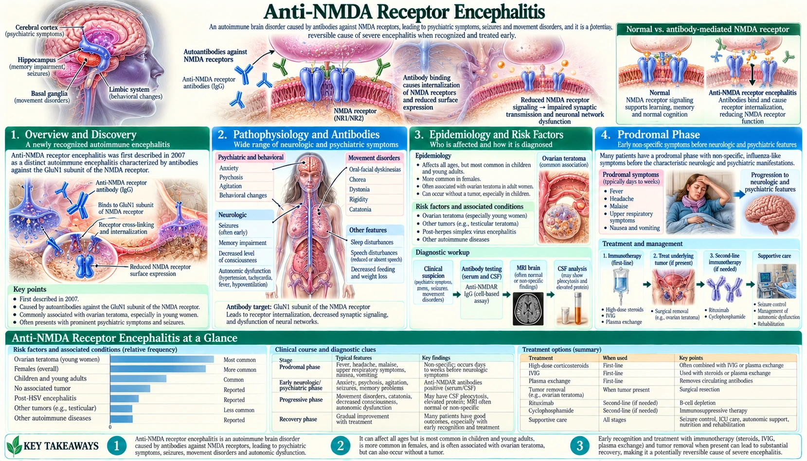
Table of Contents
- Overview and Discovery
- Pathophysiology and Antibodies
- Epidemiology and Risk Factors
- Prodromal Phase
- Psychiatric Phase
- Neurological Phase
- Diagnosis
- Electroencephalogram and Imaging Findings
- Treatment — First-Line
- Treatment — Second-Line and Tumor Removal
- Prognosis and Relapse
- Key Research Papers
- Featured Videos
- Connections
Overview and Discovery
Anti-NMDA receptor encephalitis is the most common autoimmune encephalitis identified to date. It is a severe, immune-mediated brain inflammation caused by antibodies that attack a specific receptor in the nervous system — and it is largely treatable when caught in time.
The disease was formally described by neurologist Josep Dalmau and colleagues in 2007, in a landmark case series of 12 women with psychiatric symptoms and movement disorders, 11 of whom had an ovarian teratoma and one a teratoma in the chest (mediastinum) ( PubMed PMID: 17262855). Before this discovery, countless patients were diagnosed with viral encephalitis of unknown cause, treatment-resistant schizophrenia, or conversion disorder — and many were confined to psychiatric wards without an accurate diagnosis or effective treatment.
The name has evolved over time. Early cases were called paraneoplastic anti-NMDAR encephalitis because of the strong association with ovarian teratomas — tumors that can contain neural tissue capable of triggering an immune response. As research expanded, it became clear that a substantial proportion of patients — particularly men, children, and older adults — have no identifiable tumor. The disease is now recognized as a primary autoimmune condition that can arise with or without an underlying tumor.
The causative antibodies target the GluN1 subunit (also called NR1) of the N-methyl-D-aspartate (NMDA) receptor — a glutamate receptor critical to memory, learning, synaptic plasticity, and inhibitory tone throughout the brain. Loss of N-methyl-D-aspartate receptor function, particularly in the limbic system, produces the characteristic combination of psychosis, movement disorders, and autonomic instability that defines this syndrome.
Back to Table of ContentsPathophysiology and Antibodies
The N-methyl-D-aspartate receptor is a tetrameric ion channel assembled from combinations of GluN1 (NR1), GluN2 (NR2A-D), and GluN3 subunits. The GluN1 subunit is obligatory — every functional N-methyl-D-aspartate receptor contains two GluN1 subunits. Anti-GluN1 immunoglobulin G (IgG) antibodies therefore do not attack a rare variant receptor; they target every N-methyl-D-aspartate receptor in the brain.
The antibodies cause internalization and functional loss of synaptic N-methyl-D-aspartate receptors through a process of antibody-mediated crosslinking and endocytosis. This reduces N-methyl-D-aspartate receptor density at synapses — particularly in the hippocampus, amygdala, frontal cortex, and other limbic structures where N-methyl-D-aspartate receptors are densely expressed. The damage is not structural (no complement-mediated cell death in most cases), which explains why many patients recover substantially when antibodies are removed.
The effect of N-methyl-D-aspartate receptor loss closely mimics N-methyl-D-aspartate receptor antagonists such as ketamine and phencyclidine (PCP). At subanesthetic doses, these drugs produce the same syndrome: acute psychosis, hallucinations, agitation, movement stereotypies, and dissociation. This pharmacological parallel was an important early clue to the mechanism.
A critical downstream effect involves GABAergic interneurons. These inhibitory neurons are particularly dependent on tonic N-methyl-D-aspartate receptor activation for their function. When N-methyl-D-aspartate receptors are lost, interneuron activity falls, disinhibiting downstream pyramidal and dopaminergic neurons. This disinhibition amplifies the apparent psychosis and movement disorder beyond simple receptor loss at excitatory synapses.
The immune response is primarily antibody-mediated rather than T-cell cytotoxic. Immunoglobulin G antibodies (predominantly IgG1) in cerebrospinal fluid (CSF) and serum are the diagnostic marker and the pathogenic agent. This is critical therapeutically: because neurons are not being killed by cytotoxic T cells, the damage is largely reversible with immunotherapy, even after months of severe illness.
In patients with ovarian teratoma, the prevailing model is that the teratoma contains neural tissue — neurons, glial cells, and myelin — that expresses N-methyl-D-aspartate receptors outside the normal immune-privilege of the blood-brain barrier. This triggers a systemic immune response against GluN1 epitopes that then cross-reacts with N-methyl-D-aspartate receptors in the brain. In patients without a tumor, the trigger for immune sensitization is less well understood; post-infectious autoimmunity (particularly after herpes simplex virus encephalitis) is one recognized mechanism.
Back to Table of ContentsEpidemiology and Risk Factors
Anti-NMDA receptor encephalitis affects people of all ages and backgrounds, but the epidemiology shows strong demographic patterns that reflect its underlying biology.
Sex and age: The overall female-to-male ratio is approximately 4:1. Peak onset is in the 20s and 30s, though the disease occurs from infancy to old age. The strong female predominance reflects the ovarian teratoma association; in children under 12 and in men, the sex ratio is closer to equal.
Incidence: Annual incidence is estimated at approximately 1.5 per million in population-based studies, making it a rare but not vanishingly rare condition. It is almost certainly underdiagnosed — recognition of the syndrome has improved dramatically since 2007, and incidence estimates have risen accordingly. In one California study, anti-NMDAR encephalitis was more common than any individual viral encephalitis cause.
Ovarian teratoma: Present in 50–60% of women under 45 with anti-NMDAR encephalitis. The teratoma may be small (a few millimeters) and only detectable by magnetic resonance imaging (MRI) after ultrasound is negative — systematic imaging is essential. Men rarely have teratomas but can have thymomas, mediastinal teratomas, or small cell lung cancer as associated tumors.
Children and adolescents: Represent a substantial portion of cases. In children, the disease often presents with more prominent movement disorders and seizures relative to the psychiatric features seen in adults. Teratoma is uncommon in girls under 12.
Post-HSV trigger: Anti-NMDAR encephalitis can develop 2–6 weeks after herpes simplex virus (HSV) encephalitis, presumably because HSV-induced brain inflammation releases sequestered NMDAR antigens that then drive an autoimmune response. This is an important diagnostic consideration in any patient who deteriorates or fails to improve after acyclovir treatment for HSV encephalitis.
Ethnicity: The disease occurs across all ethnic groups. Higher reported rates in Black and Asian women in some cohorts may reflect differential teratoma prevalence rather than immune susceptibility differences.
Back to Table of ContentsProdromal Phase
Before the dramatic neurological syndrome becomes apparent, most patients experience a prodromal phase lasting one to two weeks that is easy to overlook or misattribute.
The prodrome resembles a viral illness: headache, low-grade fever, fatigue, nausea, and upper respiratory symptoms. Some patients also notice subtle behavioral changes — mild mood shifts, irritability, difficulty concentrating, or unusual forgetfulness. Sleep disturbance is reported in some cases.
The prodrome is often so unremarkable that patients, families, and clinicians do not recall it as significant once the full neurological picture emerges. It is frequently reconstructed retrospectively after diagnosis. At the time, patients are typically managed as having a minor viral illness and sent home.
This early phase matters for two reasons. First, it sets the stage for initial misdiagnosis of the full syndrome as continued viral illness. Second, in research cohorts, the prodromal symptoms correlate with the timing of antibody production and central nervous system (CNS) inflammation — the immune response is already underway before the classic syndrome appears.
No specific intervention during the prodromal phase has been shown to prevent progression, but awareness of this pattern may prompt earlier diagnostic workup in patients who then develop psychiatric or neurological symptoms in the weeks following a febrile illness.
Back to Table of ContentsPsychiatric Phase
The psychiatric phase is the most diagnostically treacherous stage of anti-NMDA receptor encephalitis. It mimics primary psychiatric illness so completely that most patients are admitted to psychiatric facilities before the neurological diagnosis is considered.
Typical features include abrupt-onset psychosis — vivid visual and auditory hallucinations, paranoid delusions, and agitation emerging over days, not weeks. This rapid onset is atypical for primary psychotic disorders like schizophrenia, which usually develop gradually over months. Behavioral disinhibition is common: patients may be sexually disinhibited, aggressive, or profoundly disorganized in ways that seem out of character.
Memory deficits are nearly universal in this phase, though they may be difficult to assess in a psychotic patient. Short-term memory is particularly affected, reflecting hippocampal N-methyl-D-aspartate receptor loss. Some patients have insight early on — they recognize that something is neurologically wrong with them, not just psychiatrically.
Patients in this phase are routinely started on antipsychotic medications. These drugs have limited efficacy against the psychosis (which is not dopamine-mediated in the usual sense) and can worsen autonomic instability, lower seizure threshold, and increase the risk of neuroleptic malignant syndrome — a particular danger as the disease progresses into the neurological phase.
Median time from symptom onset to diagnosis was historically more than three months in early cohorts. This delay is associated with worse outcomes. Recognition of the syndrome has shortened this window considerably — emergency physicians, psychiatrists, and neurologists are now more likely to consider anti-NMDAR encephalitis in a young patient with new-onset psychosis plus atypical features (memory loss, movement abnormalities, seizure, fever, or rapid escalation).
Key clinical red flags in the psychiatric setting that clinical reviews list as reasons for cerebrospinal fluid antibody testing: new-onset psychosis in a young woman with no psychiatric history, seizures in a psychiatric patient, catatonia or abnormal movements developing alongside psychosis, autonomic instability, or failure to respond to multiple antipsychotic agents.
Back to Table of ContentsNeurological Phase
As the disease progresses, neurological features emerge and dominate the clinical picture. Most patients require intensive care unit admission during this phase.
Seizures occur in 70–80% of patients. They can be focal or generalized, and status epilepticus — continuous or rapidly recurring seizures — is a life-threatening complication. The seizures can be pharmacologically difficult to control, reflecting widespread cortical dysfunction rather than a discrete seizure focus.
Movement disorders are among the most distinctive features of anti-NMDAR encephalitis. Orofacial dyskinesias — stereotyped, repetitive movements of the mouth, lips, jaw, and tongue (lip-smacking, jaw-clenching, tongue protrusion, chewing movements) — are nearly pathognomonic. These movements are rhythmic, difficult for the patient to suppress, and can persist even when the patient is otherwise unresponsive. Choreoathetosis (irregular, flowing involuntary movements of the limbs and trunk) and dystonia (sustained abnormal postures) are also common.
Decreased consciousness progresses along a spectrum: from hyperkinetic agitation through catatonia (motionless unresponsiveness with preserved arousal) to unresponsive wakefulness resembling a vegetative state. Mutism is common — patients who appear conscious may stop speaking entirely.
Autonomic instability is potentially fatal and is the leading cause of intensive care unit (ICU) admission. Features include: alternating tachycardia and bradycardia; hyperthermia (temperatures up to 40–41°C) not attributable to infection; profuse diaphoresis; hypersalivation; blood pressure lability ranging from hypertensive crisis to hypotension; and urinary incontinence. This reflects N-methyl-D-aspartate receptor loss in hypothalamic and brainstem autonomic centers.
Central hypoventilation — failure of the brainstem respiratory drive — requires mechanical ventilation in approximately 75% of intensive care unit patients. This is not peripheral respiratory failure; the lungs are normal. The brainstem circuitry that drives breathing is dysfunctional.
The autonomic and movement disorder phases frequently overlap, creating a clinical picture that has historically been described as resembling a continuous drug-induced delirium. Recognition that this syndrome — psychosis, movement disorders, autonomic failure, and hypoventilation in a young woman — constitutes a discrete and treatable disease entity was the central contribution of Dalmau's 2007 work.
Back to Table of ContentsDiagnosis
Diagnosis rests on clinical recognition combined with antibody testing. No single test is sufficient in isolation; the diagnosis is confirmed by the clinical syndrome plus positive antibody testing.
Antibody testing: The gold standard is detection of anti-GluN1 immunoglobulin G antibodies in cerebrospinal fluid (CSF) by cell-based assay. Cerebrospinal fluid sensitivity exceeds 95% in patients with the full clinical syndrome. Serum antibody testing is less sensitive (approximately 85%) — some patients are seronegative but cerebrospinal fluid-positive. For this reason serum and cerebrospinal fluid are usually tested together. The cell-based assay, which uses live cells transfected to express GluN1, is significantly more accurate than tissue-based immunohistochemistry assays.
Cerebrospinal fluid analysis: Typically shows lymphocytic pleocytosis (white cell count up to 100 cells/mm³, predominantly lymphocytes), mildly elevated protein, and normal glucose. Cerebrospinal fluid is normal in approximately 20% of patients — a normal cerebrospinal fluid does not exclude the diagnosis, and the antibody test still applies.
Brain magnetic resonance imaging: Normal in approximately 50% of patients. When abnormal, findings include FLAIR and T2 hyperintensity in the hippocampi, insula, frontal cortex, and basal ganglia. Magnetic resonance imaging changes are more often seen in patients with longer or more severe disease. A normal magnetic resonance imaging does not exclude anti-NMDAR encephalitis, and antibody testing does not depend on it.
Tumor search: In women, the standard work-up is pelvic ultrasound and/or magnetic resonance imaging to detect ovarian teratoma; if ultrasound is negative, contrast-enhanced pelvic magnetic resonance imaging is the usual next step. In men and older women, computed tomography (CT) of chest, abdomen, and pelvis is appropriate to look for thymoma or other tumors. The tumor search is not a one-time evaluation — a negative initial search warrants follow-up imaging at 6 and 12 months, as small teratomas can be missed.
Differential diagnosis: Viral encephalitis (HSV, CMV, Epstein-Barr virus, EBV), other autoimmune encephalitides (anti-LGI1, anti-CASPR2, anti-GABA-B), primary psychiatric disorders (schizophrenia, bipolar disorder with psychosis), neuroleptic malignant syndrome, malignant catatonia, and drug intoxication. The combination of psychiatric symptoms, movement disorders, autonomic instability, and cerebrospinal fluid pleocytosis in a young person is the pattern that clinical reviews describe as calling for anti-NMDAR antibody testing before psychiatric diagnoses are made.
Back to Table of ContentsElectroencephalogram and Imaging Findings
Electroencephalography (electroencephalogram, EEG) and neuroimaging provide supporting evidence for the diagnosis and help gauge disease severity, though neither is required for diagnosis when antibody testing is positive.
Electroencephalogram findings: The most common electroencephalogram pattern is diffuse slow background activity — generalized delta or theta waves replacing the normal alpha-dominant background. This reflects widespread cortical dysfunction rather than a focal lesion. Electroencephalogram may also show epileptiform discharges (sharp waves, spike-wave complexes) or frank seizure activity.
The extreme delta brush (EDB) pattern is a distinctive electroencephalogram finding in anti-NMDAR encephalitis, although its first describers wrote that how specific it is remains unclear. First described by Schmitt and colleagues in 2012 ( PubMed PMID: 22933737), it consists of rhythmic delta activity (1–3 Hz) with superimposed fast activity (10–30 Hz beta frequency bursts) riding on the crest of each delta wave — superficially resembling the delta brush pattern seen in premature neonates. Extreme delta brush occurs in approximately 30% of adult patients with anti-NMDAR encephalitis, most often during the unresponsive phase. In that first series, patients with the pattern had a more prolonged illness, with longer hospital stays (a mean of 128 against 43 days) and longer electroencephalogram monitoring. While not universal, the authors wrote that its presence should raise consideration of this syndrome.
Magnetic resonance imaging findings in detail: When present (in approximately 50% of cases), magnetic resonance imaging abnormalities on FLAIR and T2-weighted sequences reflect the distribution of N-methyl-D-aspartate receptor density and limbic involvement. The medial temporal lobes (hippocampus and parahippocampal gyrus) are most commonly affected. The insula, frontal and parietal cortex, basal ganglia, thalamus, brainstem, and cerebellum can also show signal changes. Contrast enhancement is rare. Leptomeningeal enhancement occurs occasionally.
FDG-PET: Research studies have shown that FDG-PET can detect metabolic abnormalities even when magnetic resonance imaging is normal. Early in the disease, frontal hypometabolism is characteristic; later, paradoxical frontal hypermetabolism may occur alongside occipital hypometabolism. FDG-PET is not a standard clinical requirement but can be diagnostically useful in ambiguous cases.
The clinical teaching point is this: a normal magnetic resonance imaging does not exclude anti-NMDAR encephalitis, and diagnosis does not depend on magnetic resonance imaging abnormalities appearing. The antibody test is the definitive investigation.
Back to Table of ContentsTreatment — First-Line
Treatment of anti-NMDAR encephalitis is two-pronged: remove the antibody-producing source (the tumor, if present) and suppress the antibody-producing immune response. The sooner treatment begins, the better the outcome.
First-line immunotherapy consists of one or more of the following, often given in combination:
- Intravenous methylprednisolone (IVMP): 1,000 mg per day for 5 days. High-dose corticosteroids suppress broad B-cell and T-cell activity and reduce central nervous system inflammation. Often followed by an oral prednisone taper over weeks to months.
- Intravenous immunoglobulin (IVIG): 0.4 g/kg/day for 5 days (total dose 2 g/kg). Mechanisms include Fc receptor blockade, complement inhibition, and accelerated antibody catabolism. Well-tolerated and can be given simultaneously with IVMP.
- Plasma exchange (PLEX): 5–7 exchanges over 10–14 days. Directly removes pathogenic immunoglobulin G antibodies from circulation and cerebrospinal fluid (via equilibration). Most rapidly effective at antibody removal but requires central venous access and carries procedural risks.
Most centers use IVMP and IVIG together as initial therapy, adding PLEX if the response is inadequate or the patient is critically ill. There is no randomized controlled trial comparing these agents head-to-head; treatment protocols are based on observational cohort data and expert consensus.
Concurrent supportive management is essential:
- Antiepileptic drugs (AEDs) for seizures — often multiple agents are required.
- Benzodiazepines for agitation and catatonia — generally better tolerated than antipsychotics.
- Antipsychotics are described as poorly effective in this disease and as raising the risk of autonomic complications, so clinical reviews describe using them sparingly.
- Intensive care unit-level monitoring and ventilatory support for central hypoventilation.
- Careful hemodynamic monitoring for autonomic instability — pacemaker placement is occasionally required for recurrent symptomatic bradycardia.
Response timeline: Unlike antibody-mediated diseases that respond within days to plasma exchange, anti-NMDAR encephalitis typically requires weeks before clinical improvement becomes apparent. This slow response reflects the time needed for antibody clearance from the central nervous system compartment and recovery of N-methyl-D-aspartate receptor surface expression. Prolonged intensive care unit stays (weeks to months) do not preclude an excellent eventual recovery.
Escalation threshold: If there is no meaningful clinical improvement after 2 weeks of first-line therapy, treatment protocols move to second-line agents without further delay. Early escalation is associated with better outcomes.
Back to Table of ContentsTreatment — Second-Line and Tumor Removal
Approximately 45–55% of patients require second-line immunotherapy because of insufficient response to first-line treatment. Early escalation to second-line agents improves long-term outcomes.
Rituximab is the preferred second-line agent. It is a chimeric anti-CD20 monoclonal antibody that depletes circulating B cells — the cells that produce the pathogenic anti-GluN1 antibodies. Dosing regimens vary: 375 mg/m² weekly for 4 doses, or 1,000 mg given twice, two weeks apart. B-cell depletion lasts approximately 6 months after treatment. Titulaer and colleagues (2013, PMID: 23290630) demonstrated in a large observational cohort that early use of rituximab (with or without cyclophosphamide) was independently associated with improved outcomes. Rituximab does not cross the blood-brain barrier but reduces peripheral antibody production, allowing intrathecal antibody levels to fall.
Cyclophosphamide is an alkylating agent with broad immunosuppressive effects on both B and T lymphocytes. Dose: 750 mg/m² intravenous (IV) monthly for 3–6 cycles. Often combined with rituximab in the second-line setting, particularly in patients with severe disease or slow response to rituximab alone. Long-term risks (bladder toxicity, infection, secondary malignancy) are weighed against the severity of the neurological disease.
Ovarian teratoma removal is not just a secondary intervention — it is arguably the most important treatment in women with teratoma-associated disease. Removal of the antigenic stimulus (the teratoma containing the N-methyl-D-aspartate receptor-expressing neural tissue) removes the ongoing driver of the immune response. Clinical studies consistently show that patients who undergo early teratoma removal have significantly better outcomes than those in whom removal is delayed.
Key principles for tumor removal:
- Patients whose tumour was treated early had better outcomes and fewer neurological relapses than the rest in a 2008 series of 100 patients (Dalmau and colleagues, Lancet Neurology, listed under Key Research Papers); in practice the teratoma is removed, often by laparoscopic surgery, as soon as the patient is medically stable.
- The ovary containing the teratoma is removed; the contralateral ovary is preserved unless bilateral teratomas are present.
- Neurological improvement typically follows weeks after tumor removal, not immediately.
- Teratoma removal does not replace immunotherapy — both are required.
- In patients with no identifiable tumor, full first- and second-line immunotherapy is still effective.
Maintenance immunotherapy: After the acute episode, some patients benefit from long-term maintenance immunosuppression with mycophenolate mofetil or azathioprine to prevent relapse, particularly those who required second-line treatment or have had a relapse.
Back to Table of ContentsPrognosis and Relapse
The prognosis of anti-NMDAR encephalitis is substantially better than its severity during acute illness would suggest. The antibody-mediated, non-cytotoxic mechanism means that neuronal loss is limited compared to many other forms of encephalitis, and recovery can occur even after months of severe disability.
Functional outcomes: More than 75% of patients achieve a good functional outcome (modified Rankin Scale score 0–2, indicating no significant disability) by 24 months after onset. This statistic is remarkable given that most of these patients spent weeks to months in the intensive care unit, intubated, with severe movement disorders and autonomic instability.
Recovery trajectory: Recovery is slow and not linear. Improvements in consciousness, autonomic stability, and movement disorders typically precede recovery of higher cognitive functions. Memory, executive function, behavioral regulation, and cognitive endurance (the capacity to sustain mental effort) are often the last domains to fully recover. Many patients experience cognitive fatigue — the sense of mental exhaustion after normal tasks — for months to years into recovery, even when formal cognitive testing is near-normal.
Relapse: In a 2013 cohort of 577 patients, the risk of relapse was 12% within 2 years of the initial episode (Titulaer and colleagues). Relapses are usually less severe than the initial presentation (67% of relapses in that cohort) and often respond faster to immunotherapy. Risk factors for relapse include: no immunotherapy or inadequate initial treatment, no tumor removal (when a tumor was present), and male sex. Long-term maintenance immunosuppression reduces relapse risk in high-risk patients.
Mortality: Approximately 5–7% of patients die during the acute illness, predominantly from intensive care unit complications — ventilator-associated pneumonia, autonomic crisis, pulmonary embolism, or multiorgan failure. Death directly from brain disease is uncommon. Mortality is strongly associated with delay to diagnosis and treatment.
Predictors of poor outcome: Delay to treatment, requirement for intensive care unit admission, failure to respond to first-line immunotherapy, absence of tumor identification and removal (in tumor-associated cases), and high antibody titers at diagnosis correlate with worse outcomes. Conversely, early initiation of immunotherapy — even before full diagnostic confirmation — is consistently associated with better outcomes in observational studies.
Long-term quality of life: The majority of patients who survive the acute illness eventually return to independent function, work, and school. However, subtle neuropsychological impairments — particularly in memory, processing speed, and behavioral regulation — can persist for years and may not be apparent on standard clinical examination. Formal neuropsychological testing and ongoing rehabilitation support are appropriate for many recovered patients.
Back to Table of ContentsKey Research Papers
The following peer-reviewed publications represent the foundational and landmark evidence base for anti-NMDA receptor encephalitis. All citations include PubMed links for direct access.
- Dalmau J et al. (2007) Paraneoplastic anti-N-methyl-D-aspartate receptor encephalitis associated with ovarian teratoma. Ann Neurol. 61(1):25–36. — PubMed PMID: 17262855
- Dalmau J et al. (2008) Anti-NMDA-receptor encephalitis: case series and analysis of the effects of antibodies. Lancet Neurol. — PubMed PMID: 18851928
- Titulaer MJ et al. (2013) Treatment and prognostic factors for long-term outcome in patients with anti-NMDA receptor encephalitis: an observational cohort study. Lancet Neurol. — PubMed PMID: 23290630
- Irani SR et al. (2010) N-methyl-aspartate antibody encephalitis: temporal progression of clinical and paraclinical observations in a predominantly non-paraneoplastic disorder of both sexes. Brain. — PubMed PMID: 20511282
- Florance NR et al. (2009) Anti-N-methyl-D-aspartate receptor (NMDAR) encephalitis in children and adolescents. Ann Neurol. 66(1):11–18. — PubMed PMID: 19670433
- Schmitt SE et al. (2012) Extreme delta brush: a unique EEG pattern in adults with anti-NMDA receptor encephalitis. Neurology. 79(11):1094–1100. — PubMed PMID: 22933737
- Gresa-Arribas N et al. (2014) Antibody titres at diagnosis and during follow-up of anti-NMDA receptor encephalitis: a retrospective study. Lancet Neurol. — PubMed PMID: 24360484
- Prüss H et al. (2012) N-methyl-D-aspartate receptor antibodies in herpes simplex encephalitis. Ann Neurol. — PubMed PMID: 23280840
- Kayser MS & Dalmau J (2016) Anti-NMDA receptor encephalitis, autoimmunity, and psychosis. Schizophr Res. 176(1):36–40. — PubMed PMID: 25458857
- Finke C et al. (2012) Cognitive deficits following anti-NMDA receptor encephalitis. J Neurol Neurosurg Psychiatry. — PubMed PMID: 21933952
- Dalmau J et al. (2011) Clinical experience and laboratory investigations in patients with anti-NMDAR encephalitis. Lancet Neurol. — PubMed PMID: 21163445
- Planagumà J et al. (2015) Human N-methyl D-aspartate receptor antibodies alter memory and behaviour in mice. Brain. — PubMed PMID: 25392198
Featured Videos
Lectures, case studies, and patient perspectives on anti-NMDA receptor encephalitis from neurologists and immunologists.
Back to Table of Contents