Necrotizing Fasciitis
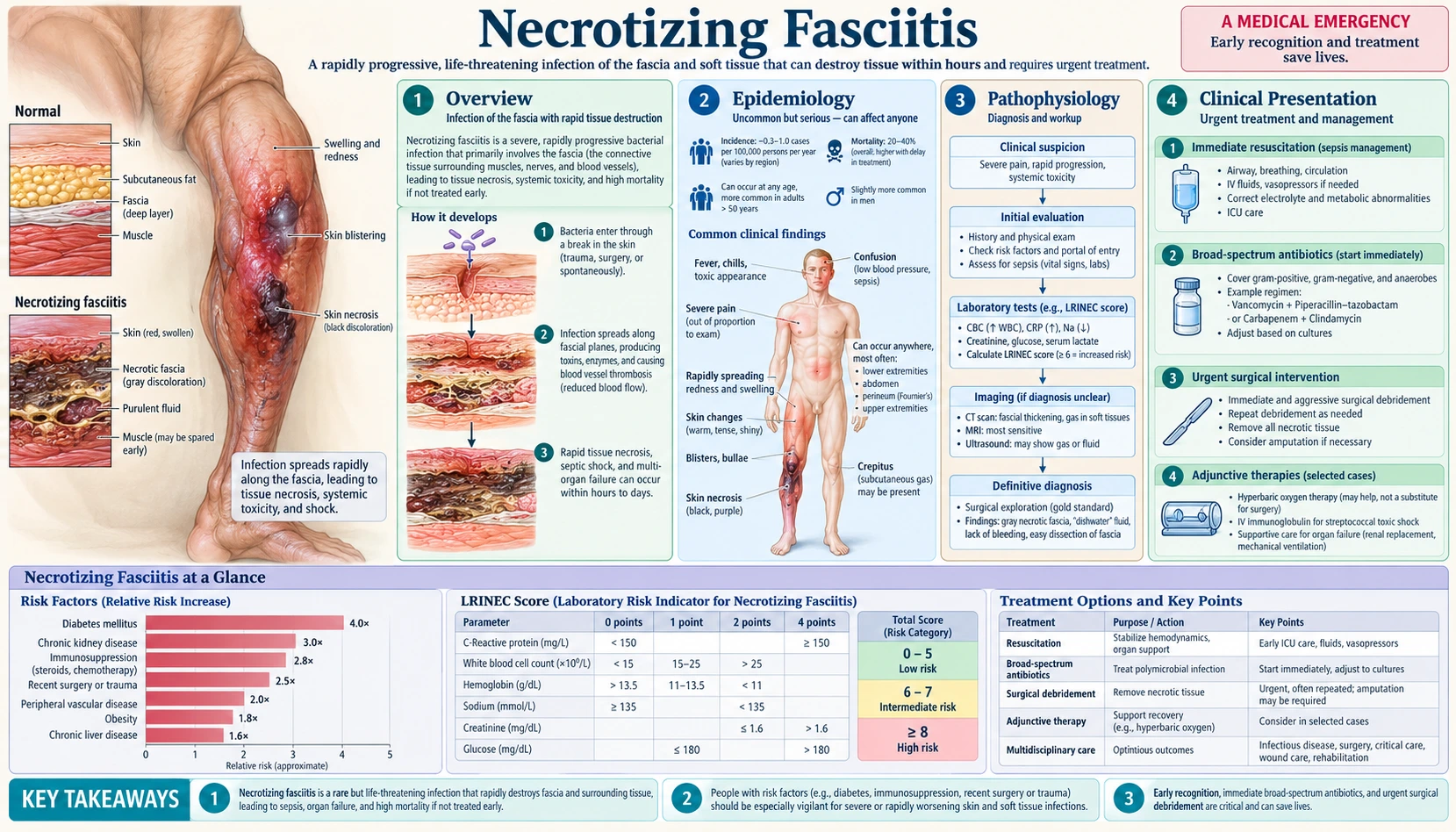
Necrotizing fasciitis is a rare, life-threatening bacterial infection of the soft tissue beneath the skin that spreads along the fascia, the sheets of connective tissue around muscles. It is sometimes called “flesh-eating bacteria” in the press. Infections are classed as polymicrobial (Type I, a mixture of organisms) or monomicrobial (Type II, typically Group A Streptococcus or, less commonly, Staphylococcus aureus), and severe pain out of proportion to what the skin shows is described as the most consistent finding (PubMed PMID: 28779832). Reviews describe the mainstay of treatment as early and complete surgical debridement combined with antibiotics, close monitoring and physiologic support (PubMed PMID: 17278065).
Table of Contents
1. Overview
Necrotizing fasciitis (NF) belongs to the group of necrotizing soft-tissue infections (NSTIs). A 2014 radiology review describes it as a rapidly spreading, progressive necrosis of the deep fascia and subcutaneous tissue that is often underestimated because the early stages lack specific clinical findings (PubMed PMID: 24469151). In a Singapore series of 89 consecutive patients, only 13 had a diagnosis of necrotizing fasciitis at the time of admission, and treatment with antibiotics before admission often masked the severity of the underlying infection (PubMed PMID: 12925624).
Type I: Polymicrobial (Mixed) Infection
A 2017 review classes NSTIs into two main types. Type I, the most common, describes polymicrobial infections and accounts for an estimated 70–80% of cases (PubMed PMID: 28779832). A 2005 clinical review notes that most cultures yield a mixture of aerobic and anaerobic organisms (PubMed PMID: 15817551). In the Singapore series, polymicrobial synergistic infection was the most common cause (48 of 89 patients, 53.9%), with streptococci and Enterobacteriaceae the most common isolates, and diabetes mellitus was the most common comorbidity (63 patients, 70.8%) (PubMed PMID: 12925624). NSTI of the perineum, Fournier’s gangrene, is generally polymicrobial (PubMed PMID: 28779832).
Type II: Monomicrobial Infection
Type II infections, an estimated 20–30% of cases in the same review, are monomicrobial and typically involve Group A Streptococcus (GAS, Streptococcus pyogenes) or, less commonly, Staphylococcus aureus (PubMed PMID: 28779832). In a Cleveland series of 65 patients, S. pyogenes was isolated in only 17% of infections but accounted for 53% of monomicrobial infections (PubMed PMID: 7748037). Community-associated methicillin-resistant S. aureus (MRSA) was described as an emerging cause in Los Angeles: 14 such patients were identified in 2003–2004, 4 of them (29%) with no serious coexisting conditions or risk factors (PubMed PMID: 15814880). Patients often have some prior history of trauma, which may be trivial, such as an insect bite, scratch or abrasion (PubMed PMID: 15817551). GAS pyrogenic exotoxins act as superantigens, and the 2017 review reports streptococcal toxic shock syndrome (STSS) in up to half of NSTI cases due to GAS (PubMed PMID: 28779832).
Fournier’s Gangrene
Fournier’s gangrene is a necrotizing infection of the perineum, genital and perianal area. It is named after the French physician Jean-Alfred Fournier, who described it in 1883; a review dates the first description to 1764 (PubMed PMID: 17034687). A short review notes that a similar condition may occasionally affect the female genitalia (PubMed PMID: 17034687). In the Maryland series of 198 patients with NSTI, the perineum was the most common site of origin (36% of cases) (PubMed PMID: 8916882). The testis and epididymis tend to be spared, although bilateral testicular gangrene has been reported (PubMed PMID: 17323114).
2. Epidemiology
The CDC describes necrotizing fasciitis caused by Group A Streptococcus as rare, says anyone can get it, and states that even with treatment up to 1 in 5 people died from the infection (CDC: About necrotizing fasciitis). Its clinical guidance puts the mortality of streptococcal necrotizing fasciitis in the most recent 5 years at around 15 to 20% (CDC: Clinical guidance for necrotizing fasciitis).
Other figures, by source:
- Incidence: the 2017 review gives the US incidence of NSTI due to invasive GAS as 0.4 per 100,000 (PubMed PMID: 28779832); the 2005 BMJ review estimates about 500 new cases a year in the United Kingdom and notes that the true incidence is difficult to record (PubMed PMID: 15817551).
- Mortality across all types: a 2020 systematic review of 109 studies (6,051 patients) found that 1,277 patients died (21.1%), and that average reported mortality stayed around 20% over the past 20 years (PubMed PMID: 31921330). Single-center series reported 29% (65 patients) (PubMed PMID: 7748037) and 25.3% (198 patients) (PubMed PMID: 8916882).
- Conditions that raise risk: the CDC lists conditions that weaken the immune system, including cancer, liver cirrhosis, diabetes and kidney disease; the BMJ review lists advanced age, chronic renal failure, peripheral vascular disease, diabetes mellitus and drug misuse (PubMed PMID: 15817551). Among the 14 Los Angeles patients with community-associated MRSA necrotizing fasciitis, 43% had current or past injection-drug use (PubMed PMID: 15814880).
- Factors linked to death: a 2026 meta-analysis of 41 cohort studies (168,261 patients) found moderate- or high-certainty associations with higher mortality for older age, chronic liver disease, chronic kidney disease, a high Charlson Comorbidity Index and immunosuppression, and for hypotension, bacteremia, acute kidney injury, coagulopathy, thrombocytopenia and shock (PubMed PMID: 42138515). In the Maryland series, diabetes did not predispose patients to death except together with renal dysfunction or peripheral vascular disease (PubMed PMID: 8916882). In the Cleveland series, S. pyogenes infection was not associated with increased mortality (18%) (PubMed PMID: 7748037).
3. Pathophysiology
Spread along the fascia
The main imaging finding described in the 2014 radiology review is thickening of the deep fasciae due to fluid accumulation and reactive hyperemia; the necrosis involves the deep fascia and the subcutaneous tissue (PubMed PMID: 24469151).
Gas in the tissues
Gas within the necrotized fasciae is characteristic but may be lacking (PubMed PMID: 24469151). The BMJ review reports crepitus in only 37% and soft-tissue air on plain radiograph in 57% of patients, and states that their absence does not exclude the diagnosis (PubMed PMID: 15817551).
Toxin-mediated disease in Type II GAS
The 2017 review describes GAS pyrogenic exotoxins acting as superantigens that bind to antigen-presenting cells and cause rapid proliferation of T cells, leading to toxic shock syndrome (PubMed PMID: 28779832). In the Canadian IVIG study, IVIG therapy enhanced the ability of patient plasma to neutralize bacterial mitogenicity and reduced T-cell production of interleukin-6 and tumor necrosis factor alpha (PubMed PMID: 10825042).
Skin changes as the infection progresses
The BMJ review describes the skin becoming increasingly tense and erythematous with indistinct margins, changing color from red-purple to dusky blue before necrosis and bullae form, the bullae eventually becoming hemorrhagic; it also lists skin anesthesia among the clinical findings (PubMed PMID: 15817551). The CDC’s clinical guidance describes the overlying skin turning dusky, cutaneous ischemia and bullae within 24 to 48 hours.
4. Clinical Presentation
Early presentation
The CDC’s clinical guidance describes necrotizing fasciitis as usually beginning with pain in the affected area accompanied by erythema, heat, swelling and tenderness. The 2017 review calls pain out of proportion to examination findings the most consistent finding (PubMed PMID: 28779832), and the BMJ review writes that severe pain disproportionate to local findings together with systemic toxicity should raise suspicion of the condition (PubMed PMID: 15817551). The CDC’s patient page lists early symptoms as a red, warm or swollen area of skin that spreads quickly, fever, and severe pain, including beyond the area that is red, warm or swollen.
How often individual signs are present
A 2019 meta-analysis of 23 studies (5,982 patients) pooled the accuracy of single signs: fever had a sensitivity of 46.0% and specificity of 77.0%, hemorrhagic bullae 25.2% and 95.8%, and hypotension 21.0% and 97.7%. The authors concluded that the absence of any one physical examination feature, such as fever or hypotension, is not sufficient to rule out NSTI (PubMed PMID: 29672405).
Late and systemic signs
Reported later findings include dusky or blue skin, hemorrhagic bullae and necrosis (PubMed PMID: 15817551), crepitus (PubMed PMID: 15817551), and, in patients presenting at an advanced stage, signs of systemic shock and sepsis (PubMed PMID: 15817551). The CDC’s patient page lists later symptoms as changes in skin color, diarrhea or nausea, dizziness, fatigue, pus or oozing from the infected area, and ulcers, blisters or black spots on the skin.
5. Diagnosis
The BMJ review states that the diagnosis remains a clinical one, with findings from all investigations used together rather than any single test (PubMed PMID: 15817551). The 2019 meta-analysis concluded that, given the poor sensitivity of the tests, a high clinical suspicion warrants early surgical consultation (PubMed PMID: 29672405), and the CDC’s clinical guidance states that imaging should never delay surgical exploration.
LRINEC Score
The Laboratory Risk Indicator for Necrotizing Fasciitis (LRINEC) score was developed by Wong et al. (2004) in a retrospective study at two teaching hospitals in Singapore, with a developmental cohort of 314 patients (89 with necrotizing fasciitis, 225 with severe cellulitis or abscesses) and a validation cohort of 140 (PubMed PMID: 15241098). It combines six routine tests; the scoring table as reproduced in a 2017 systematic review (PubMed PMID: 28462647):
- C-reactive protein >150 → 4 points
- White cells (109/L): 15–25 → 1 point; >25 → 2 points
- Hemoglobin (g/dL): 11–13.5 → 1 point; <11 → 2 points
- Sodium <135 mmol/L → 2 points
- Creatinine >141 μmol/L → 2 points
- Glucose >10 → 1 point
In the original study a cut-off of 6 points gave a positive predictive value of 92.0% and a negative predictive value of 96.0%, with an area under the ROC curve of 0.980 in the developmental and 0.976 in the validation cohort (PubMed PMID: 15241098).
Later studies found lower accuracy. The 2019 meta-analysis found that LRINEC ≥6 had a pooled sensitivity of 68.2% and specificity of 84.8%, and LRINEC ≥8 a sensitivity of 40.8% and specificity of 94.9%; the authors concluded that LRINEC had poor sensitivity and should not be used to rule out NSTI (PubMed PMID: 29672405). In a 10-year emergency-department review, 30 of 47 classifiable patients with necrotizing fasciitis (63.8%) scored as low risk, while 10.7% of 948 patients with cellulitis scored as moderate or high risk (PubMed PMID: 28611889). A 2017 systematic review of 16 studies (846 patients) reported a mean score of 6.06 in patients with necrotizing fasciitis and an area under the ROC curve of 0.927 (PubMed PMID: 28462647). The 2017 review adds that a multicenter prospective evaluation found that a cut-off of 6 failed to discriminate (PubMed PMID: 28779832).
CT Scanning
In the 2019 meta-analysis, computed tomography had a sensitivity of 88.5% and a specificity of 93.3%, against 48.9% and 94.0% for plain radiography; the authors concluded that CT is superior to plain radiography (PubMed PMID: 29672405). The 2017 review describes lack of fascial enhancement on contrast CT, with involvement of the fascia, as more specific for NSTI (PubMed PMID: 28779832).
MRI
The radiology review describes thickening of the deep fasciae as the main finding, best seen on MRI (PubMed PMID: 24469151). The 2017 review notes that MRI can be overly sensitive as well as time-consuming and can delay necessary surgery (PubMed PMID: 28779832).
Bedside Ultrasound
In a prospective Taipei emergency-department study of 62 patients with clinically suspected necrotizing fasciitis (17 confirmed), ultrasound, using diffuse subcutaneous thickening with a fluid layer more than 4 mm deep along the deep fascia as the criterion, had a sensitivity of 88.2% and a specificity of 93.3% (PubMed PMID: 12460854).
The Finger Test
The radiology review lists bedside tests, including the “finger test” and biopsy, among adjuncts described for early recognition (PubMed PMID: 24469151). The 2017 review describes a positive finger sign as a finger inserted along the fascial planes easily dissecting the overlying tissue without resistance (PubMed PMID: 28779832), and the BMJ review describes “murky dishwater fluid” in the wound on exploration of the deep fascia (PubMed PMID: 15817551).
6. Treatment: Surgical Debridement
The CDC’s clinical guidance names early and aggressive surgical exploration and debridement of necrotic tissue as the primary treatment, and the 2014 IDSA guideline recommends prompt surgical consultation for patients with aggressive infections with signs of systemic toxicity or suspected necrotizing fasciitis (PubMed PMID: 24973422). Time to surgery has been studied repeatedly:
- In the Cleveland series of 65 patients (1989–1994), the average time from admission to operation was 90 hours in nonsurvivors and 25 hours in survivors (PubMed PMID: 7748037).
- In the Singapore series of 89 patients, only a delay in surgery of more than 24 hours was correlated with increased mortality on multivariate analysis (relative risk 9.4) (PubMed PMID: 12925624).
- The 2020 meta-analysis found lower mortality with surgery within 6 hours of presentation than with later surgery (19% vs 32%; OR 0.43, 95% CI 0.26–0.70; 10 studies) and with surgery within 12 hours (OR 0.41, 95% CI 0.27–0.61; 16 studies). Patient delay before presentation did not significantly affect mortality, and none of the time-related variables significantly reduced the amputation rate (PubMed PMID: 31921330).
Extent of debridement
The Maryland authors concluded that NSTIs are best treated by early and repeated extensive debridement together with broad-spectrum antibiotics (PubMed PMID: 8916882), and the BMJ review describes aggressive surgical debridement (PubMed PMID: 15817551).
Return to the operating room
The 2017 review describes re-exploration in the operating room typically within 24–48 hours, with an average of 3–4 debridement procedures before further dressing changes at the bedside (PubMed PMID: 28779832); the BMJ review also describes further surgical exploration 24–48 hours later (PubMed PMID: 15817551). The Cleveland series averaged 3.3 operative debridements per patient (PubMed PMID: 7748037).
Wound management between debridements
The 2017 review describes vacuum-assisted closure devices (negative pressure wound therapy) as widely used to provide consistent nursing care, suction soft-tissue edema and promote granulation tissue (PubMed PMID: 28779832).
Fournier’s gangrene: surgical specifics
In an Ecuadorian series of 51 patients with anoperineal and genital necrotizing infection, orchidectomy and diversion colostomy were performed in 10 patients, the average hospital stay was 32 days and 8 patients died (15.5%) (PubMed PMID: 16336801). The BMJ review describes a defunctioning colostomy as an option considered when wounds are regularly contaminated with feces (PubMed PMID: 15817551).
Skin grafting and reconstruction
The BMJ review describes wound coverage by split-thickness skin grafting or tissue transfer, and vacuum-assisted closure therapy to promote healing and help close wounds (PubMed PMID: 15817551).
7. Antibiotic Therapy
A 2007 review describes the mainstay of treatment as early and complete surgical debridement combined with antimicrobial therapy, close monitoring and physiologic support (PubMed PMID: 17278065).
Empiric broad-spectrum regimen
The 2014 IDSA guideline states that empiric antibiotic treatment should be broad, giving as examples vancomycin or linezolid plus piperacillin-tazobactam or a carbapenem, or plus ceftriaxone and metronidazole (PubMed PMID: 24973422). The 2017 review lists an MRSA-active agent (vancomycin, daptomycin, linezolid or ceftaroline) together with a broad-spectrum gram-negative agent, with metronidazole or clindamycin added if the regimen lacks anaerobic activity (PubMed PMID: 28779832). The Los Angeles MRSA authors concluded that where community-associated MRSA is endemic, empirical treatment of suspected necrotizing fasciitis should include antibiotics active against it (PubMed PMID: 15814880).
The role of clindamycin for GAS
The 2014 IDSA guideline recommends penicillin plus clindamycin for documented Group A streptococcal necrotizing fasciitis (PubMed PMID: 24973422), and the CDC’s clinical guidance describes high-dose penicillin and clindamycin, noting that clindamycin interferes with toxin production. The evidence behind the combination:
- Animal study: in a 1988 mouse model of streptococcal myositis, penicillin was ineffective unless given at the time of bacterial injection, while clindamycin-treated mice had survival rates of 100%, 100%, 80% and 70% when treatment was delayed 0, 2, 6 and 16.5 hours; the authors suggested that, unlike penicillin, clindamycin is not affected by the “Eagle effect” (PubMed PMID: 3292661).
- Human cohort: in 1,079 patients with invasive GAS infection treated with beta-lactams in US hospitals, in-hospital mortality among propensity-matched patients was 6.5% with adjunctive clindamycin and 11.0% without (adjusted OR 0.44) (PubMed PMID: 33333013).
- Meta-analysis: in STSS, clindamycin treatment was associated with lower mortality (OR 0.14), with low certainty of evidence (PubMed PMID: 36456018).
The 2017 review describes clindamycin as having an effect independent of inoculum size or infection stage and potential antitoxin properties, and as thought to lessen the severity of shock by decreasing toxin production (PubMed PMID: 28779832).
Duration
The 2017 review reports that guidelines suggest continuing appropriate antibiotics for a minimum of 48–72 hours after fever and other systemic signs of infection have resolved and the patient is hemodynamically stable, and notes much variability in the choice and duration of antibiotics in practice (PubMed PMID: 28779832).
8. Adjunctive Therapies: IVIG and Hyperbaric Oxygen
Intravenous Immunoglobulin (IVIG) for Streptococcal Toxic Shock
IVIG is pooled polyspecific immunoglobulin G. Its proposed role in streptococcal toxic shock rests on neutralizing bacterial exotoxins; in the Canadian study, IVIG increased the ability of patient plasma to neutralize bacterial mitogenicity (PubMed PMID: 10825042).
The studies, in order:
- Kaul et al. (1999), published in Clinical Infectious Diseases: 21 consecutive patients with STSS treated with a median IVIG dose of 2 g/kg were compared with 32 earlier patients who did not receive it. Thirty-day survival was 67% versus 34%, and on multivariate analysis the odds ratio for survival with IVIG was 8.1 (95% CI 1.6–45). This was an observational comparison, not a randomized trial (PubMed PMID: 10825042).
- Darenberg et al. (2003): a European randomized, double-blind, placebo-controlled trial in STSS was stopped early because of slow recruitment, with 21 patients enrolled (10 IVIG, 11 placebo). Mortality at 28 days was 3.6-fold higher in the placebo group, but the primary end point did not reach statistical significance; organ-failure scores fell significantly on days 2 and 3 with IVIG (PubMed PMID: 12884156).
- INSTINCT (2017): a randomized, blinded, placebo-controlled trial from Copenhagen in 100 intensive-care patients with NSTI, given 25 g of IVIG or saline daily for the first 3 days of ICU admission. It found no apparent effect on self-reported physical function at 6 months (median physical component score 36 vs 31; p = 0.81) (PubMed PMID: 28421246).
- Kadri et al. (2017): among 4,127 patients with debrided necrotizing fasciitis and vasopressor-dependent shock at US hospitals, 164 (4%) received IVIG; in-hospital mortality did not differ between matched groups (27.3% vs 23.6%; adjusted OR 1.00) (PubMed PMID: 28034881).
- Meta-analyses in clindamycin-treated STSS: one found mortality fell from 33.7% to 15.7% with IVIG across one randomized and four nonrandomized studies (PubMed PMID: 29788397); another found an association between IVIG and lower mortality (OR 0.34, 95% CI 0.15–0.75), with low certainty of evidence (PubMed PMID: 36456018).
The STSS studies report lower mortality with IVIG, mostly from nonrandomized data, while the INSTINCT trial and the US cohort, both in broader NSTI populations, found no benefit. A 2007 review describes the effect of IVIG as controversial (PubMed PMID: 17278065).
Hyperbaric Oxygen (HBO)
Hyperbaric oxygen therapy is the administration of 100% oxygen in a pressure chamber at more than one atmosphere absolute. A 2015 Cochrane review found no randomized or pseudo-randomized trials meeting its inclusion criteria and could neither support nor refute HBO in necrotizing fasciitis (PubMed PMID: 25879088). The 2017 review states that no prospective literature supports its use and that society guidelines do not recommend its routine use (PubMed PMID: 28779832); the 2014 IDSA guideline’s section on gas gangrene states that HBO therapy is not recommended because it has not been proven to benefit the patient (PubMed PMID: 24973422). The Maryland authors, in a retrospective series, wrote that HBO appeared to offer the advantage of earlier wound closure (PubMed PMID: 8916882), and the BMJ review describes the role of HBO as controversial (PubMed PMID: 15817551).
9. Complications and Recovery
Acute complications
The 2026 meta-analysis identified hypotension, bacteremia, acute kidney injury, coagulopathy, thrombocytopenia and shock as illness factors associated with higher mortality (PubMed PMID: 42138515). The 2017 review reports streptococcal toxic shock syndrome in up to half of NSTI cases due to GAS (PubMed PMID: 28779832).
Surgical complications
- Amputation: 12 of 65 patients in the Cleveland series needed amputation to control the infection (PubMed PMID: 7748037); the BMJ review notes that amputation may be necessary for necrotizing infections of the extremities (PubMed PMID: 15817551).
- Prolonged wound care: survivors often endure multiple surgeries and prolonged hospitalization and rehabilitation (PubMed PMID: 33303335).
Long-term recovery
In a Dutch qualitative study of 25 survivors (median 5 years after diagnosis), initial misdiagnosis was common, and survivors described long-term physical consequences (scarring, cognitive impairment, fatigue, sleeping problems, recurrent infections), psychological consequences (traumatic stress symptoms, fear of relapse, adjusting to an altered appearance, sexual issues) and social consequences; family members were also strongly affected (PubMed PMID: 36840905). In a survey of 44 Dutch survivors who had been in intensive care, quality of life was lower than in reference populations in several domains, especially physical functioning (PubMed PMID: 35678866).
10. Prevention and Known Risk Factors
The sources describe risk factors rather than a proven prevention method:
- Entry through the skin: the CDC’s patient page states that the bacteria most commonly enter through a break in the skin (burns, cuts and scrapes, insect bites, puncture wounds, surgical wounds) and that infection can also follow blunt trauma, and the BMJ review notes that patients often report prior, sometimes trivial, trauma (PubMed PMID: 15817551).
- Spread of Group A Streptococcus: the CDC’s clinical guidance describes standard infection-control practices, including hand hygiene and respiratory etiquette, as reducing the spread of GAS.
- Underlying conditions: the conditions listed by the CDC and the BMJ review, chiefly diabetes, kidney and liver disease, cancer, peripheral vascular disease, advanced age and drug misuse, are covered in the Epidemiology section above.
- NSAID use (debated): in a French pharmacovigilance case-control study of 38 NSTI cases (mostly children; 25 exposed to ibuprofen, 24 with chickenpox), the adjusted odds ratio for NSAID exposure was 31.38 (95% CI 6.40–153.84); the authors noted the limits of a spontaneous-reporting system, could not say whether NSAIDs raise the risk in all patients, and suggested they may mask symptoms and delay diagnosis (PubMed PMID: 18261144). A sub-Saharan African case-control study of lower-limb necrotizing fasciitis found NSAID use at symptom onset to be a risk factor (OR 7.85) (PubMed PMID: 33558036). In STSS, a meta-analysis was uncertain whether NSAIDs increase the odds of death (very low certainty) (PubMed PMID: 36456018). Neither design can establish cause and effect.
Key Research Papers
- Wong CH, Khin LW, Heng KS, Tan KC, Low CO. (2004). The LRINEC (Laboratory Risk Indicator for Necrotizing Fasciitis) score: a tool for distinguishing necrotizing fasciitis from other soft tissue infections. Crit Care Med 32(7):1535-41 — PubMed PMID: 15241098
- McHenry CR, Piotrowski JJ, Petrinic D, Malangoni MA. (1995). Determinants of mortality for necrotizing soft-tissue infections. Ann Surg 221(5):558-63 — PubMed PMID: 7748037
- Kaul R, McGeer A, Norrby-Teglund A, et al. (1999). Intravenous immunoglobulin therapy for streptococcal toxic shock syndrome — a comparative observational study. Clin Infect Dis 28(4):800-7 — PubMed PMID: 10825042
- Anaya DA, Dellinger EP. (2007). Necrotizing soft-tissue infection: diagnosis and management. Clin Infect Dis 44(5):705-10 — PubMed PMID: 17278065
- Elliott DC, Kufera JA, Myers RA. (1996). Necrotizing soft tissue infections. Risk factors for mortality and strategies for management. Ann Surg 224(5):672-83 — PubMed PMID: 8916882
- Miller LG, Perdreau-Remington F, Rieg G, et al. (2005). Necrotizing fasciitis caused by community-associated methicillin-resistant Staphylococcus aureus in Los Angeles. N Engl J Med 352(14):1445-53 — PubMed PMID: 15814880
- Sarani B, Strong M, Pascual J, Schwab CW. (2009). Necrotizing fasciitis: current concepts and review of the literature. J Am Coll Surg 208(2):279-88 — PubMed PMID: 19228540
- Stevens DL, Bryant AE. (2017). Necrotizing soft-tissue infections. N Engl J Med 377(23):2253-65 — PubMed PMID: 29211672
- Hasham S, Matteucci P, Stanley PR, Hart NB. (2005). Necrotising fasciitis. BMJ 330(7495):830-3 — PubMed PMID: 15817551
- Paz Maya S, Dualde Beltrán D, Lemercier P, Leiva-Salinas C. (2014). Necrotizing fasciitis: an urgent diagnosis. Skeletal Radiol 43(5):577-89 — PubMed PMID: 24469151
- Wong CH, Chang HC, Pasupathy S, et al. (2003). Necrotizing fasciitis: clinical presentation, microbiology, and determinants of mortality. J Bone Joint Surg Am 85(8):1454-60 — PubMed PMID: 12925624
- Nawijn F, Smeeing DPJ, Houwert RM, Leenen LPH, Hietbrink F. (2020). Time is of the essence when treating necrotizing soft tissue infections: a systematic review and meta-analysis. World J Emerg Surg 15:4 — PubMed PMID: 31921330
- Fernando SM, Tran A, Cheng W, et al. (2019). Necrotizing soft tissue infection: diagnostic accuracy of physical examination, imaging, and LRINEC score: a systematic review and meta-analysis. Ann Surg 269(1):58-65 — PubMed PMID: 29672405
- Neeki MM, Dong F, Au C, et al. (2017). Evaluating the Laboratory Risk Indicator to differentiate cellulitis from necrotizing fasciitis in the emergency department. West J Emerg Med 18(4):684-689 — PubMed PMID: 28611889
- Bechar J, Sepehripour S, Hardwicke J, Filobbos G. (2017). Laboratory risk indicator for necrotising fasciitis (LRINEC) score for the assessment of early necrotising fasciitis: a systematic review of the literature. Ann R Coll Surg Engl 99(5):341-346 — PubMed PMID: 28462647
- Yen ZS, Wang HP, Ma HM, Chen SC, Chen WJ. (2002). Ultrasonographic screening of clinically-suspected necrotizing fasciitis. Acad Emerg Med 9(12):1448-51 — PubMed PMID: 12460854
- Stevens DL, Bisno AL, Chambers HF, et al. (2014). Practice guidelines for the diagnosis and management of skin and soft tissue infections: 2014 update by the Infectious Diseases Society of America. Clin Infect Dis 59(2):e10-52 — PubMed PMID: 24973422
- Bonne SL, Kadri SS. (2017). Evaluation and management of necrotizing soft tissue infections. Infect Dis Clin North Am 31(3):497-511 — PubMed PMID: 28779832
- Stevens DL, Bryant AE, Goldstein EJ. (2021). Necrotizing soft tissue infections. Infect Dis Clin North Am 35(1):135-155 — PubMed PMID: 33303335
- Stevens DL, Gibbons AE, Bergstrom R, Winn V. (1988). The Eagle effect revisited: efficacy of clindamycin, erythromycin, and penicillin in the treatment of streptococcal myositis. J Infect Dis 158(1):23-8 — PubMed PMID: 3292661
- Babiker A, Li X, Lai YL, et al. (2021). Effectiveness of adjunctive clindamycin in β-lactam antibiotic-treated patients with invasive β-haemolytic streptococcal infections in US hospitals: a retrospective multicentre cohort study. Lancet Infect Dis 21(5):697-710 — PubMed PMID: 33333013
- Darenberg J, Ihendyane N, Sjölin J, et al. (2003). Intravenous immunoglobulin G therapy in streptococcal toxic shock syndrome: a European randomized, double-blind, placebo-controlled trial. Clin Infect Dis 37(3):333-40 — PubMed PMID: 12884156
- Madsen MB, Hjortrup PB, Hansen MB, et al. (2017). Immunoglobulin G for patients with necrotising soft tissue infection (INSTINCT): a randomised, blinded, placebo-controlled trial. Intensive Care Med 43(11):1585-1593 — PubMed PMID: 28421246
- Kadri SS, Swihart BJ, Bonne SL, et al. (2017). Impact of intravenous immunoglobulin on survival in necrotizing fasciitis with vasopressor-dependent shock: a propensity score-matched analysis from 130 US hospitals. Clin Infect Dis 64(7):877-885 — PubMed PMID: 28034881
- Parks T, Wilson C, Curtis N, Norrby-Teglund A, Sriskandan S. (2018). Polyspecific intravenous immunoglobulin in clindamycin-treated patients with streptococcal toxic shock syndrome: a systematic review and meta-analysis. Clin Infect Dis 67(9):1434-1436 — PubMed PMID: 29788397
- Bartoszko JJ, Elias Z, Rudziak P, et al. (2022). Prognostic factors for streptococcal toxic shock syndrome: systematic review and meta-analysis. BMJ Open 12(12):e063023 — PubMed PMID: 36456018
- Levett D, Bennett MH, Millar I. (2015). Adjunctive hyperbaric oxygen for necrotizing fasciitis. Cochrane Database Syst Rev 1:CD007937 — PubMed PMID: 25879088
- Kruger N, Durr K, Fernando SM, et al. (2026). Prognostic factors associated with mortality among patients with necrotizing soft-tissue infection: a systematic review and meta-analysis. Crit Care Med 54(7):1779-1787 — PubMed PMID: 42138515
- Vaz I. (2006). Fournier gangrene. Trop Doct 36(4):203-4 — PubMed PMID: 17034687
- Sierra-Luzuriaga G, Sierra-Montenegro E, Cruz-Lavallen V. (2005). Synergistic necrotizing fasciitis of the anoperineal and external genitalia: progression to Fournier’s gangrene? Cir Cir 73(5):369-73 (in Spanish) — PubMed PMID: 16336801
- Gupta A, Dalela D, Sankhwar SN, et al. (2007). Bilateral testicular gangrene: does it occur in Fournier’s gangrene? Int Urol Nephrol 39(3):913-5 — PubMed PMID: 17323114
- Souyri C, Olivier P, Grolleau S, Lapeyre-Mestre M. (2008). Severe necrotizing soft-tissue infections and nonsteroidal anti-inflammatory drugs. Clin Exp Dermatol 33(3):249-55 — PubMed PMID: 18261144
- Pitché P, Diata AB, Faye O, et al. (2021). Risk factors associated with necrotizing fasciitis of the lower limbs: a multicenter case-control study. Ann Dermatol Venereol 148(3):161-164 — PubMed PMID: 33558036
- Suijker J, Stoop M, Meij-de Vries A, et al. (2023). The impact of necrotizing soft tissue infections on the lives of survivors: a qualitative study. Qual Life Res 32(7):2013-2024 — PubMed PMID: 36840905
- van Stigt SFL, Schrooten TKJ, Knubben M, Tan ECTH. (2022). Impact of severe necrotizing fasciitis on quality of life in the Netherlands. Eur J Trauma Emerg Surg 48(6):4805-4811 — PubMed PMID: 35678866
PubMed Topic Searches
The following links open live PubMed queries covering the major clinical dimensions of necrotizing fasciitis. Each search opens in a new tab and returns the most current peer-reviewed literature.
- PubMed: Necrotizing fasciitis surgery outcomes
- PubMed: LRINEC score NF diagnosis
- PubMed: Group A Streptococcus toxic shock treatment
- PubMed: Fournier’s gangrene debridement
- PubMed: NF CT imaging diagnosis
- PubMed: IVIG streptococcal toxic shock
- PubMed: Clindamycin toxin inhibition GAS
- PubMed: NF amputation outcomes
- PubMed: Hyperbaric oxygen NF evidence
- PubMed: NF MRSA polymicrobial
- PubMed: NF diabetes risk
- PubMed: NF mortality ICU
- PubMed: Prognostic factors in necrotizing fasciitis
Connections
- Infectious Disease
- Cellulitis
- Sepsis
- Strep Throat
- C. difficile
- Tetanus
- Meningitis
- Osteomyelitis
- All Conditions
- Invasive GAS: Necrotizing Fasciitis and Streptococcal Toxic Shock — the Group A Streptococcus side of Type II necrotizing fasciitis, from M protein virulence to toxic shock.