Glucose-6-Phosphate Dehydrogenase (G6PD) Deficiency
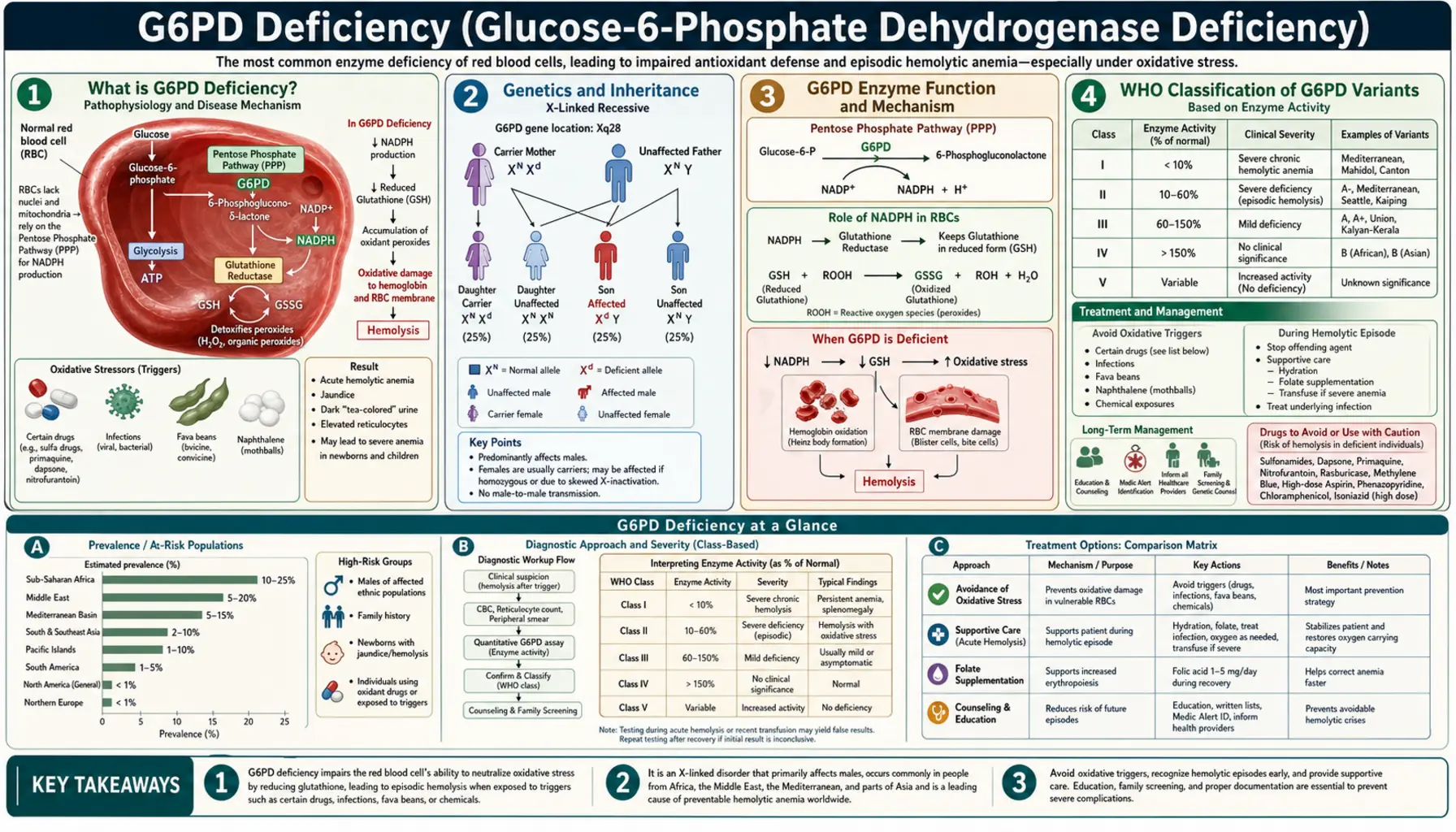
Table of Contents
- What Glucose-6-Phosphate Dehydrogenase Deficiency Is
- Genetics and Inheritance
- Glucose-6-Phosphate Dehydrogenase Enzyme Function and Mechanism
- World Health Organization Classification of Glucose-6-Phosphate Dehydrogenase Variants
- Triggers for Acute Hemolytic Episodes
- Clinical Manifestations
- Diagnosis
- Treatment and Management
- Global Burden and Malaria Protection
- Living with Glucose-6-Phosphate Dehydrogenase Deficiency
- Key Research Papers
- Connections
- Featured Videos
What Glucose-6-Phosphate Dehydrogenase Deficiency Is
Glucose-6-phosphate dehydrogenase (G6PD) deficiency is the most common enzyme deficiency in the world, affecting an estimated 400 to 500 million people globally. That makes it more common than any other inherited enzyme disorder, more common than sickle cell disease, and roughly comparable in global prevalence to the total number of people with diabetes. Yet most people with glucose-6-phosphate dehydrogenase deficiency live their entire lives without knowing they have it, because the condition is almost completely silent between episodes.
The deficiency affects a single enzyme — glucose-6-phosphate dehydrogenase — that red blood cells (RBCs) depend on to protect themselves from oxidative damage. When everything is calm, the enzyme's absence causes no problem. But when the body faces an oxidative stress — a serious infection, a particular medication, or even a plate of fava beans — the unprotected red blood cells rupture in a process called hemolysis. The result can range from a mild, self-resolving episode of fatigue and dark urine to a medical emergency requiring blood transfusion.
Glucose-6-phosphate dehydrogenase deficiency is most prevalent in sub-Saharan Africa, the Mediterranean basin, the Middle East, and Southeast Asia — a distribution that closely mirrors historical malaria transmission zones. This overlap is not coincidental: the same genetic variants that impair RBC oxidative defense also confer partial protection against Plasmodium falciparum, the deadliest malaria parasite. Glucose-6-phosphate dehydrogenase deficiency is a classic example of a balanced polymorphism, where a genetic disadvantage in one context becomes a survival advantage in another.
Genetics and Inheritance
Glucose-6-phosphate dehydrogenase deficiency follows an X-linked recessive inheritance pattern. The gene encoding glucose-6-phosphate dehydrogenase sits on the long arm of the X chromosome at position Xq28, a densely gene-rich region near the telomere. Because males have only one X chromosome (they are hemizygous), any mutation in their single glucose-6-phosphate dehydrogenase gene directly results in deficiency — there is no second copy to compensate. This is why the condition is dramatically more common and more severe in males than in females.
Females carry two X chromosomes and are therefore more likely to be heterozygous carriers — possessing one normal and one deficient copy of the gene. Most heterozygous females have intermediate enzyme activity and remain asymptomatic. However, a fraction of heterozygous females become fully affected through a process called X-inactivation (or lyonization). During early embryonic development, each cell randomly inactivates one of its two X chromosomes. If a female's cells preferentially inactivate the X chromosome carrying the normal glucose-6-phosphate dehydrogenase allele — a phenomenon called skewed X-inactivation — her red blood cells will predominantly express the deficient gene, leaving her as clinically vulnerable as an affected male.
Females who are homozygous (carrying two deficient alleles) are as severely affected as males. While this requires inheriting a deficient allele from both parents, it is not rare in high-prevalence populations where consanguineous marriage is common.
The glucose-6-phosphate dehydrogenase gene itself spans approximately 18 kilobases and contains 13 exons. More than 200 different mutations are known, according to a 2020 review in Blood — about half of them common in particular populations and about half rare, sporadic ones — and the vast majority are missense mutations that reduce enzyme activity or stability rather than eliminating it entirely. The clinical consequences depend almost entirely on which mutation is present, since different mutations produce different levels of residual enzyme activity.
Glucose-6-Phosphate Dehydrogenase Enzyme Function and Mechanism
To understand why glucose-6-phosphate dehydrogenase deficiency causes problems specifically in red blood cells, it helps to understand what the enzyme normally does and why RBCs are uniquely vulnerable without it.
Glucose-6-phosphate dehydrogenase is the first and rate-limiting enzyme of the pentose phosphate pathway (PPP), also called the hexose monophosphate shunt. In this pathway, glucose-6-phosphate is oxidized, and the energy released is captured not as adenosine triphosphate (ATP) (as in glycolysis) but as reduced nicotinamide adenine dinucleotide phosphate (NADPH) — a reducing molecule that serves as the cell's primary antioxidant currency.
Reduced nicotinamide adenine dinucleotide phosphate's key role in RBCs is to regenerate reduced glutathione (GSH) — the cell's master antioxidant. Glutathione exists in two forms: reduced (GSH, active) and oxidized (GSSG, inactive). The enzyme glutathione reductase uses reduced nicotinamide adenine dinucleotide phosphate to continuously convert GSSG back to reduced glutathione, keeping the cell's antioxidant defenses armed. When oxidative stress occurs — from reactive oxygen species generated during infections, drug metabolism, or certain foods — reduced glutathione neutralizes those threats before they can damage cellular proteins and lipids.
Here is the critical vulnerability of red blood cells: RBCs have no mitochondria and no nucleus. They cannot carry out aerobic respiration, they cannot synthesize new proteins, and — crucially — they have no alternative source of reduced nicotinamide adenine dinucleotide phosphate. Every other cell type in the body can compensate for low PPP activity using mitochondrial sources of reducing power. RBCs cannot. The pentose phosphate pathway is their only source of reduced nicotinamide adenine dinucleotide phosphate, making glucose-6-phosphate dehydrogenase the sole guardian of their oxidative defense.
In glucose-6-phosphate dehydrogenase-deficient RBCs exposed to oxidative stress, the cascade of injury proceeds as follows:
- Oxidative stress depletes reduced nicotinamide adenine dinucleotide phosphate faster than glucose-6-phosphate dehydrogenase can replenish it.
- Without reduced nicotinamide adenine dinucleotide phosphate, glutathione reductase cannot regenerate reduced glutathione; the cell's antioxidant pool collapses.
- Reactive oxygen species oxidize hemoglobin, causing it to denature and precipitate inside the RBC. These precipitates are visible under the microscope as Heinz bodies — named after the physician who first described them, not the condiment.
- Heinz-body-laden RBCs become rigid and are caught in the spleen. Splenic macrophages "bite" off the Heinz body-containing membrane portions, producing bite cells and blister cells on peripheral smear.
- The damaged, rigid RBCs are either removed by the spleen (extravascular hemolysis) or rupture directly in the bloodstream (intravascular hemolysis), releasing hemoglobin into the plasma and causing hemoglobinuria — the characteristic dark urine of a glucose-6-phosphate dehydrogenase hemolytic episode.
World Health Organization Classification of Glucose-6-Phosphate Dehydrogenase Variants
The World Health Organization's older five-class system, endorsed by a WHO Working Group in 1985 and published in 1989, classified glucose-6-phosphate dehydrogenase variants based on enzyme activity and clinical severity; WHO replaced it in 2022 (see below), and the class numbers used elsewhere on this page (Class I to V) are from this older system. Understanding the class of a patient's variant is clinically important because it predicts their risk for chronic hemolysis versus only episodic hemolysis, and guides decisions about trigger avoidance and monitoring.
Class I — Severe deficiency with chronic nonspherocytic hemolytic anemia (CNSHA): Enzyme activity is less than 10% of normal, and hemolysis is ongoing even without an identifiable trigger. Patients with Class I variants suffer from chronic anemia, persistent jaundice, splenomegaly, and gallstones (from chronic bilirubin overproduction). Class I variants are relatively rare and tend to involve mutations in structurally critical regions of the enzyme. Examples include glucose-6-phosphate dehydrogenase Hektoen and glucose-6-phosphate dehydrogenase Bangkok.
Class II — Severe deficiency (<10% activity) with episodic hemolysis only: Despite very low enzyme activity, patients with Class II variants are entirely asymptomatic between episodes. The most clinically important Class II variant is glucose-6-phosphate dehydrogenase Mediterranean, caused by a c.563C>T mutation that produces a serine-to-phenylalanine substitution at position 188 (S188F). Glucose-6-phosphate dehydrogenase Mediterranean is the predominant variant across the Mediterranean basin, Middle East, and Indian subcontinent. Patients with this variant are at high risk for severe hemolysis when exposed to fava beans, primaquine, or infection.
Class III — Moderate deficiency (10–60% activity) with episodic hemolysis: This is the most common class worldwide. The defining example is glucose-6-phosphate dehydrogenase A- (also written glucose-6-phosphate dehydrogenase A-minus), the predominant variant in people of African descent. Glucose-6-phosphate dehydrogenase A- is caused by two concurrent mutations: V68M (p.Val68Met) and N126D (p.Asn126Asp). The enzyme produced by glucose-6-phosphate dehydrogenase A- has near-normal catalytic activity but is dramatically unstable — it degrades rapidly as red blood cells age. This means that young red blood cells (reticulocytes) in people with glucose-6-phosphate dehydrogenase A- have almost normal enzyme activity, while older cells (which circulate for weeks) become severely deficient. This age-dependent vulnerability has an important clinical consequence: when a hemolytic episode is triggered in a glucose-6-phosphate dehydrogenase A- patient, the older, deficient RBCs are destroyed, but the younger RBCs survive. The episode is therefore self-limiting, and the patient recovers even without treatment as the bone marrow releases a wave of young, enzyme-replete reticulocytes. This contrasts sharply with glucose-6-phosphate dehydrogenase Mediterranean, where even reticulocytes are severely deficient.
Class IV — Normal activity (60–100%): No clinical significance. Includes the normal glucose-6-phosphate dehydrogenase B (the wild-type reference) and glucose-6-phosphate dehydrogenase A+ (a common polymorphism in Africa with normal activity).
Class V — Increased activity (>150%): Rare variants with supranormal enzyme activity. No established clinical consequence.
The 2022 WHO classification. In 2022 a WHO Global Malaria Programme technical panel revised the scheme, and the WHO Malaria Policy Advisory Group endorsed it in March 2022. A 2023 analysis prepared for this revision gathered enzyme results from 2,291 men: the average remaining activity of 16 common variants ranged from 1.9% to 33% of normal, values overlapped widely between variants, and they did not cluster above or below 10% — the line that had separated Class II from Class III. The panel therefore merged the old Class II and Class III into one group. The current classes are:
- Class A — median activity under 20% of normal, with chronic hemolytic anemia.
- Class B — median activity under 45%; all the common variants (including the Mediterranean and A- variants), which carry the risk of neonatal jaundice and of acute hemolytic anemia triggered by certain medicines, fava beans or infection.
- Class C — median activity over 60%, no hemolysis.
- Class U — uncertain clinical significance, a temporary class for variants with too little information.
The old Class V was dropped because it rested on a single case report. WHO notes that severe, life-threatening hemolysis can occur with any Class B variant, that the classes describe variants rather than individuals (a person's activity can sit above or below their variant's median), and that its advice on primaquine and tafenoquine depends on each person's measured enzyme activity, not on the class.
Triggers for Acute Hemolytic Episodes
The practical management of glucose-6-phosphate dehydrogenase deficiency is largely about identifying and avoiding the triggers that can convert a silent condition into a medical emergency. Triggers are oxidative stressors that overwhelm the already-limited antioxidant capacity of glucose-6-phosphate dehydrogenase-deficient red blood cells.
Infections are a recognized trigger for acute hemolytic episodes; a 2020 review in Blood describes acute hemolysis triggered by fava beans, by a number of drugs (such as primaquine and rasburicase), or, more rarely, by infection. Infections are the single most common trigger for acute hemolytic episodes, accounting for the majority of crises seen in clinical practice. Any significant infection — bacterial pneumonia, viral hepatitis, typhoid fever, influenza — can provoke hemolysis through the massive oxidative burst generated by activated immune cells. In glucose-6-phosphate dehydrogenase-deficient children, pallor and dark urine after a febrile illness are warning signs of hemolysis rather than simply the infection itself.
Antimalarial drugs are historically the most important pharmacological triggers and were instrumental in glucose-6-phosphate dehydrogenase deficiency's original discovery during the Korean War, when American soldiers of African descent developed hemolysis after receiving primaquine prophylaxis. Today:
- Primaquine and tafenoquine — both 8-aminoquinoline antiparasitic drugs used to eliminate dormant malaria liver stages (hypnozoites) — are the most potent hemolytic triggers. Tafenoquine is particularly dangerous because its long half-life (about 15 days, per its FDA label) means hemolysis cannot be terminated quickly by stopping the drug. The FDA label states that glucose-6-phosphate dehydrogenase testing must be performed before tafenoquine is prescribed, and contraindicates it in anyone with glucose-6-phosphate dehydrogenase deficiency or unknown status; its clinical trials excluded patients whose enzyme activity was below 70% of the site's normal median.
- Dapsone — used for leprosy, malaria prophylaxis, and Pneumocystis pneumonia prevention — carries significant hemolytic risk and is one of the seven medicines with solid evidence against their use in the 2010 evidence-based review described below.
- Chloroquine and artemisinin derivatives are generally safe at therapeutic doses; some references list chloroquine as a cause of mild hemolysis at high doses, and the 2010 evidence-based review did not find evidence against its use in normal therapeutic doses.
What the tafenoquine label says. Tafenoquine was first approved in the United States in 2018 for the radical cure (prevention of relapse) of Plasmodium vivax malaria in people aged 16 and older, as a single 300 mg dose given with chloroquine. Besides contraindicating it in glucose-6-phosphate dehydrogenase deficiency or unknown status, the FDA label contraindicates breastfeeding when the infant is glucose-6-phosphate dehydrogenase deficient or of unknown status, states that it is not recommended during pregnancy because it may cause hemolytic anemia in a deficient fetus, and reports declines in hemoglobin in some glucose-6-phosphate dehydrogenase-normal patients in its trials. A 2024 WHO paper adds that, unlike primaquine, tafenoquine cannot be discontinued once started.
How strong the evidence is for each medicine. A 2010 evidence-based review of medicines and glucose-6-phosphate dehydrogenase deficiency (Youngster and colleagues, Drug Safety) found solid evidence to prohibit only seven medicines in current use: dapsone, methylene blue (methylthioninium chloride), nitrofurantoin, phenazopyridine, primaquine, rasburicase and toluidine blue (tolonium chloride). For all other medicines it found no evidence against their use in normal therapeutic doses, and it concluded that many compounds have probably been wrongly cited as causing hemolysis because they were given to patients already having an infection-related hemolytic episode. Tafenoquine was approved after that review. Drug lists in other sources therefore mix firm risks with medicines only some references name.
Other medications of concern:
- Cotrimoxazole (trimethoprim-sulfamethoxazole) — listed by some references as a hemolytic risk because of its sulfonamide component, particularly in Class II patients; the 2010 evidence-based review did not find evidence against its use in normal therapeutic doses.
- Nitrofurantoin — used for urinary tract infections; one of the seven medicines with solid evidence against their use in glucose-6-phosphate dehydrogenase deficiency in the 2010 evidence-based review.
- Nalidixic acid — an older quinolone antibiotic (not a fluoroquinolone), listed by some references; modern fluoroquinolones (ciprofloxacin, levofloxacin) are generally considered safe.
- Rasburicase — a uricase enzyme used to prevent tumor lysis syndrome — is an absolute contraindication in glucose-6-phosphate dehydrogenase deficiency. Rasburicase generates hydrogen peroxide as a byproduct, causing catastrophic and potentially fatal hemolysis. Glucose-6-phosphate dehydrogenase testing is required before any rasburicase use.
- Methylene blue — used to treat methemoglobinemia — paradoxically depends on reduced nicotinamide adenine dinucleotide phosphate for its mechanism and is ineffective in glucose-6-phosphate dehydrogenase-deficient patients; it may actually worsen hemolysis.
- Phenazopyridine (urinary analgesic) is one of the seven medicines in the 2010 evidence-based review; aspirin at high doses is listed by some references.
- Toluidine blue (tolonium chloride) — the seventh medicine on that review's list.
Fava beans (Vicia faba) — commonly known as broad beans — contain two glycosides, vicine and convicine. When these are split, they release their aglycones, divicine and isouramil — the potent oxidants, directly toxic to glucose-6-phosphate dehydrogenase-deficient RBCs, that are the causative agents of favism. A 2016 laboratory study of faba bean found that, once released by the enzyme β-glucosidase, divicine and isouramil broke down almost completely within 60 minutes in bean extract and within 120 minutes in isolated fractions at body temperature (37 °C, pH 5). The hemolytic reaction to fava beans is called favism and is one of the oldest recognized food-drug-gene interactions in history, described in ancient Greek texts. Favism is particularly severe in Class II patients (glucose-6-phosphate dehydrogenase Mediterranean) and in young children. Even inhalation of fava bean pollen has been reported to trigger episodes. Fava beans are a dietary staple in Mediterranean and Middle Eastern cuisines — their avoidance can be a significant dietary and cultural challenge for affected patients.
Naphthalene — found in traditional moth balls — is a well-documented trigger, particularly relevant for neonates exposed to naphthalene-containing products used in some cultural traditions to store baby clothes. Neonatal hemolysis from naphthalene exposure can be severe.
Clinical Manifestations
The clinical presentation of glucose-6-phosphate dehydrogenase deficiency spans a wide spectrum from lifelong complete silence to potentially fatal hemolytic crisis, depending primarily on the patient's World Health Organization (WHO) variant class and the trigger encountered.
Asymptomatic phase: The overwhelming majority of people with glucose-6-phosphate dehydrogenase deficiency are entirely asymptomatic between episodes. They have normal hemoglobin, normal bilirubin, and normal reticulocyte counts. A routine complete blood count gives no hint of the underlying deficiency. This prolonged silence between episodes is both the condition's most reassuring feature and its most dangerous: patients who do not know their diagnosis cannot take precautions.
Acute hemolytic episode: When a trigger is encountered, symptoms typically begin within 24 to 72 hours and include:
- Malaise, fatigue, and pallor from acute anemia as red cells are destroyed faster than they can be replaced.
- Back pain and abdominal pain — often cramping and diffuse — from the systemic hemolytic process.
- Jaundice (yellowing of the skin and sclera) from the bilirubin released as hemoglobin is broken down. Jaundice may appear within hours of onset.
- Dark urine — classically described as "coca-cola colored" or "port wine" urine — from hemoglobinuria (free hemoglobin in the urine). This is a hallmark sign that intravascular hemolysis is occurring.
- Tachycardia and dyspnea in severe cases as the body compensates for falling oxygen-carrying capacity.
In glucose-6-phosphate dehydrogenase A- (Class III) patients, the episode is characteristically self-limiting. As older, enzyme-deficient RBCs are destroyed, younger reticulocytes — which have higher enzyme activity — are relatively spared. The bone marrow mounts a reticulocyte response, and the hemoglobin nadir is typically reached by day 7–8, after which the patient recovers spontaneously even without treatment. Hemoglobin rarely falls below 8 g/dL in uncomplicated A- episodes.
In glucose-6-phosphate dehydrogenase Mediterranean (Class II) patients, episodes are far more severe because even reticulocytes are enzyme-deficient. Without a protected younger RBC cohort, hemolysis can be catastrophic, with hemoglobin falling precipitously to transfusion-requiring levels.
Neonatal jaundice: glucose-6-phosphate dehydrogenase deficiency is one of the most important causes of neonatal hyperbilirubinemia worldwide, particularly in Mediterranean, African, and Asian populations. Unlike ABO or Rh hemolytic disease of the newborn, the jaundice in glucose-6-phosphate dehydrogenase deficiency is not purely hemolytic — it also reflects impaired hepatic bilirubin conjugation (neonatal livers are already immature at conjugating bilirubin, and glucose-6-phosphate dehydrogenase deficiency further compromises this capacity). Neonatal glucose-6-phosphate dehydrogenase-related jaundice can progress to kernicterus — bilirubin deposition in the brain causing permanent neurological damage — if not treated promptly with phototherapy or exchange transfusion. In populations where glucose-6-phosphate dehydrogenase deficiency is prevalent, universal newborn screening for jaundice is a public health imperative.
Chronic nonspherocytic hemolytic anemia (CNSHA): Restricted to Class I variants, CNSHA is a state of ongoing hemolysis requiring chronic management: regular monitoring, folate supplementation, transfusion support during crises, and sometimes splenectomy. Gallstones develop early from chronic bilirubin overproduction and may require cholecystectomy.
Diagnosis
Accurate diagnosis of glucose-6-phosphate dehydrogenase deficiency requires understanding both the appropriate tests and their important limitations — particularly the critical pitfall of testing during an acute hemolytic episode.
Fluorescent spot test (FST) — screening: The most widely used screening test for glucose-6-phosphate dehydrogenase deficiency. A blood spot on filter paper is incubated with glucose-6-phosphate and NADP+; G6PD-sufficient samples produce reduced nicotinamide adenine dinucleotide phosphate, which fluoresces under ultraviolet (UV) light, while glucose-6-phosphate dehydrogenase-deficient samples do not. The FST is inexpensive, rapid, and reliable for identifying hemizygous males and homozygous females. However, it has poor sensitivity for heterozygous females with intermediate activity levels and may produce false normal results in these individuals.
Quantitative glucose-6-phosphate dehydrogenase assay — confirmatory: The gold standard for diagnosis is a spectrophotometric measurement of glucose-6-phosphate dehydrogenase enzymatic activity expressed as units per gram of hemoglobin (U/gHb). The normal reference range is approximately 4.6–13.5 U/gHb. Class I variants typically show <1 U/gHb; Class II, 1–3 U/gHb; Class III, 3–8 U/gHb. The test is reported relative to hemoglobin to correct for the number of red cells in the sample.
The acute episode pitfall — a critical diagnostic trap: Testing glucose-6-phosphate dehydrogenase activity during or immediately after a hemolytic episode routinely yields falsely normal results in glucose-6-phosphate dehydrogenase A- patients. The mechanism is straightforward: the oldest, most glucose-6-phosphate dehydrogenase-deficient RBCs are selectively destroyed during hemolysis, leaving behind a population enriched in younger reticulocytes with relatively higher enzyme activity. A blood sample taken at the hemoglobin nadir may show enzyme activity in the low-normal range even in a clearly glucose-6-phosphate dehydrogenase-deficient patient. For this reason, confirmatory quantitative glucose-6-phosphate dehydrogenase testing is deferred until 2–3 months after an acute episode, long enough for the reticulocyte response to settle and the full population of aging, enzyme-deficient RBCs to re-accumulate. This pitfall is less relevant for glucose-6-phosphate dehydrogenase Mediterranean patients, whose reticulocytes are also enzyme-deficient, but it has caused missed diagnoses in countless glucose-6-phosphate dehydrogenase A- patients in emergency settings.
Peripheral blood smear findings: During an acute episode, a skilled hematology review of the peripheral smear reveals characteristic changes:
- Heinz bodies — visible only with special supravital stains (crystal violet or brilliant cresyl blue), not on a standard Wright-Giemsa smear. They appear as dark purple inclusions abutting the RBC membrane.
- Bite cells (degmacytes) — RBCs that appear to have a "bite" taken out of them, created when splenic macrophages remove Heinz body-laden membrane segments.
- Blister cells — RBCs with an apparently empty vacuole under the membrane where Heinz bodies have been removed.
- Polychromasia — a bluish tint to some RBCs reflecting reticulocytosis (the bone marrow's compensatory response).
Newborn screening: Many countries with high glucose-6-phosphate dehydrogenase prevalence, including Greece, Cyprus, Hong Kong, Singapore, and Saudi Arabia, incorporate glucose-6-phosphate dehydrogenase deficiency into their universal newborn screening programs. In the United States, newborn screening for glucose-6-phosphate dehydrogenase is not universal but is offered in some states.
Treatment and Management
There is no cure for glucose-6-phosphate dehydrogenase deficiency, and most patients never require medical treatment. Management centers on education, trigger avoidance, and prompt response to acute episodes when they occur.
Patient education — the cornerstone of management: Education after a diagnosis of glucose-6-phosphate dehydrogenase deficiency covers:
- The foods involved (fava beans above all; some sources also list blueberries, soya products, and tonic water [contains quinine] as lower-level concerns, though fava beans are the only clearly documented dietary trigger in clinical practice).
- A list of the medicines that carry risk, and those named only by some sources.
- Warning signs of a hemolytic episode (dark urine, jaundice, rapid fatigue), which hospitals treat as urgent.
- Why every healthcare provider and pharmacist needs to know the diagnosis before a new medication is prescribed.
Medical alert identification: A medical alert bracelet or card for people with Class I or Class II variants states their diagnosis and the absolute contraindications (rasburicase, tafenoquine, methylene blue). In an emergency, this information can prevent inadvertent administration of a triggering drug.
Management of acute hemolytic episodes:
- Remove the trigger: The offending drug is stopped immediately, and any precipitating infection is treated.
- Hydration: Intravenous fluids to maintain urine output and protect the kidneys from hemoglobin precipitation in the tubules (which can cause acute kidney injury).
- Blood transfusion: Indicated when hemoglobin falls below approximately 7 g/dL or when the fall is rapid regardless of absolute level, or when the patient has cardiovascular compromise. Packed RBCs from a glucose-6-phosphate dehydrogenase-normal donor provide functional cells unaffected by the trigger.
- Folate supplementation: During a hemolytic episode, the bone marrow's accelerated erythropoiesis can deplete folate stores. Supplementation with 1–5 mg folic acid daily supports recovery.
- Phototherapy or exchange transfusion for neonatal hyperbilirubinemia, following standard thresholds based on gestational age and bilirubin level.
Primaquine dosing strategy in glucose-6-phosphate dehydrogenase A- patients: Radical cure of vivax or ovale malaria requires elimination of dormant liver stages (hypnozoites), which requires primaquine. For Class III (G6PD A-) patients, a supervised weekly high-dose primaquine regimen (0.75 mg/kg weekly for 8 weeks) is an alternative to the standard daily regimen, taking advantage of the self-limiting hemolysis in A- patients while allowing bone marrow recovery between doses. This strategy is not appropriate for Class I or II patients.
Management of CNSHA (Class I variants): These patients require hematology specialist care. Splenectomy may reduce transfusion requirements by removing the primary site of extravascular hemolysis. Folate supplementation is ongoing. All trigger medications and foods are kept away, as even a small additional oxidative burden can precipitate a crisis in an already-hemolyzed patient.
Emerging therapies: Gene therapy trials targeting glucose-6-phosphate dehydrogenase deficiency are in early-stage development, offering the prospect of a one-time curative treatment for Class I CNSHA patients. Small-molecule pharmacological chaperones that stabilize mutant glucose-6-phosphate dehydrogenase protein are also under investigation.
Global Burden and Malaria Protection
Glucose-6-phosphate dehydrogenase deficiency is distributed across the globe in a pattern that reflects ancient and ongoing selection pressure from malaria. The highest allele frequencies — often 15–25% of males in affected populations — are found in sub-Saharan Africa, the Mediterranean basin, the Middle East, the Indian subcontinent, and Southeast Asia. In some hyperendemic malaria regions, such as parts of West Africa and Papua New Guinea, glucose-6-phosphate dehydrogenase deficiency allele frequencies reach 30% or higher.
The malaria-protective mechanism of glucose-6-phosphate dehydrogenase deficiency has been studied for decades and is now reasonably well characterized. Glucose-6-phosphate dehydrogenase-deficient RBCs appear to resist Plasmodium falciparum infection through multiple mechanisms:
- The parasite requires reduced nicotinamide adenine dinucleotide phosphate for its own metabolism; reduced nicotinamide adenine dinucleotide phosphate availability in deficient cells impairs parasite growth.
- Glucose-6-phosphate dehydrogenase-deficient RBCs expressing ring-stage parasites are removed from circulation by the spleen more rapidly than normal infected cells, limiting parasite multiplication.
- The oxidative environment inside deficient cells may directly damage the parasite.
The main study behind the protection figure (Ruwende and colleagues, Nature 1995) was two large case-control studies of over 2,000 African children: the common African variant, glucose-6-phosphate dehydrogenase A-, was associated with a 46–58% reduction in the risk of severe malaria in both girls carrying one copy (heterozygous females) and boys with their single X copy (hemizygous males), though males experience the tradeoff of greater hemolytic risk. The authors' mathematical model suggested that a counterbalancing disadvantage of the deficiency has slowed its rise in malaria-endemic regions. This protection rivals the protection conferred by sickle cell trait (HbAS), making glucose-6-phosphate dehydrogenase deficiency one of the most powerful malaria-resistance polymorphisms in the human genome.
This protective relationship has major implications for malaria eradication programs. The most effective drugs for clearing malaria liver stages — primaquine and tafenoquine — are precisely the drugs most dangerous to glucose-6-phosphate dehydrogenase-deficient individuals. Mass drug administration programs using these agents depend on glucose-6-phosphate dehydrogenase testing to keep from triggering hemolytic crises at population scale. The development of rapid, field-deployable glucose-6-phosphate dehydrogenase diagnostic tests has become a public health priority for the World Health Organization's malaria elimination agenda.
Living with Glucose-6-Phosphate Dehydrogenase Deficiency
For the vast majority of people with glucose-6-phosphate dehydrogenase deficiency, the condition is compatible with a completely normal life, with one important caveat: it depends on the person and their healthcare providers knowing about it. The practical points are straightforward.
The variant class matters. Under the older system, Class III (G6PD A-) patients have a much more forgiving margin than Class I or II patients, but every deficient class carries a risk from triggers; under the 2022 WHO system the former Class II and III variants form one group (Class B), and WHO notes that severe hemolysis can occur with any of them.
A known diagnosis protects in emergencies. In an emergency room or surgical setting, a provider who doesn't know the diagnosis may administer rasburicase (for hyperuricemia from tumor lysis) or prescribe methylene blue (for methemoglobinemia) — both potentially catastrophic in glucose-6-phosphate dehydrogenase deficiency. A medical alert bracelet or a note in emergency contact information is how many people make the diagnosis known.
Fava beans are the clearest dietary trigger. In Mediterranean and Middle Eastern households where ful medames, bessara, or other fava-bean dishes are cultural staples, avoidance can be socially difficult. Favism episodes can be severe — even fatal in young children — and no tolerance-building or desensitization exists.
Children and schools. School nurses and teachers who know about a child's deficiency can watch for the two main risks: fava beans (many school lunch programs in Mediterranean-origin communities serve them) and naphthalene exposure from mothball-stored clothing.
Pregnancy. Glucose-6-phosphate dehydrogenase-deficient mothers carrying glucose-6-phosphate dehydrogenase-deficient fetuses may experience fetal or neonatal hemolysis. When neonatologists know that a glucose-6-phosphate dehydrogenase-deficient mother is delivering, the newborn can be monitored for jaundice and treated promptly.
Key Research Papers
- Nkhoma ET, Poole C, Vannappagari V, Hall SA, Beutler E. The global prevalence of glucose-6-phosphate dehydrogenase deficiency: a systematic review and meta-analysis. Blood Cells Mol Dis. 2009;42(3):267–278. — PubMed PMID: 19233695
- Cappellini MD, Fiorelli G. Glucose-6-phosphate dehydrogenase deficiency. Lancet. 2008;371(9606):64–74. — PubMed PMID: 18177777
- Luzzatto L, Mehta A, Vulliamy T. Glucose-6-phosphate dehydrogenase deficiency. In: Scriver CR et al., eds. The Metabolic and Molecular Bases of Inherited Disease. 8th ed. McGraw-Hill; 2001. — Search PubMed
- Howes RE, Piel FB, Patil AP, et al. G6PD deficiency prevalence and estimates of affected populations in malaria endemic countries: a geostatistical model-based map. PLoS Med. 2012;9(11):e1001339. — PubMed PMID: 23152723
- Youngster I, Arcavi L, Schechmaster R, et al. Medications and glucose-6-phosphate dehydrogenase deficiency: an evidence-based review. Drug Saf. 2010;33(9):713–726. — PubMed PMID: 20701405
- Beutler E. G6PD deficiency. Blood. 1994;84(11):3613–3636. — PubMed PMID: 7949118
- WHO Working Group. Glucose-6-phosphate dehydrogenase deficiency. Bull World Health Organ. 1989;67(6):601–611. — PubMed PMID: 2633878
- Ruwende C, Khoo SC, Snow RW, et al. Natural selection of hemi- and heterozygotes for G6PD deficiency in Africa by resistance to severe malaria. Nature. 1995;376(6537):246–249. — PubMed PMID: 7617034
- Luzzatto L, Ally M, Notaro R. Glucose-6-phosphate dehydrogenase deficiency. Blood. 2020;136(11):1225–1240. — PubMed PMID: 32702756
- Minucci A, Moradkhani K, Hwang MJ, et al. Glucose-6-phosphate dehydrogenase (G6PD) mutations database: review of the "old" and update of the new mutations. Blood Cells Mol Dis. 2012;48(3):154–165. — PubMed PMID: 22293322
- Belfield KD, Tichy EM. Review and drug therapy implications of glucose-6-phosphate dehydrogenase deficiency. Am J Health Syst Pharm. 2018;75(3):97–104. — PubMed PMID: 29305344
- Recht J, Ashley EA, White NJ. Safety of 8-aminoquinoline antimalarial medicines. WHO Evidence Review Paper. 2014. (A World Health Organization report, not indexed in PubMed.)
- Nannelli C, Bosman A, Cunningham J, Dugué PA, Luzzatto L. Genetic variants causing G6PD deficiency: Clinical and biochemical data support new WHO classification. Br J Haematol. 2023;202(5):1024–1032. — PubMed PMID: 37415281
- Luzzatto L, Bancone G, Dugué PA, et al. New WHO classification of genetic variants causing G6PD deficiency. Bull World Health Organ. 2024;102(8):615–617. — PubMed PMID: 39070600
- Pulkkinen M, Zhou X, Lampi AM, Piironen V. Determination and stability of divicine and isouramil produced by enzymatic hydrolysis of vicine and convicine of faba bean. Food Chem. 2016;212:10–19. — PubMed PMID: 27374500
- US Food and Drug Administration. Tafenoquine tablets, prescribing information (initial U.S. approval 2018; label revised August 2023). DailyMed. — DailyMed label
Connections
- Hematology
- Anemia
- Hemolytic-Uremic Syndrome
- Hereditary Spherocytosis
- Thalassemia
- Paroxysmal Nocturnal Hemoglobinuria
- Sickle Cell Disease
- Aplastic Anemia
- Thrombocytopenia
- Complete Blood Count
- Iron
- Folate (B9)
- Jaundice
- Malaria — glucose-6-phosphate dehydrogenase variants confer partial resistance; antimalarials trigger hemolysis.