Turner Syndrome
- Overview and Epidemiology
- Cytogenetics and Variants
- Physical Features
- Cardiovascular Complications
- Gonadal Dysgenesis and Fertility
- Growth and Growth Hormone Therapy
- Cognitive and Behavioral Profile
- Estrogen Replacement Therapy
- Long-Term Monitoring and Surveillance
- Key Research Papers
- Connections
Overview and Epidemiology
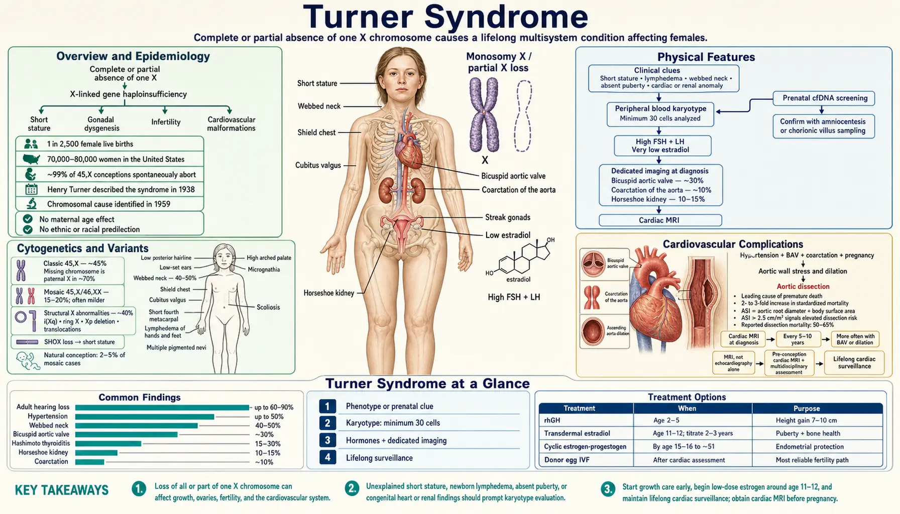
Turner syndrome (also called Monosomy X) is a chromosomal condition that affects females exclusively, arising from the complete or partial absence of one of the two X chromosomes normally present in every female cell. It is the most common sex chromosome abnormality in females, occurring in approximately 1 in 2,500 female live births; the 2024 international clinical practice guideline gives a frequency of 25 to 50 per 100,000 females. In the United States an estimated 70,000–80,000 women are living with the condition.
That 2024 guideline updates and extends the earlier guidelines, including the 2017 international guideline it supersedes. It came out of a consensus meeting in Aarhus, Denmark, in June 2023, was started by the European Society for Endocrinology and the Pediatric Endocrine Society together with many other medical societies, and adds new advances in diagnosis and genetics, estrogen treatment, fertility, other illnesses that come with the condition (co-morbidities), and thinking skills and mental health (neurocognition and neuropsychology).
The syndrome is remarkably lethal in utero: roughly 99% of 45,X conceptions spontaneously abort, making it one of the most lethal chromosomal abnormalities ever conceived. Only those fetuses that retain enough functional cells survive to term, which is why the clinical spectrum seen at birth represents the fortunate minority of an intrinsically severe abnormality.
The condition was first described by American endocrinologist Henry Turner in 1938, when he reported a cohort of women with short stature, sexual infantilism, cubitus valgus, and webbed neck. The underlying chromosomal cause — loss of an entire X chromosome — was not identified until 1959 by Charles Ford and colleagues using newly developed karyotyping techniques.
The hallmark features are short stature, gonadal dysgenesis, infertility, and cardiovascular malformations. Intelligence is usually in the normal range. There is no ethnic or racial predilection, and unlike trisomies such as Down syndrome, Turner syndrome is not associated with advanced maternal age — the chromosomal loss occurs with roughly equal probability at any maternal age.
Cytogenetics and Variants
Turner syndrome is not a single chromosomal event but a family of karyotypic abnormalities, all sharing functional haploinsufficiency of X-linked genes that normally escape X-inactivation.
- Classic 45,X (Monosomy X): Approximately 45% of Turner syndrome cases carry a single X chromosome with no second sex chromosome at all. In roughly 70% of these cases the missing chromosome is the paternal X, indicating that the error most frequently arises during paternal meiosis or early post-zygotic division. There is no maternal age effect.
- Mosaic 45,X/46,XX: Seen in 15–20% of cases. A proportion of cells carry a normal 46,XX karyotype alongside a monosomic 45,X line. The phenotype is typically milder; some mosaic individuals experience spontaneous puberty, and 2–5% conceive naturally.
- Structural X abnormalities (~40%): The most common structural variant is isochromosome Xq (i(Xq)) — duplication of the long arm with loss of the short arm. Other variants include ring chromosome X, terminal deletions of the short arm (Xp), and translocations. Loss of the short arm consistently causes short stature and typical Turner features because it removes one copy of the SHOX gene (short stature homeobox gene) located in the pseudoautosomal region 1 (PAR1) of the X chromosome.
SHOX haploinsufficiency underlies the short stature and skeletal dysplasia seen in virtually all Turner variants. SHOX normally escapes X-inactivation; losing one copy disrupts chondrocyte proliferation and enchondral bone growth. Importantly, SHOX haploinsufficiency also causes short stature in Leri–Weill dyschondrosteosis, confirming its direct role.
Karyotyping from peripheral blood lymphocytes (minimum 30 cells analyzed) is the diagnostic standard. Prenatal diagnosis is increasingly made via cell-free fetal DNA (cfDNA) screening, confirmed by amniocentesis or chorionic villus sampling.
Physical Features
Turner syndrome has a recognizable but variable physical phenotype. Not every feature is present in every individual, and mosaic cases can be remarkably subtle.
- Short stature: The most consistent feature. Untreated adult height averages approximately 143 cm (4 feet 8 inches) in Western European populations, roughly 20 cm below the female average. Height loss begins gradually in early childhood and accelerates at the expected age of the adolescent growth spurt, which is absent.
- Webbed neck (pterygium colli): Neck skin folds extending from the mastoid to the acromion, caused by resolution of a fetal nuchal cystic hygroma. Present in about 40–50% of cases.
- Low posterior hairline and low-set ears: Remnants of the resolved nuchal hygroma.
- Lymphedema: Puffy hands and feet at birth, often the presenting sign in a newborn girl. Results from impaired lymphatic development secondary to the fetal cystic hygroma.
- Shield chest: Broad, flat chest with widely-spaced nipples — a classic but not universal finding.
- Cubitus valgus: Increased carrying angle at the elbow; often the feature that prompts referral in older children.
- Short fourth metacarpal: The "Drummond metacarpal sign" — shortening of the fourth (and sometimes fifth) metacarpal, visible on hand X-ray.
- Multiple pigmented nevi, nail dysplasia, high arched palate, micrognathia, scoliosis.
Structural abnormalities of internal organs are common: bicuspid aortic valve in ~30%, coarctation of the aorta in ~10%, and horseshoe kidney in 10–15%. These require dedicated imaging at diagnosis, not just clinical examination.
Cardiovascular Complications
Cardiovascular disease is the leading cause of premature death in Turner syndrome, accounting for a two- to three-fold increase in standardized mortality compared with the general female population. The 2024 international guideline states that people with Turner syndrome require lifelong cardiovascular surveillance, at a frequency set by their risk factors for aortic dissection.
The most common structural defect is bicuspid aortic valve (BAV), present in approximately 30% of individuals. BAV predisposes to aortic stenosis, regurgitation, and — critically — progressive dilation of the ascending aorta. Coarctation of the aorta occurs in ~10% and independently raises blood pressure and aortic wall stress. Partial anomalous pulmonary venous return is also more prevalent than in the general population.
Aortic dissection is the most feared complication. It can occur even with only mild aortic dilation and at younger ages than in other aortic syndromes. Key risk factors include hypertension, BAV, coarctation, and — importantly — pregnancy. The aortic size index (ASI), calculated as aortic root diameter divided by body surface area, is the preferred metric because Turner women tend to have a small body surface area (BSA); an ASI > 2.5 cm/m² signals elevated dissection risk. Reported dissection mortality ranges from 50–65%.
Magnetic resonance imaging (MRI) of the thoracic aorta (not echocardiography alone, which underestimates ascending aortic dimensions in Turner syndrome) is the standard surveillance modality. Estrogen replacement does not fully protect against aortic dilation. The 2024 international guideline recommends cardiovascular magnetic resonance imaging (CMR) in all adolescents and adults newly diagnosed with Turner syndrome, ideally within 12 months, in addition to or instead of the first echocardiogram (in young children it can wait until it can be done without general anesthesia). For people with no significant heart disease at the first screening, it recommends a heart ultrasound (transthoracic echocardiography) at age 9–11 years, after growth is complete or at transfer to adult care, and at least every 5–10 years in adults, with more frequent imaging when BAV, dilation or other risk factors are present. Elective surgical repair thresholds vary by guideline but generally align with BAV-specific aortic aneurysm criteria rather than Marfan thresholds; the 2024 guideline describes weighing elective aortic surgery in adults with moderate dilation (ASI above 2.3 cm/m²) plus at least one other risk factor, and evaluating for surgery when dilation is severe (ASI above 2.5 cm/m²) on its own.
Gonadal Dysgenesis and Fertility
Ovarian failure is nearly universal in classic 45,X Turner syndrome. In utero, ovarian follicles form normally but undergo accelerated atresia; by birth or early childhood the ovaries have been replaced by streak gonads — thin fibrous bands of connective tissue incapable of producing estrogen or maturing eggs.
The hormonal consequence is hypergonadotropic hypogonadism: the pituitary senses the absence of ovarian estrogen and releases very high levels of follicle-stimulating hormone (FSH) and luteinizing hormone (LH), which remain persistently elevated while estradiol remains very low. This pattern, often detected on newborn screening or at the expected time of puberty, is a reliable biochemical marker.
Primary amenorrhea occurs in approximately 90% of 45,X women. The 10% who experience spontaneous puberty are almost exclusively mosaic (45,X/46,XX) individuals with residual functional ovarian tissue. Of those who menstruate spontaneously, premature ovarian insufficiency (POI) eventually develops in the large majority.
Fertility options:
- Donor egg in vitro fertilization (IVF) is the most reliable path to parenthood. Because the uterus is structurally normal and responds well to estrogen priming, success rates with donor eggs are comparable to those in women with other causes of POI. The 2024 international guideline lists egg or embryo donation among the family-building options, alongside fertility preservation, foster care, adoption, surrogacy and the choice to remain childless.
- Oocyte cryopreservation may be offered to mosaic girls with documented ovarian reserve, ideally before further follicle depletion occurs. The 2024 international guideline recommends hormone stimulation of the ovaries followed by egg freezing as the main fertility-preservation option for those with fertility potential who have started their periods and are mature enough to understand the procedure, in experienced centres, and recommends that it not be offered to girls who have not started their periods. The guideline notes that the live-birth rate after egg freezing in Turner syndrome is unknown (one case report of a live birth) and that freezing ovarian tissue remains experimental. A 2016 review with practical guidelines reported that egg freezing has been performed in girls with Turner syndrome as young as 13 and ovarian tissue freezing in children before puberty, but that how well these approaches work in this group is unknown.
- Spontaneous conception (2–5% of mosaic cases): These pregnancies carry a high rate of miscarriage and an elevated risk of chromosomally abnormal offspring, and genetic counseling is offered. The 2024 international guideline puts the chance of spontaneous pregnancy in Turner syndrome as a whole at about 10%, more often with a mosaic karyotype.
Cardiac risk during pregnancy is substantial — aortic dissection risk increases during the hemodynamic stress of pregnancy. The 2024 international guideline recommends thorough heart screening and counselling by maternal–fetal medicine specialists and cardiologists experienced in Turner syndrome before a pregnancy is planned, especially if egg or embryo donation is considered. Those with BAV, aortic dilation, or coarctation may be advised against carrying a pregnancy.
Growth and Growth Hormone Therapy
Short stature is the single most consistent feature of Turner syndrome across all karyotypic variants. The mean untreated adult height in Western populations is approximately 143 cm (± 7 cm), roughly 20 cm below the female population average. Height deficit arises from SHOX haploinsufficiency compounded by the absence of the pubertal estrogen-driven growth spurt.
Recombinant human growth hormone (rhGH) therapy is the standard of care and is U.S. Food and Drug Administration-approved specifically for Turner-associated short stature. Turner girls are not classically growth hormone (GH) deficient — they have normal or low-normal GH secretion — but are GH-responsive because GH receptor signaling downstream of SHOX is intact.
Key practical points:
- Start early: Treatment is typically initiated between ages 2 and 5 years, as soon as height begins to fall off the normal growth curve. Earlier start correlates with greater final height gain.
- Dosing: Standard doses are higher than those used for GH deficiency (0.35–0.375 mg/kg/week, given daily subcutaneously).
- Oxandrolone adjunct: Low-dose oxandrolone (anabolic steroid, 0.03–0.05 mg/kg/day) added from about age 9 years provides an additional ~2–3 cm of final height over GH alone, without significant virilization at these doses.
- Duration: GH is continued until near-final height (bone age approximately 14–15 years) or until growth velocity falls below 2 cm/year.
- Outcome: With early, optimized treatment, final height gain of approximately 7–10 cm above untreated prediction is achievable. Most treated women reach approximately 150–152 cm.
GH therapy at standard doses has not been associated with significant adverse effects in Turner syndrome. Glucose metabolism is monitored during treatment, given the baseline insulin resistance present in the condition.
Cognitive and Behavioral Profile
Turner syndrome is not associated with intellectual disability. The majority of affected women have intelligence in the normal range, with a mean full-scale IQ typically between 90 and 100. Most complete secondary and post-secondary education, hold employment, and live independently.
However, a consistent and well-characterized specific cognitive profile distinguishes Turner syndrome from other conditions and from the general female population:
- Relative weaknesses: Visuospatial processing, nonverbal memory, mental rotation, arithmetic and mathematical reasoning, executive function (planning, cognitive flexibility), and processing speed.
- Relative strengths: Verbal ability, reading, phonological processing, and verbal memory. Many Turner women have notably strong social-verbal skills.
Attention difficulties are common; the rate of attention-deficit/hyperactivity disorder (ADHD) diagnosis is elevated compared with age-matched peers. Anxiety disorders are also more prevalent, and some women describe difficulties with social cognition and interpreting social cues — though a formal autism spectrum diagnosis is uncommon.
The cognitive profile is thought to reflect the haploinsufficiency of X-linked genes that normally contribute to visuospatial and executive networks — in particular, the paternal X chromosome (which is more often the missing one) may carry imprinted genes influencing social cognition and frontal-lobe function.
Practical implications: neuropsychological testing at school entry, targeted learning-support for math and visual tasks, and treatment of attention-deficit/hyperactivity disorder and anxiety when present are the cornerstones of cognitive management. These specific difficulties sit alongside overall intelligence in the normal range. The 2024 international guideline recommends that cognitive and neuropsychological evaluations and behavioral, social and emotional screening be part of care across the lifespan.
Estrogen Replacement Therapy
Because approximately 90% of Turner women do not undergo spontaneous puberty, estrogen replacement therapy (ERT) is essential — both to induce the physical changes of puberty and to prevent the long-term consequences of estrogen deficiency (osteoporosis, cardiovascular risk, cognitive effects, and impaired quality of life).
When to start: The 2024 international guideline recommends starting low-dose estrogen between ages 11 and 12 years if follicle-stimulating hormone is raised on at least two measurements in a row, timed to coincide with the normal onset of puberty in the general population. Delaying past age 14 is associated with reduced bone accrual and psychosocial disadvantage.
How to initiate: Treatment begins with the lowest available dose of transdermal estradiol (patch or gel, preferred over oral estrogen for its more physiological pharmacokinetics and avoidance of first-pass hepatic effects). The dose is gradually increased over 2–3 years, mimicking the slow rise of estradiol through normal puberty. Rushing induction compromises final adult height by advancing bone age prematurely.
Maintenance phase: Once puberty induction is complete (typically by age 15–16), cyclic combined estrogen-progestogen is prescribed to produce regular withdrawal bleeds and prevent endometrial hyperplasia. The uterus responds normally to hormonal cycling.
Goals of ERT beyond puberty:
- Breast development, pubic and axillary hair growth, and uterine development sufficient to sustain a donor-egg pregnancy.
- Bone mineral density accrual and maintenance (prevents osteoporosis).
- Cardiovascular risk reduction (low estrogen is a major risk factor for premature atherosclerosis).
- Quality of life, libido, mood, and cognitive function.
ERT is not a fertility treatment — it does not restore ovarian function. The 2024 international guideline cites expert opinion to continue treatment until the average age of menopause (mean age 51–53 years). Premature discontinuation is associated with accelerated bone loss and increased cardiovascular and cognitive risk. ERT is distinct from menopausal hormone therapy in that it replaces what was never present, not what was lost.
Long-Term Monitoring and Surveillance
Turner syndrome is a lifelong condition requiring structured, multidisciplinary surveillance. Many complications emerge or worsen in adulthood and are preventable or manageable with timely detection. Transition from pediatric to adult care is a critical and often poorly executed step, usually to an adult endocrinologist familiar with Turner syndrome; the 2024 international guideline recommends an intentional, defined, individualized transition pathway beginning in early adolescence.
Annual surveillance typically includes:
- Blood pressure: Hypertension is common (up to 50% of adults) even without coarctation, and is the most modifiable risk factor for aortic dissection.
- Thyroid function (thyroid-stimulating hormone (TSH), free thyroxine (free T4)): Autoimmune thyroiditis (Hashimoto's) occurs in 15–30% of women with Turner syndrome — a dramatically elevated rate compared with the general population.
- Hearing: Sensorineural hearing loss is progressive and affects up to 60–90% of adults. Hearing aids are used when indicated; the 2024 international guideline recommends hearing tests every 2–3 years in childhood and adolescence, every 5 years in adults, and whenever hearing loss is suspected.
- Glucose and lipids: Insulin resistance and dyslipidemia are common, even in lean women. The 2024 international guideline recommends diabetes screening with hemoglobin A1c or fasting glucose every 1–2 years starting at age 10–12 years, or sooner if symptoms appear.
- Renal function: Horseshoe kidney and other renal anomalies may predispose to hypertension and recurrent urinary tract infections.
- Celiac disease screening (immunoglobulin A (IgA) anti-tissue transglutaminase (anti-tTG) antibodies): Prevalence is approximately 2–4 times higher than in the general population.
Interval surveillance (less than annual):
- Cardiac magnetic resonance imaging at diagnosis, then heart imaging at least every 5–10 years in adults without significant heart disease (the 2024 guideline names echocardiography for this interval), or more frequently with BAV, aortic dilation, or hypertension.
- Dual-energy X-ray absorptiometry (DEXA) scan for bone mineral density; the 2024 international guideline recommends one after growth is complete but before age 21, then every 5–10 years throughout adulthood. Low bone density is near-universal without adequate ERT.
- Ophthalmologic evaluation: Strabismus, amblyopia, and ptosis are more common than in the general population.
- Psychoeducational evaluation in childhood; psychological support for anxiety and body-image concerns at any age.
- Genetic counseling for all women considering pregnancy — to address fetal chromosomal risk, miscarriage rates, and cardiac clearance.
Key Research Papers
- Bondy CA; Turner Syndrome Study Group (2007). Care of girls and women with Turner syndrome: a guideline of the Turner Syndrome Study Group. J Clin Endocrinol Metab. — PubMed PMID: 17047017
- Sybert VP, McCauley E (2004). Turner's syndrome. N Engl J Med. — PubMed PMID: 15371580
- Davenport ML (2010). Approach to the patient with Turner syndrome. J Clin Endocrinol Metab. — PubMed PMID: 20375216
- Gravholt CH et al. (2024). Clinical practice guidelines for the care of girls and women with Turner syndrome. Eur J Endocrinol. 190(6):G53–G151. — PubMed PMID: 38748847
- Gravholt CH et al. (2017). Clinical practice guidelines for the care of girls and women with Turner syndrome: proceedings from the 2016 Cincinnati International Turner Syndrome Meeting. Eur J Endocrinol. (Earlier edition, replaced by the 2024 guideline.) — PubMed PMID: 28705803
- Swerdlow AJ et al. (2001). Mortality and cancer incidence in persons with numerical sex chromosome abnormalities: a cohort study. Ann Hum Genet. — PubMed PMID: 11427177
- Mazzanti L, Cacciari E (1998). Congenital heart disease in patients with Turner's syndrome. J Pediatr. — PubMed PMID: 9821430
- Ranke MB, Saenger P (2001). Turner's syndrome. Lancet. — PubMed PMID: 11498234
- Morgan T (2007). Turner syndrome: diagnosis and management. Am Fam Physician. — PubMed PMID: 17708142
- Oktay K et al. (2016). Fertility preservation in women with Turner syndrome: a comprehensive review and practical guidelines. J Pediatr Adolesc Gynecol. — PubMed PMID: 26485320
- Gravholt CH et al. (2019). Turner syndrome: mechanisms and management. Nat Rev Endocrinol. — PubMed PMID: 31213699
Connections
- Genetics
- Down Syndrome
- Klinefelter Syndrome
- Reproductive Medicine
- Cardiology — Aortic Complications
- Endocrinology — Gonadal Dysgenesis
- Nephrology — Horseshoe Kidney
- Lab Tests — Karyotype