Acute Cholangitis
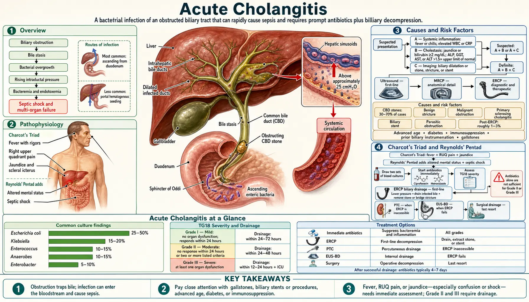
Table of Contents
- Overview
- Pathophysiology
- Causes and Risk Factors
- Charcot's Triad and Reynolds' Pentad
- Tokyo Guidelines Severity Grading (TG18)
- Diagnosis
- Antibiotic Therapy
- Biliary Drainage — ERCP and Alternatives
- Prognosis and Complications
- Key Research Papers
- Connections
- Featured Videos
Overview
Acute cholangitis — also called ascending cholangitis — is a bacterial infection of the biliary tract that arises almost always from biliary obstruction. When bile cannot drain normally, stasis creates the perfect environment for bacterial overgrowth; bacteria ascend from the duodenum through the sphincter of Oddi, infect the stagnant bile, and can rapidly spread into the bloodstream.
Historically this was a death sentence. Jean-Martin Charcot described the classic clinical triad in 1877, yet even after recognition the condition carried close to 100% mortality until the development of endoscopic biliary drainage in the 1970s–1980s transformed it from a surgical catastrophe into a manageable emergency. Today, with prompt antibiotics and biliary decompression, overall mortality ranges from 2–10%. Patients who develop the full Reynolds' Pentad — septic shock plus altered mental status on top of Charcot's Triad — still carry mortality exceeding 50% without urgent drainage.
The Tokyo Guidelines, most recently updated in 2018 (TG18), provide internationally adopted diagnostic criteria and a three-tier severity grading system that drives treatment decisions worldwide.
Pathophysiology
Under normal conditions the biliary tree is sterile. Bile flow itself provides mechanical clearance, and bile salts have mild antibacterial properties. Obstruction disrupts both mechanisms simultaneously: bile stasis allows bacteria to colonize and multiply unchecked while rising intraductal pressure — above approximately 25 cmH2O — drives bacteria and their endotoxins across the biliary epithelium directly into the hepatic sinusoids and then the systemic circulation.
Two routes of bacterial entry are recognized:
- Ascending from the duodenum: the most common path — enteric bacteria migrate retrograde through the sphincter of Oddi, particularly when sphincter competence is impaired by stones, stents, or prior sphincterotomy.
- Hematogenous seeding via the portal circulation: less common but clinically important in patients with portal hypertension or bowel ischemia.
The microbiological profile reflects the gut flora. Escherichia coli accounts for 25–50% of isolates, Klebsiella species for 15–20%, and Enterobacter for 5–10%. Gram-positive Enterococcus contributes 10–15%. Anaerobes — Bacteroides, Clostridium — appear in 10–15% of cultures, most often in patients who have undergone prior biliary surgery or endoscopic sphincterotomy. Infections are polymicrobial in 30–40% of cases. Bacteremia is detected in blood cultures in 50–70% of patients when drawn before antibiotics.
The systemic inflammatory response triggered by biliary bacteremia and endotoxemia can escalate rapidly to septic shock and multi-organ failure, which is why biliary decompression — not antibiotics alone — is the cornerstone of treatment in moderate and severe disease.
Causes and Risk Factors
Any condition that obstructs bile flow can precipitate acute cholangitis. The most important causes are:
- Choledocholithiasis — common bile duct stones are the leading cause in Western countries, responsible for 30–70% of cases. Stones create both mechanical obstruction and a nidus for bacterial colonization.
- Benign biliary strictures — most arise after cholecystectomy (clip migration, ischemic injury to the bile duct) or after prior ERCP with inadvertent ductal damage.
- Malignant obstruction — cholangiocarcinoma (bile duct cancer), carcinoma of the head of the pancreas, and ampullary carcinoma all obstruct the common bile duct (CBD). Malignant cholangitis carries a worse prognosis because the obstruction cannot be definitively relieved without surgery, and stents inevitably clog.
- Primary sclerosing cholangitis (PSC) — diffuse inflammatory stricturing of the intra- and extrahepatic bile ducts leads to recurrent episodes of cholangitis; long-term prophylactic antibiotics are required in selected patients.
- Biliary stents — both plastic and metal stents become colonized over time and can clog, causing cholangitis weeks to months after elective stent placement. A classic scenario: a patient with a pancreatic cancer stent presents with fever and jaundice several months later.
- Parasitic biliary obstruction — Clonorchis sinensis (Chinese liver fluke) and Ascaris lumbricoides (roundworm) are important causes in East and Southeast Asia.
- Post-ERCP cholangitis — occurs in roughly 1–3% of ERCP procedures, typically from inadequate drainage of a segment after contrast injection.
Key risk factors include advanced age, diabetes, immunosuppression (organ transplant, human immunodeficiency virus (HIV), chemotherapy), prior biliary instrumentation, and the presence of gallstones.
Charcot's Triad and Reynolds' Pentad
Two classic clinical syndromes define the spectrum of acute cholangitis, and both carry the names of the physicians who first described them.
Charcot's Triad (1877)
Jean-Martin Charcot described the combination of:
- Fever with rigors — temperature typically exceeding 38.5°C, often with dramatic chills
- Right upper quadrant (RUQ) pain — colicky or constant, sometimes radiating to the right shoulder
- Jaundice — scleral icterus and yellowing of skin from conjugated hyperbilirubinemia
The triad is highly specific for cholangitis when all three elements are present, but it is seen in only 50–70% of patients. Many — especially the elderly or immunosuppressed — present with an incomplete picture: fever without pain, or jaundice without fever. Relying on the full triad to diagnose cholangitis will miss a significant proportion of cases.
Reynolds' Pentad (1959)
B.M. Reynolds and E.L. Dargan added two more features to Charcot's Triad to define suppurative cholangitis — the condition in which pus is present under pressure within the common bile duct:
- All three elements of Charcot's Triad, plus:
- Altered mental status — confusion, lethargy, or obtundation from septic encephalopathy
- Septic shock — hypotension with evidence of poor tissue perfusion
Reynolds' Pentad signals a biliary emergency. The common bile duct is essentially a pressurized sewer; bacteria and endotoxins pour into the systemic circulation faster than the body can contain the response. Without immediate biliary decompression, mortality exceeds 50%. Every minute spent on optimization without draining the duct is a minute spent allowing the sepsis cascade to deepen.
Tokyo Guidelines Severity Grading (TG18)
The Tokyo Guidelines were first published in 2007 (TG07), revised in 2013 (TG13), and most recently updated in 2018 (TG18). They provide a three-tier severity system that directly determines the urgency of biliary drainage.
Grade I — Mild
The patient meets criteria for acute cholangitis but does not meet the criteria for Grade II or Grade III at diagnosis (the 2013 criteria, kept in TG18). The TG18 management guideline states that for mild cholangitis, initial treatment including antibiotics is sufficient in most cases and most patients do not need biliary drainage; drainage is considered if the patient does not respond to initial treatment. (The original 2007 Tokyo Guidelines graded severity partly by response to initial treatment: Grade I was cholangitis that responded to it.)
Grade II — Moderate
The patient has no organ dysfunction but has two or more of the following findings at presentation (in the 2007 guidelines, Grade II was instead defined as cholangitis that did not respond to initial medical treatment):
- Leukocytosis above 12,000/mm3 or leukopenia below 4,000/mm3
- High fever (≥39°C)
- Age ≥75 years
- Serum bilirubin ≥5 mg/dL
- Serum albumin less than 0.7 times the lower limit of normal
For Grade II, TG18 indicates early endoscopic or percutaneous biliary drainage. In the large Japan–Taiwan case series behind TG18, Grade II patients treated with early or urgent drainage had significantly lower 30-day mortality than those who were not.
Grade III — Severe
The patient has dysfunction in at least one organ system:
- Cardiovascular: hypotension requiring vasopressors
- Neurological: altered level of consciousness
- Respiratory: PaO2/FiO2 ratio below 300
- Renal: oliguria or serum creatinine above 2.0 mg/dL
- Hepatic: PT-INR above 1.5
- Hematological: platelet count below 100,000/mm3
For Grade III, TG18 calls for respiratory and circulatory support, with biliary drainage performed as soon as possible after the patient's general condition has been improved by initial treatment; such patients are managed in an intensive care unit (ICU). Mortality without drainage in Grade III exceeds 50%. The goal is to achieve hemodynamic stability sufficient to safely perform ERCP, then decompress the duct as rapidly as possible.
Diagnosis
The TG18 diagnostic criteria provide a structured framework that avoids over-reliance on the classic triad. Diagnosis proceeds through three criteria categories:
- Criterion A — Systemic inflammation: fever or chills, OR laboratory evidence (elevated white blood cell count or C-reactive protein (CRP), or other inflammatory markers)
- Criterion B — Cholestasis: jaundice (serum bilirubin ≥2 mg/dL) OR abnormal liver function tests (alkaline phosphatase (ALP), gamma-glutamyl transferase (GGT), aspartate aminotransferase (AST), or alanine aminotransferase (ALT) more than 1.5 times the upper limit of normal)
- Criterion C — Imaging: biliary dilatation on imaging, OR evidence of an etiology such as a stone, stricture, or stent on ultrasound, computed tomography (CT), or MRCP
Suspected cholangitis: A + B, or A + C. Definite cholangitis: A + B + C. The diagnosis also requires other causes of infection to be excluded (pneumonia, urinary tract infection, appendicitis).
Laboratory Findings
- Leukocytosis (or leukopenia in severe sepsis)
- Elevated C-reactive protein, procalcitonin, and erythrocyte sedimentation rate (ESR)
- Conjugated hyperbilirubinemia
- Elevated alkaline phosphatase and gamma-glutamyl transferase — cholestatic pattern
- Transaminase elevation (aspartate aminotransferase/alanine aminotransferase) — hepatocyte involvement in severe cases
- Blood cultures positive in 50–70% — always draw two sets before starting antibiotics
- Coagulation studies: elevated PT in hepatic dysfunction
Imaging
- Abdominal ultrasound: first-line — identifies biliary dilatation, gallbladder stones, and pericholecystic changes. Common bile duct stones are visible in only 50% (gas from duodenum obscures the distal common bile duct).
- MRCP (magnetic resonance cholangiopancreatography): non-invasive gold standard for anatomical detail — excellent for common bile duct stone location, stricture characterization, and PSC staging. No radiation, no contrast injection into the duct.
- Computed tomography abdomen with contrast: useful for complications (liver abscess, pneumobilia, malignancy staging), but inferior to MRCP for intrinsic biliary pathology.
- ERCP (endoscopic retrograde cholangiopancreatography): simultaneously diagnostic and therapeutic — the procedure of choice when common bile duct stones are strongly suspected and biliary drainage is needed. Contrast injected under fluoroscopy shows the entire biliary tree.
- PTC (percutaneous transhepatic cholangiography): when ERCP is inaccessible (surgically altered anatomy, complete duodenal obstruction, failed cannulation). Under ultrasound or fluoroscopic guidance, a needle punctures a dilated peripheral bile duct; a drain is left in place.
Antibiotic Therapy
TG18 states that initial medical treatment, including antibiotics, starts immediately once the diagnosis is made, with blood cultures drawn first. In acute cholangitis, every hour of delay before antibiotics worsens outcome. The choice of regimen is driven by disease severity, whether the infection is community-acquired or healthcare-associated, and local resistance patterns.
Mild to Moderate — Community-Acquired
- Ciprofloxacin + metronidazole — covers gram-negative bacilli and anaerobes; widely used, good biliary penetration
- Amoxicillin-clavulanate — broad-spectrum beta-lactam/inhibitor combination; convenient single agent
- Cefazolin + metronidazole — adequate for mild disease in low-resistance environments
- Ampicillin-sulbactam — another acceptable beta-lactam/inhibitor option
Moderate to Severe and Healthcare-Associated
- Piperacillin-tazobactam — preferred broad-spectrum choice; excellent gram-negative, gram-positive, and anaerobic coverage
- Meropenem or imipenem-cilastatin — reserved for suspected ESBL-producing organisms, prior antibiotic exposure, or healthcare-associated infection with high local resistance rates
- Vancomycin added when methicillin-resistant Staphylococcus aureus (MRSA) is a concern (healthcare-associated, prior methicillin-resistant Staphylococcus aureus history)
Duration and De-escalation
After successful biliary drainage — defined as resolution of fever and normalization of inflammatory markers — antibiotic duration is typically 4–7 days. Prolonged courses are not necessary and contribute to resistance. Blood culture sensitivities guide de-escalation to the narrowest effective agent within 48–72 hours.
A critical principle: antibiotics alone are not sufficient for Grade II or Grade III cholangitis. They suppress bacteremia and reduce systemic inflammation, but the infected, obstructed bile duct remains a continuous source unless mechanically drained. Mortality does not fall to acceptable levels with antibiotics alone in moderate or severe disease.
Biliary Drainage — ERCP and Alternatives
Biliary decompression is the definitive treatment for acute cholangitis. The goal is to lower intraductal pressure, drain infected bile, and remove or bypass the obstruction.
ERCP — The Gold Standard
Endoscopic retrograde cholangiopancreatography is the first-line procedure for biliary drainage in acute cholangitis. A side-viewing duodenoscope is advanced to the ampulla of Vater; the biliary orifice is cannulated, and contrast is injected under fluoroscopy. The endoscopist can then:
- Perform endoscopic sphincterotomy — cutting the sphincter of Oddi to widen the common bile duct orifice
- Extract common bile duct stones with a balloon catheter or Dormia basket
- Place a plastic or self-expanding metal stent to bridge a stricture and maintain drainage
- Place a nasobiliary drain — a tube left in the duct and exiting through the nose for external drainage and irrigation
Timing of ERCP follows TG18 severity:
- Grade I: most patients do not need drainage; it is considered when there is no response to initial treatment
- Grade II: early drainage
- Grade III: as soon as possible after the patient's general condition has been improved by initial treatment and respiratory/circulatory support
After successful stone extraction, elective laparoscopic cholecystectomy is recommended for patients whose stones originated from the gallbladder, to prevent recurrent choledocholithiasis.
PTC — Percutaneous Transhepatic Cholangiography
When ERCP is inaccessible — surgically altered anatomy (Roux-en-Y gastric bypass, Whipple procedure), complete duodenal obstruction, or failed endoscopic cannulation — interventional radiology performs PTC under ultrasound or fluoroscopic guidance. A needle is advanced through the abdominal wall and liver parenchyma into a dilated peripheral bile duct; a guidewire is advanced and an external drain is placed. A subsequent internal-external drain or permanent metal stent can be placed in a staged fashion once the acute infection is controlled.
EUS-Guided Biliary Drainage (EUS-BD)
An emerging technique that has become a viable alternative when ERCP fails. Under endoscopic ultrasound guidance, the endoscopist can create a controlled fistula between the bile duct and an adjacent gastrointestinal lumen:
- Choledochoduodenostomy (EUS-CDS): direct puncture from the duodenal bulb into the common bile duct — best for distal obstruction
- Hepaticogastrostomy (EUS-HGS): puncture from the stomach into the left intrahepatic duct — used when the duodenum is inaccessible
EUS-BD offers a single-session approach that avoids the risks of PTC (bleeding, bile leak, drain dislodgment) and is now available at specialized centers worldwide.
Surgical Decompression
Open or laparoscopic surgical drainage is a last resort in acute cholangitis — reserved for cases where endoscopic and percutaneous approaches have failed and the obstruction is amenable to surgical correction. Operative morbidity and mortality are substantially higher in the acute setting than in elective cases. Procedures include T-tube choledochostomy for bile duct decompression and common bile duct exploration for stone removal.
Definitive Treatment of the Underlying Cause
Draining the duct controls the acute episode; it does not treat the underlying cause. After recovery from the acute illness:
- Common bile duct stones: complete stone clearance at ERCP + cholecystectomy (if gallbladder stones are the source)
- Benign stricture: balloon dilation + stenting; surgery if endoscopic management fails
- Malignant obstruction: palliative metal stenting for unresectable disease; surgical resection with curative intent for operable disease
- PSC: ursodeoxycholic acid, endoscopic dilation of dominant strictures, liver transplantation in end-stage disease
Prognosis and Complications
The prognosis of acute cholangitis has improved dramatically over the past four decades as endoscopic drainage has replaced emergency surgery as the primary treatment modality. Overall in-hospital mortality with modern treatment is 2–10%. However, outcomes vary sharply by severity grade.
Prognostic Factors
Independent predictors of poor outcome include:
- Age over 70 years
- Malignant biliary obstruction (versus stone disease)
- Presence of hepatic abscess
- Acute renal failure at admission
- Septic shock requiring vasopressors
- Prior episode of acute cholangitis
- Liver cirrhosis or portal hypertension
- Delay to biliary drainage
Major Complications
- Septic shock and multi-organ failure: the primary cause of death in acute cholangitis. Cardiovascular collapse → renal failure → hepatic failure → respiratory failure. Intensive care unit admission and organ-supportive care are required in Grade III disease.
- Pyogenic liver abscess: bacteria seeded into the liver via the portal circulation form intrahepatic collections. Diagnosis by computed tomography or ultrasound. Management requires systemic antibiotics for 4–6 weeks plus percutaneous or surgical drainage for abscesses above 3–5 cm. Untreated, mortality approaches 100%.
- Biliary septicemia: overwhelming bacteremia leading to disseminated intravascular coagulation (DIC), profound hypotension, and death — the terminal event in untreated suppurative cholangitis.
- Biliary stricture: recurrent episodes of cholangitis cause progressive inflammation and fibrosis of the bile duct wall, leading to secondary strictures that themselves predispose to further cholangitis — a vicious cycle.
- Secondary biliary cirrhosis: chronic biliary obstruction, if not relieved, causes portal fibrosis progressing to cirrhosis with portal hypertension, varices, and hepatic failure. This is particularly relevant in PSC.
- Post-ERCP pancreatitis: occurs in 3–5% of therapeutic ERCP procedures; usually self-limited but can be severe.
- Recurrent cholangitis: especially in patients with PSC, biliary-enteric anastomoses, or indwelling stents; long-term prophylactic antibiotics (e.g., ciprofloxacin 500 mg daily) are considered in patients with multiple recurrences.
Key Research Papers
- Miura F, et al. Tokyo Guidelines 2018: initial management of acute biliary infection and flowchart for acute cholangitis. J Hepatobiliary Pancreat Sci. 2018;25(1):31–40. — PubMed PMID: 28941329
- Kiriyama S, et al. Tokyo Guidelines 2018: diagnostic criteria and severity grading of acute cholangitis (with videos). J Hepatobiliary Pancreat Sci. 2018;25(1):17–30. — PubMed PMID: 29032610
- Mukai S, et al. Indications and techniques of biliary drainage for acute cholangitis in updated Tokyo Guidelines 2018. J Hepatobiliary Pancreat Sci. 2017;24(10):537–549. — PubMed PMID: 28834389
- Wada K, et al. Diagnostic criteria and severity assessment of acute cholangitis: Tokyo Guidelines. J Hepatobiliary Pancreat Surg. 2007;14(1):52–58. — PubMed PMID: 17252297
- Reynolds BM, Dargan EL. Acute obstructive cholangitis; a distinct clinical syndrome. Ann Surg. 1959;150(2):299–303. — PubMed PMID: 13670595
- Gomi H, et al. TG13 antimicrobial therapy for acute cholangitis and cholecystitis. J Hepatobiliary Pancreat Sci. 2013;20(1):60–70. — PubMed PMID: 23340954
- Bonnel DH, et al. Placement of metallic stents for treatment of postoperative biliary strictures: long-term outcome in 25 patients. AJR Am J Roentgenol. 1997;169(6):1517–1522. — PubMed PMID: 9393155
- PubMed search: acute cholangitis biliary drainage outcomes
Connections
- Gastroenterology
- Choledocholithiasis
- Gallstones
- Acute Cholecystitis
- Primary Sclerosing Cholangitis
- Pancreatitis
- Gallbladder Disease
Featured Videos
Featured videos will be added shortly.