Calciphylaxis (Calcific Uremic Arteriolopathy)
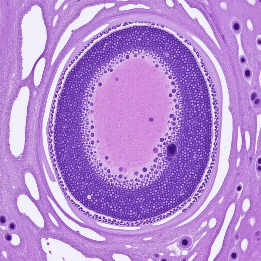
Calciphylaxis — formally called Calcific Uremic Arteriolopathy (CUA) — is a rare, life-threatening vascular disorder in which calcium deposits accumulate inside the small blood vessels of the skin, triggering tissue death and excruciatingly painful ulcers that almost inevitably become infected. It strikes primarily people on long-term dialysis for kidney failure, though it can occur without kidney disease in certain metabolic conditions. Despite aggressive treatment, one-year mortality exceeds 40–80%, with sepsis from infected skin wounds the leading killer — making early recognition and rapid, coordinated intervention the only meaningful chance of survival.
- Calciphylaxis Explained
- How Calciphylaxis Develops
- Who Is at Risk
- Symptoms and Skin Findings
- Diagnosing Calciphylaxis
- Non-Uremic Calciphylaxis
- Treatment Approaches
- Sodium Thiosulfate and Emerging Therapies
- Prognosis and Quality of Life
- Key Research Papers
- Connections
- Featured Videos
Calciphylaxis Explained
Calciphylaxis is a catastrophic vascular disease of the small arterioles supplying the skin and fat tissue. The defining event is calcification — calcium crystals depositing directly inside the walls of tiny blood vessels — combined with blood clot formation inside those vessels. The result is that patches of skin and fat are literally cut off from their blood supply, starved of oxygen, and begin to die. The dying tissue becomes a portal for bacteria, and overwhelming infection — sepsis — is what kills most patients.
The condition predominantly affects people with end-stage renal disease (ESRD) on hemodialysis or peritoneal dialysis. In these patients, the kidneys have lost the ability to regulate calcium, phosphorus, and parathyroid hormone — creating the perfect chemical storm for vascular calcification. In United States national dialysis records, recorded cases rose from 3.7 to 5.7 per 10,000 people on long-term hemodialysis from 2007 onward (Nigwekar 2014), though the true incidence is likely higher because mild or early cases are often misdiagnosed as peripheral arterial disease, diabetic ulcers, or wound complications.
The name "calciphylaxis" — coined by Hans Selye in the 1960s — reflects the original (now outdated) theory that calcium acted like a trigger for an anaphylactic-like sensitization response. The more accurate modern term, Calcific Uremic Arteriolopathy, describes what actually happens: uremia (kidney failure) creates conditions that cause arterioles (small arteries) to calcify. Despite the rebranding in academic literature, "calciphylaxis" remains the name most clinicians and patients use.
One-year mortality runs 40–80% across published series — among the highest of any dermatological condition. Patients with proximal lesions (thighs, abdomen, buttocks) have substantially worse outcomes than those with distal lesions (lower legs). Sepsis — most often from Staphylococcus aureus or gram-negative organisms colonizing necrotic wounds — is the immediate cause of death in the majority of cases. Without recognition and treatment, death can occur within weeks to months of the first skin lesion appearing.
How Calciphylaxis Develops
Calciphylaxis is not simply "too much calcium in the blood." It results from a fundamental breakdown of the biological systems that normally prevent calcium from depositing inside blood vessel walls — a failure of calcification inhibition combined with a shift of vascular smooth muscle cells toward a bone-forming state.
The Calcification Inhibitors That Fail
Healthy arteries resist calcification through three major inhibitory proteins:
- Fetuin-A (alpha-2-Heremans-Schmid glycoprotein): a liver-derived protein that circulates in blood and physically blocks calcium-phosphate crystal formation. In dialysis patients and those with chronic inflammation or malnutrition, fetuin-A levels are dramatically reduced. Without fetuin-A, calcium-phosphate crystals nucleate and grow unchecked inside vessel walls.
- Matrix Gla Protein (MGP): produced by vascular smooth muscle cells within artery walls, MGP is the most potent local inhibitor of vascular calcification. The critical catch: MGP requires vitamin K-dependent carboxylation to be activated. In its uncarboxylated, inactive form, MGP cannot stop calcium deposition. Warfarin — by blocking vitamin K recycling — prevents MGP carboxylation and is the single most important modifiable risk factor for calciphylaxis. Patients on warfarin who develop calciphylaxis frequently have dramatically elevated levels of uncarboxylated, inactive MGP in their blood vessels.
- Osteoprotegerin (OPG): a decoy receptor that inhibits osteoclast activation. In uremia, OPG levels are impaired and the RANK-RANKL pathway that drives bone resorption — and, paradoxically, vascular calcification — becomes dysregulated.
The Calcium-Phosphorus Product
In healthy kidneys, excess phosphorus is cleared in the urine. In dialysis patients, phosphorus accumulates between sessions. When the product of serum calcium (mg/dL) multiplied by serum phosphorus (mg/dL) exceeds approximately 55–72 mg²/dL², the solution becomes supersaturated and calcium-phosphate crystals precipitate out into soft tissues and blood vessel walls. This is essentially the same chemistry as a supersaturated salt solution forming crystals — except it is happening inside arteries.
Vascular Smooth Muscle Transdifferentiation
Under the chemical stress of uremia, high phosphorus, and inflammatory cytokines, vascular smooth muscle cells undergo a transformation — they begin expressing genes normally reserved for bone-forming osteoblasts (Runx2, osteopontin, alkaline phosphatase). These osteoblast-like vascular cells actively deposit hydroxyapatite crystals into the vessel wall, turning arterioles into calcified, rigid tubes that can no longer dilate to deliver blood to the skin.
Intimal Hyperplasia, Microthrombosis, and Ischemia
Calcium deposits in the vessel wall trigger a secondary response: fibro-intimal hyperplasia (the inner lining of the vessel thickens reactively), which further narrows the already-calcified lumen. The damaged endothelium activates clotting, and microthrombi form inside the tiny arterioles supplying the skin's fat layer (subcutaneous adipose tissue). When these vessels clot off, the fat lobules they supply undergo ischemic necrosis. The skin above — now without its underlying blood supply — becomes violaceous, then necrotic, forming the characteristic black eschar. Secondary infection of the necrotic wound is the rule, not the exception.
Who Is at Risk
Calciphylaxis clusters in specific patient populations. Recognizing the risk profile allows clinicians to maintain a high index of suspicion when the first skin changes appear:
Primary Risk Factors
- End-stage renal disease on dialysis: The overwhelming majority of calciphylaxis cases (approximately 90–95%) occur in patients on hemodialysis or peritoneal dialysis. Longer dialysis vintage and inadequate dialysis are additional risk elevators within this group.
- Warfarin use: Warfarin is the single most important modifiable risk factor. By blocking vitamin K-dependent carboxylation of Matrix Gla Protein, warfarin leaves MGP inactive and unable to prevent vascular calcification. Multiple studies show that warfarin use dramatically increases calciphylaxis risk in dialysis patients, and discontinuing warfarin is a cornerstone of treatment. Reviews describe switching people who take warfarin for atrial fibrillation or venous thromboembolism to another blood thinner (apixaban, rivaroxaban, or low-molecular-weight heparin) when calciphylaxis develops.
- Secondary hyperparathyroidism with elevated parathyroid hormone (PTH): Elevated parathyroid hormone drives bone turnover and releases calcium and phosphorus into the blood, worsening the mineral imbalance that fuels calcification. Very high parathyroid hormone levels (above 400–600 pg/mL) are a consistent risk factor in dialysis-associated calciphylaxis.
- Repeated insulin injections into the skin: A study of 1,030 people on hemodialysis who developed calciphylaxis, each matched with two who did not (Nigwekar 2016), found that people with diabetes who injected insulin had higher odds of calciphylaxis on the lower belly and upper thighs, rising with the number of injections a day (odds ratio 3.74 for more than three injections a day). The authors read this as a dose effect of repeated skin injury. In the same study, diabetes, higher body mass index, higher blood calcium, phosphorus and parathyroid hormone, and treatment with nutritional vitamin D, cinacalcet and warfarin were also linked to later calciphylaxis, which appeared a median of 925 days after hemodialysis began.
Additional Risk Factors
- Obesity: Body mass index above 30 is consistently associated with calciphylaxis risk, possibly because obese adipose tissue has relatively poor vascular supply and is more vulnerable to ischemia when arterioles calcify.
- Hypoalbuminemia: Low serum albumin (below 3.5 g/dL) reflects malnutrition, inflammation, and reduced fetuin-A production — all of which impair calcification inhibition. Albumin below 3.0 g/dL at calciphylaxis onset is associated with markedly worse prognosis.
- High calcium-phosphate product: Product exceeding 55–72 mg²/dL² indicates a supersaturated mineral state that promotes crystal precipitation into vessel walls.
- Female sex: Women are affected at roughly two to three times the rate of men in most series, for reasons that are not fully understood but may relate to fat distribution, hormonal effects on vascular calcification, and higher rates of autoimmune and inflammatory conditions.
- Diabetes mellitus: Diabetes adds vascular injury, advanced glycation end-products, and impaired wound healing that compound the damage once calciphylaxis lesions develop.
- Calcium-based phosphate binders: Calcium carbonate and calcium acetate — historically the most commonly used phosphate binders in dialysis — increase total calcium load and may contribute to the supersaturated state. Non-calcium-based binders (sevelamer, lanthanum carbonate) are the ones reviews describe for high-risk patients.
- Active vitamin D analogs (calcitriol, paricalcitol): When used at high doses, these agents raise serum calcium and phosphorus, worsening the mineral imbalance.
- Poor nutritional status and inflammatory disease: Both reduce fetuin-A and albumin, stripping away two important layers of calcification protection.
- Protein C or S deficiency: Rare inherited or acquired deficiencies of these natural anticoagulants may promote the microthrombosis component of calciphylaxis.
Symptoms and Skin Findings
Calciphylaxis is above all else a disease of pain — pain that is often described as the worst the patient has ever experienced, disproportionate to what the wound looks like, and present even before visible skin breakdown occurs. Pain is so characteristic that its absence raises doubt about the diagnosis. The skin findings evolve through a predictable sequence if intervention is not prompt:
Early Stage — Livedo Reticularis and Violaceous Mottling
The first visible sign is a net-like, purplish discoloration of the skin — livedo reticularis — reflecting sluggish blood flow and early arteriolar occlusion in the skin's small vessels. The affected skin feels indurated (hard) and tender to touch. At this stage, the diagnosis is most commonly missed: livedo reticularis has many causes, and in a dialysis patient it may be attributed to vascular disease or positioning. Pain out of proportion to the skin findings is the clinical alarm signal at this stage.
Intermediate Stage — Nodules, Plaques, and Dusky Discoloration
Over days to weeks, the mottled skin progresses to raised, firm, extremely tender nodules and plaques. The color deepens from violaceous to a dusky, dark purple-black — reflecting worsening ischemia of the subcutaneous fat. The skin may feel board-like. Secondary blistering (hemorrhagic bullae) can develop over the plaques as skin dies from below.
Advanced Stage — Necrotic Ulcers with Stellate Eschar
Without treatment, the necrotic plaques ulcerate, forming the pathognomonic stellate (star-shaped) black eschar with a violaceous halo — a zone of dead, leathery black tissue surrounded by a ring of dark purple, dusky, living (but ischemic) skin. The ulcers are often irregular, coalescent, and can cover large surface areas of the thigh, abdomen, or buttocks. The odor of necrotic tissue is often present. These open wounds are an immediate gateway for bacterial invasion.
Location Matters — Proximal vs. Distal
Lesions almost always occur on areas with abundant subcutaneous fat because the subcutaneous fat lobules are where the calcified arterioles first lose perfusion:
- Proximal locations (worse prognosis): thighs, abdomen (especially the pannus in obese patients), buttocks, and breast. Proximal calciphylaxis is associated with 1-year mortality above 60–80%.
- Distal locations (somewhat better prognosis): lower legs, calves, feet, and fingers. Acral calciphylaxis (affecting digits) carries a better prognosis, though amputation may still be required for gangrenous digits.
Superinfection and Sepsis
Once ulceration occurs, wound superinfection is virtually inevitable. The organisms are typically skin commensals — Staphylococcus aureus, Pseudomonas aeruginosa, and anaerobes — that rapidly colonize the necrotic tissue. Systemic sepsis — bacteremia, fever, hemodynamic instability, multi-organ dysfunction — is the pathway to death for most calciphylaxis patients. The wounds themselves do not cause death; the resulting infection does. This is why wound management is as important as treating the underlying mineral disorder.
Diagnosing Calciphylaxis
No single test diagnoses calciphylaxis. Diagnosis relies on the combination of the right clinical context (dialysis patient or metabolic risk factors), characteristic pain and skin findings, and confirmatory tissue or imaging evidence. Skin biopsy is the most definitive test but is performed carefully — biopsy of an active lesion can impair wound healing and worsen the ulcer.
Skin Biopsy
Biopsy of the edge of an established lesion (not the necrotic center) is the gold standard. Key histological findings on hematoxylin and eosin (H&E) staining and special stains:
- Calcium deposits in arteriolar walls: von Kossa stain (turns calcium deposits black) is more sensitive than H&E for identifying calcium. Calcium deposits are seen in the media and intima of small arterioles in the dermis and subcutis.
- Fibro-intimal hyperplasia: the inner lining (intima) of calcified vessels is thickened with proliferating fibroblasts and smooth muscle cells, narrowing the lumen.
- Microthrombi: fibrin thrombi occluding the arteriolar lumen are a characteristic finding in active calciphylaxis, distinguishing it from simple vascular calcification without ischemia.
- Ischemic fat necrosis: lobules of subcutaneous fat undergo coagulative necrosis from loss of blood supply — ghost cells with preserved outlines but no viable nuclei.
- Panniculitis and mixed inflammatory infiltrate: secondary inflammation around the necrotic fat lobules.
The biopsy site is chosen carefully — the edge of a lesion with intact but discolored skin, not the ulcerated center. Biopsy of the necrotic center rarely adds diagnostic information and reliably enlarges the wound. Some experts in severe cases prefer clinical diagnosis to avoid biopsy-related wound complications entirely.
Plain X-Ray and Computed Tomography Imaging
Plain radiographs of the affected area can reveal the characteristic "eggshell" or "railroad track" pattern of vascular calcification in small arterioles — a lacy, branching pattern of calcium in the expected distribution of subcutaneous blood vessels. Computed tomography (CT) scan (without contrast) is more sensitive and can map the extent of calcification. These imaging findings are not specific for calciphylaxis (arteriosclerosis also calcifies vessels) but in the appropriate clinical context support the diagnosis.
Nuclear Bone Scan (Tc-99m MDP)
Technetium-99m methylene diphosphonate (MDP) bone scan has emerged as a useful non-invasive tool. Because Tc-99m MDP binds to calcium-containing structures, calciphylaxis lesions show characteristic increased uptake on delayed imaging. The pattern is distinctly different from bone metastases or osteomyelitis. Some centers use bone scan as a biopsy-sparing diagnostic option, particularly in patients with fragile wounds where biopsy risks are high.
Laboratory Workup
- Serum calcium, phosphorus, calcium-phosphate product
- Intact parathyroid hormone (iPTH)
- Serum albumin (reflects nutritional status and fetuin-A)
- Vitamin K status (uncarboxylated MGP if available at reference labs)
- Coagulation studies (PT/INR for warfarin confirmation; protein C and S levels)
- Inflammatory markers (C-reactive protein (CRP), erythrocyte sedimentation rate (ESR)) — usually elevated; track wound infection
- Wound cultures when ulcers present
Differential Diagnosis
- Peripheral arterial disease (peripheral artery disease, PAD) with critical limb ischemia: arterial insufficiency ulcers are typically distal (toes, heels), painfully ischemic, without the livedo reticularis / violaceous mottling pattern of proximal skin, and the ankle-brachial index (ABI) is low; calciphylaxis involves small arterioles so large-vessel ABI may be paradoxically normal or unmeasurable due to vessel stiffness
- Diabetic foot ulcers: neuropathic and pressure-based; typically painless (neuropathy); no livedo pattern; no systemic mineral disorder
- Vasculitis: palpable purpura, systemic inflammation, positive antineutrophil cytoplasmic antibody (ANCA) or complement; biopsy shows immune complex deposition or neutrophilic infiltrate without vessel-wall calcium
- Warfarin-induced skin necrosis: occurs within the first days of warfarin initiation (protein C/S depletion); ecchymotic plaques that evolve rapidly to necrosis; associated with hereditary protein C or S deficiency; distinguished by timing and biopsy (thrombi without calcification)
- Nephrogenic systemic fibrosis (NSF): gadolinium-associated skin fibrosis in chronic kidney disease (CKD) patients; woody induration without ulceration or livedo pattern
Non-Uremic Calciphylaxis
While end-stage renal disease accounts for 90–95% of calciphylaxis cases, the condition can and does develop in people with entirely normal kidney function. This is called non-uremic calciphylaxis — a label that challenges the old assumption that calciphylaxis required uremia to occur.
Non-uremic calciphylaxis accounts for approximately 5–10% of all cases in published series. It is important to recognize because these patients are managed differently — dialysis intensification is irrelevant — and because the conditions that cause it are sometimes themselves treatable, giving a better overall prognosis than the uremic form.
Causes of Non-Uremic Calciphylaxis
- Primary hyperparathyroidism: dramatically elevated parathyroid hormone drives calcium and phosphorus dysregulation even with normal kidneys. Parathyroidectomy — surgically removing the overactive parathyroid gland(s) — is both diagnostic and therapeutic in these cases, often producing dramatic calciphylaxis improvement. This stands in contrast to uremic calciphylaxis, where parathyroidectomy is only one component of management.
- Malignancy: breast cancer is the most commonly reported cancer-associated calciphylaxis; it may occur through paraneoplastic hypercalcemia, tumor-derived PTHrP (parathyroid hormone-related peptide), or direct tumor involvement of vessels. Other reported associations include multiple myeloma, leukemia, and lymphoma. Oxalate-secreting tumors (enteric hyperoxaluria from bowel cancer or bowel resection) cause calcium oxalate rather than calcium phosphate vascular deposits, creating a histologically distinct but clinically similar picture.
- Connective tissue disease: systemic lupus erythematosus, rheumatoid arthritis, and primary Sjögren syndrome — possibly through chronic inflammation driving fetuin-A depletion and vascular endothelial activation — have been reported in association with non-uremic calciphylaxis.
- Alcoholic liver disease and cirrhosis: severe liver failure reduces fetuin-A synthesis (fetuin-A is made in the liver) and impairs vitamin K-dependent protein carboxylation — mimicking some of the same calcification-inhibition failure seen in uremic patients.
- Corticosteroid use: chronic corticosteroid exposure may alter mineral metabolism and impair wound healing, contributing to calciphylaxis susceptibility.
- Warfarin use without kidney disease: even in patients with normal renal function, prolonged warfarin use can deplete MGP function sufficiently to allow vascular calcification if other risk factors are present.
Management Principles in Non-Uremic Calciphylaxis
The wound care principles are identical to uremic calciphylaxis — aggressive wound management, pain control, infection prevention, and sodium thiosulfate are all used. The critical difference is that treating the underlying cause — removing the parathyroid adenoma, treating the malignancy, discontinuing warfarin — can produce remission that is rarely achievable in uremic calciphylaxis. Prognosis in non-uremic calciphylaxis is generally better than in ESRD-associated disease, though it remains serious and requires specialist management.
Treatment Approaches
There is no U.S. Food and Drug Administration-approved treatment for calciphylaxis, and no randomized controlled trial has established a definitive regimen. Treatment is multidisciplinary — nephrology, dermatology, wound care, pain management, nutrition, and often palliative care — and is individualized to the severity of skin involvement, the patient's overall medical status, and their goals of care.
A 2019 systematic review and meta-analysis of 147 published reports (Udomkarnjananun 2019) pooled the cohort studies of the five most-used treatments. The death rate was not significantly different with sodium thiosulfate (pooled risk ratio 0.89, 95% confidence interval 0.71–1.12), cinacalcet, hyperbaric oxygen, bisphosphonates or parathyroid surgery. The authors found no significant clinical benefit for any of the five and wrote that randomized controlled trials are needed to test them.
Wound Care — The Foundation of Survival
Because sepsis from superinfected wounds is the leading cause of death, wound management is not supportive afterthought — it is life-saving intervention:
- Non-adherent dressings: silicone-based, non-adherent dressings (silicone contact-layer dressings and foam dressings) minimize trauma to fragile tissue during dressing changes. Standard gauze that sticks to the wound bed tears away tissue with each change (traumatic debridement), which is why wound-care reviews describe non-adherent dressings instead.
- Pain management: calciphylaxis pain is often refractory to standard analgesics. Multimodal approaches combining scheduled opioids, gabapentin or pregabalin (neuropathic component), ketamine infusions, and wound care pre-medication are typically required. Inadequate pain control is a major barrier to wound care compliance, and reviews describe treating it as aggressively as any other complication.
- Surgical debridement: removal of necrotic eschar reduces the bacterial burden and can accelerate healing in selected patients. The decision to debride weighs the healing potential — aggressive debridement in patients with poor wound healing capacity can worsen outcomes. Skilled wound care surgeons or plastic surgeons with calciphylaxis experience are essential.
- Hyperbaric oxygen therapy (HBO): HBO sessions (100% oxygen at 2–3 atmospheres) increase tissue oxygen delivery to ischemic wound edges, promote angiogenesis, and have antimicrobial effects. Case series show wound healing improvement in selected calciphylaxis patients. Availability is limited and sessions require patient transfer to HBO facilities; the commitment is typically 20–40 sessions over weeks to months.
- Skin substitutes and bioengineered tissue: acellular dermal matrices and other skin substitutes can be applied to debrided wounds to facilitate re-epithelialization, particularly after surgical debridement has achieved a clean wound base.
Correcting Calcium-Phosphate Mineral Disorder
- Non-calcium-based phosphate binders: sevelamer hydrochloride (or sevelamer carbonate) and lanthanum carbonate are the agents reviews prefer because they bind dietary phosphorus without adding calcium to the body. Reviews describe stopping calcium carbonate and calcium acetate, because these binders directly worsen the calcium load in an already-supersaturated patient.
- Fewer calcium-raising vitamin D analogs: calcitriol and alfacalcidol raise serum calcium and phosphorus. Paricalcitol has a somewhat more favorable mineral profile and is the analog described when one is still needed.
- More intensive dialysis: increasing dialysis frequency (daily or six-times-weekly hemodialysis) and duration removes more calcium, phosphorus, and uremic toxins per week than standard three-times-weekly regimens. Some centers transition calciphylaxis patients to daily home hemodialysis as a definitive intensification strategy.
- Dietary phosphorus restriction: limiting processed foods, phosphate food additives, colas, and high-phosphorus foods (dairy, nuts, legumes) in coordination with renal dietitian guidance.
Managing Hyperparathyroidism
Cinacalcet (a calcimimetic agent that suppresses parathyroid hormone secretion by sensitizing the parathyroid calcium-sensing receptor) can lower parathyroid hormone without raising serum calcium — making it preferable to high-dose vitamin D analogs in calciphylaxis patients with elevated parathyroid hormone. Parathyroidectomy — surgical removal of hyperplastic or adenomatous parathyroid tissue — is reserved for patients with very high parathyroid hormone (above 800–1000 pg/mL) refractory to medical management, severe bone disease, and in non-uremic cases where primary hyperparathyroidism is the driver.
Sodium Thiosulfate and Emerging Therapies
Sodium thiosulfate (STS) has emerged as the most widely used pharmacological treatment for calciphylaxis, supported by the largest body of evidence — though that evidence remains retrospective and observational rather than from randomized trials, and the 2019 meta-analysis described under Treatment Approaches found no significant difference in deaths with it. It addresses calciphylaxis through multiple mechanisms simultaneously, which may explain its clinical usefulness in a condition with multiple intersecting pathological drivers.
Sodium Thiosulfate — Mechanisms of Action
- Calcium chelation: thiosulfate forms soluble calcium thiosulfate complexes with calcium in tissues, potentially solubilizing existing calcium-phosphate deposits and preventing new ones from forming. This chelation mechanism is the most intuitive explanation for its activity in calciphylaxis.
- Antioxidant effects: sodium thiosulfate is a potent free-radical scavenger. Oxidative stress is a major driver of vascular endothelial injury in uremia, and reducing this oxidative burden may protect residual vasculature from further calcification and thrombosis.
- Vasodilatory effects: thiosulfate may promote nitric oxide release, improving vasodilation in the ischemic wound edges and increasing blood flow to skin margins around calciphylaxis lesions.
Administration and Practical Use
The standard protocol is intravenous sodium thiosulfate 25 grams administered over 30–60 minutes during the last hour of each hemodialysis session, typically three times per week for months. Treatment duration is guided by clinical response — wound healing, pain reduction, cessation of new lesion formation — and is commonly continued for 3–6 months or longer. Patients on peritoneal dialysis require IV STS administration separately, as it cannot be given intraperitoneally.
Side effects are common but usually manageable: burning and nausea during infusion are nearly universal and can be reduced by slowing the infusion rate, pre-medicating with ondansetron, and using normal saline flush. A significant concern is that STS causes metabolic acidosis by releasing acid equivalents — in dialysis patients already prone to acidosis, the serum bicarbonate is monitored and the dialysate bicarbonate concentration adjusted upward to compensate. High-anion-gap metabolic acidosis from STS accumulation has been reported in anuric patients.
Warfarin Discontinuation
For people with calciphylaxis who take warfarin, reviews describe stopping warfarin promptly and switching to another blood thinner. The alternative depends on the indication: for atrial fibrillation in dialysis patients, apixaban at reduced dosing is increasingly used (though its safety in dialysis patients is evolving); heparin-based bridging may be needed acutely. The rationale is straightforward: warfarin is inactivating MGP — the vessel wall's own calcification inhibitor — in every dialysis patient on warfarin, and continuing it during calciphylaxis treatment defeats a key therapeutic target.
Vitamin K2 Supplementation
If warfarin is not the culprit (or after warfarin is stopped), vitamin K2 supplementation specifically in the menaquinone-7 (MK-7) form has emerged as a rational therapy. Menaquinone-7 is the most bioavailable oral form of vitamin K2 for activating MGP in peripheral tissues (including vessel walls). Doses of 360–1080 micrograms per day of menaquinone-7 have been used in observational studies. Menaquinone-7 does counteract warfarin-type blood thinners: in a 2013 study of healthy volunteers kept on a vitamin K antagonist (acenocoumarol), 10 and 20 micrograms a day of menaquinone-7 lowered the international normalized ratio (INR) to a clinically relevant degree in at least 40% and 60% of them, and 45 micrograms a day lowered the group’s average INR by about 40% (Theuwissen 2013) — doses far below those above. The goal is to restore carboxylation of MGP so it can resume its role as a local inhibitor of vascular calcification. It is regarded as a rational, low-risk add-on for people not taking warfarin-type drugs and is increasingly used as adjunctive therapy even in the absence of robust randomized trial data. What is not known: whether menaquinone-7 heals calciphylaxis wounds or improves survival, since its use rests on observational experience rather than completed randomized trials.
Bisphosphonates
Pamidronate (intravenous, IV) has been used in calciphylaxis, primarily in non-uremic cases. Bisphosphonates inhibit osteoclast-mediated bone resorption and may inhibit vascular calcification through analogous mechanisms. They are not routinely used in dialysis-dependent calciphylaxis because renal clearance is required for safe use and accumulation can cause severe adynamic bone disease in renal failure. In non-uremic calciphylaxis with intact renal function, pamidronate is an option supported by case reports.
Tissue Plasminogen Activator (tPA)
Given the prominent microthrombosis component of calciphylaxis, intralesional or systemic tPA has been tried to dissolve the microthrombi occluding arterioles. Results are anecdotal; systemic thrombolysis carries significant bleeding risk in the dialysis population. Reserved for severe refractory cases.
Hexasodium Fytate — The First Phase 3 Placebo-Controlled Trial
The CALCIPHYX trial (Sinha 2024) was the first phase 3, randomized, double-blind, placebo-controlled trial in calciphylaxis. It enrolled 71 adults on maintenance hemodialysis with an open calciphylaxis wound and strong pain, who received hexasodium fytate (a drug that blocks calcium crystal formation in blood vessels) 7 mg/kg or a placebo into the vein during dialysis. After 12 weeks, wound scores and pain had improved about equally in both groups (wound score p = 0.88; pain p = 0.15). In those 12 weeks, calciphylaxis-related events leading to a hospital stay occurred in 2 of 38 people (5%) given the drug versus 11 of 33 (33%) given placebo, and deaths in 1 of 38 (3%) versus 5 of 33 (15%). Over the whole trial, including 12 weeks of open-label drug for everyone, deaths were 5% in the drug group and 21% in the placebo group. The trial was funded by the drug’s developer.
Prognosis and Quality of Life
Calciphylaxis is one of the most lethal dermatological diagnoses. Understanding prognosis is essential not only for treatment planning but for honest, compassionate conversations with patients about goals of care.
Mortality — The Hard Numbers
One-year mortality in published series ranges from 40% to 80%. In the Mayo Clinic series of 64 patients (Weenig 2007), estimated 1-year survival was 45.8%: 61.6% for the 17 patients whose dead tissue was surgically removed (debridement) versus 27.4% for the 46 who did not have it, while survival was similar in the 16 patients who had parathyroid surgery and the 47 who did not. The variance reflects patient selection, lesion location, treatment intensity, and the era of study (outcomes may be slowly improving as sodium thiosulfate use has become widespread). The most frequently cited prognostic marker is lesion location:
- Proximal lesions (thighs, abdomen, buttocks): 1-year mortality 60–80%. The large surface area of necrosis, difficulty with wound care in skin folds, and proximity to critical structures make management far more challenging.
- Distal lesions (lower legs, feet, digits): 1-year mortality 20–40%. Smaller wounds, more accessible anatomy, and the option for local debridement or amputation of gangrenous digits allow better wound control.
- Non-uremic calciphylaxis: somewhat better prognosis than ESRD-associated disease, particularly when a treatable underlying cause (primary hyperparathyroidism, malignancy in remission) can be addressed.
Predictors of Worse Prognosis
- Serum albumin below 3.0 g/dL at diagnosis
- Proximal lesion location
- Rapidly progressive wound expansion at presentation
- Development of sepsis or bacteremia
- Delay in diagnosis beyond 2 months from symptom onset
- Continued warfarin use (failure to discontinue)
- High C-reactive protein / ongoing systemic inflammation
Cause of Death
Sepsis — systemic infection originating from superinfected calciphylaxis wounds — accounts for the majority of deaths (approximately 60–80% of fatal cases). Secondary causes include progressive multiorgan failure from sepsis, and, in a subset, withdrawal of dialysis as patients reach the limits of what treatment can offer. Calciphylaxis wounds do not typically cause direct exsanguination; the death pathway is almost always infectious.
Quality of Life and Pain
For patients who survive calciphylaxis, quality of life is profoundly impaired during the active disease phase and recovery. Pain — often requiring high-dose opioids, ketamine, or nerve blocks — dominates daily experience. Dressing changes (often twice daily) are dreaded events. Mobility may be severely limited by wound location and pain. Depression and anxiety are nearly universal in calciphylaxis patients managing open necrotic wounds over months.
Wound healing, when it occurs, takes many months. Scarring is extensive. Some patients achieve full wound closure with aggressive treatment; others stabilize with partial healing and manageable chronic wounds; others deteriorate despite all interventions.
Palliative Care Integration
Calciphylaxis reviews describe bringing in palliative care early in management — not as an alternative to active treatment, but as a parallel track. Pain management, goals-of-care conversations, advance directive completion, and psychological support are components of high-quality calciphylaxis care at every stage. For patients with proximal lesions, very low albumin, and rapid wound progression despite treatment, an honest conversation about prognosis and the limits of life-prolonging treatment is an act of compassion, not defeat. Dialysis withdrawal — with transition to comprehensive comfort care — is a legitimate, respected choice that some patients make when the burden of treatment exceeds what they are willing to endure.
Key Research Papers
- Nigwekar SU, Thadhani R, Brandenburg VM. Calciphylaxis. N Engl J Med. 2018;378(18):1704–1714. — PubMed PMID: 29719190
- Nigwekar SU, Zhao S, Wenger J, et al. A nationally representative study of calcific uremic arteriolopathy risk factors. J Am Soc Nephrol. 2016;27(11):3421–3429. — PubMed PMID: 27080977
- Brandenburg VM, Cozzolino M, Ketteler M. Calciphylaxis: a still unmet challenge. J Nephrol. 2011;24(2):142–148. — PubMed PMID: 21337312
- Hayashi M, Takamatsu I, Kanno Y, et al. A case-control study of calciphylaxis in Japanese end-stage renal disease patients. Nephrol Dial Transplant. 2012;27(4):1580–1584. — PubMed PMID: 22121234
- Weenig RH, Sewell LD, Davis MD, et al. Calciphylaxis: natural history, risk factor analysis, and outcome. J Am Acad Dermatol. 2007;56(4):569–579. — PubMed PMID: 17141359
- Seethapathy H, Brandenburg VM, Sinha S, et al. Review: update on the management of calciphylaxis. QJM. 2019;112(1):29–34. — PubMed PMID: 30304522
- Udomkarnjananun S, Kongnatthasate K, Praditpornsilpa K, et al. Treatment of calciphylaxis in CKD: a systematic review and meta-analysis. Kidney Int Rep. 2019;4(2):231–244. — PubMed PMID: 30775620
- An J, Devaney B, Ooi KY, et al. Hyperbaric oxygen in the treatment of calciphylaxis: a case series and literature review. Nephrology (Carlton). 2015;20(7):444–450. — PubMed PMID: 25707425
- Ketteler M, Schlieper G, Floege J. Calcification and cardiovascular health: new insights into an old phenomenon. Hypertension. 2006;47(6):1027–1034. — PubMed PMID: 16618842
- Nigwekar SU, Bloch DB, Nazarian RM, et al. Vitamin K-dependent carboxylation of matrix Gla protein influences the risk of calciphylaxis. J Am Soc Nephrol. 2017;28(6):1717–1722. — PubMed PMID: 28049648
- Nigwekar SU, Solid CA, Ankers E, et al. Quantifying a rare disease in administrative data: the example of calciphylaxis. J Gen Intern Med. 2014;29 Suppl 3:S724–S731. — PubMed PMID: 25029979
- Theuwissen E, Teunissen KJ, Spronk HM, et al. Effect of low-dose supplements of menaquinone-7 (vitamin K2) on the stability of oral anticoagulant treatment: dose-response relationship in healthy volunteers. J Thromb Haemost. 2013;11(6):1085–1092. — PubMed PMID: 23530987
- Sinha S, Nigwekar SU, Brandenburg V, et al. Hexasodium fytate for the treatment of calciphylaxis: a randomised, double-blind, phase 3, placebo-controlled trial with an open-label extension. EClinicalMedicine. 2024;75:102784. — PubMed PMID: 39252867
Connections
- Dermatology
- Pyoderma Gangrenosum — another rare, painful, necrotic skin ulcer with high mortality; important differential diagnosis for calciphylaxis; distinguished by absence of vascular calcification and response to steroids
- Lichen Sclerosus — chronic inflammatory skin disease; shares the feature of skin induration and tissue damage but lacks vascular calcification, ischemia, or the dialysis context
- Stevens-Johnson Syndrome — severe drug-induced skin reaction with epidermal necrosis; shares life-threatening severity but distinct pathophysiology (immune-drug reaction vs. vascular calcification) and treatment
- Bullous Pemphigoid — autoimmune blistering disease most common in the elderly; blistering skin lesions in a similar patient age group; distinguished by immunoglobulin G (IgG) autoantibodies against basement membrane zone proteins and absence of vascular calcification
- Calcium — the mineral whose dysregulated deposition inside blood vessels is the central pathological event in calciphylaxis; understanding calcium homeostasis is essential to understanding the disease
- Phosphorus — elevated serum phosphorus, retained because of failed kidneys, drives the supersaturated calcium-phosphate product that precipitates vascular crystal deposition
- Vitamin K — vitamin K-dependent carboxylation of Matrix Gla Protein is the primary mechanism protecting vessel walls from calcification; warfarin's blockade of vitamin K is the dominant modifiable risk factor for calciphylaxis