⚠ Evidence warning: Clinical trials or other evidence do not support the claims that topical tretinoin helps prevent actinic keratoses and that tirbanibulin ointment performs as well as 5-fluorouracil cream. This page keeps it as a report of what people, websites or other media say.
Actinic Keratosis

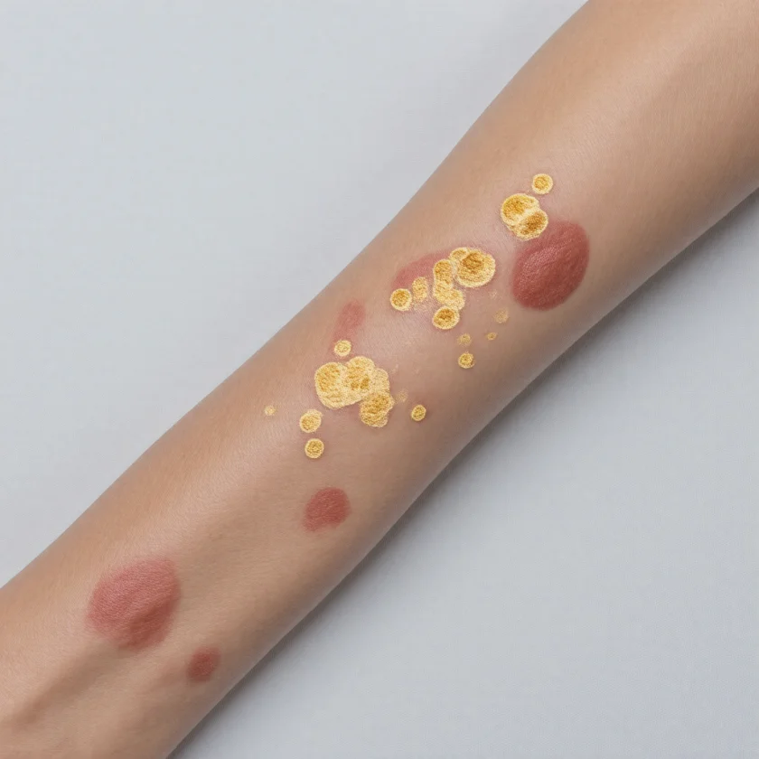
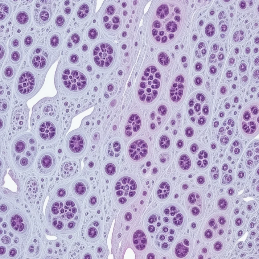
Table of Contents
Actinic Keratosis Explained
Actinic keratosis (AK) is the most common precancerous skin lesion, affecting an estimated 58 million Americans. Also known as solar keratosis, it results from cumulative ultraviolet (UV) radiation causing DNA damage and keratinocyte dysplasia — abnormal squamous cell growth that remains confined to the epidermis. The term "senile keratosis" is now considered obsolete.
AK is best understood as squamous cell carcinoma (SCC) in situ of the epidermis, though its exact classification remains debated. Some authorities consider it a true precancer; others view it as an early form of SCC itself. Either way, common dermatology teaching is the same: treat all AKs, because appearance alone does not show which lesion will progress.
The risk of an individual AK lesion progressing to invasive SCC is uncertain. Older estimates ran from approximately 0.1–10% per year per lesion, and a 2000 review of five studies reported a range of 0.025% to 16% per year. A 2013 systematic review found 0% to 0.075% per lesion-year, up to 0.53% per lesion in people with earlier nonmelanoma skin cancer, and concluded that no reliable estimate can yet be given. Regression also occurs — AK is a dynamic lesion, not a static one. However, patients with AKs carry a 6- to 10-fold increased population risk of developing SCC compared with the general population.
A critical concept in understanding AK is field cancerization: ultraviolet-damaged skin harbors multiple pre-malignant clones scattered across a broad area. This explains why AKs tend to appear in clusters on chronically sun-exposed skin, and why new lesions continue to emerge even after visible lesions are treated. AKs do not arise in isolation — they are the visible tip of a large iceberg of subclinical ultraviolet-induced DNA damage in the surrounding skin.
AK affects primarily fair-skinned individuals and is most prevalent in Australia, where lifetime cumulative sun exposure is high. In adults over 70 in Australia, prevalence exceeds 80%. In the United States, AK prevalence is estimated at roughly 10% in people in their 40s, rising steadily with age.
Pathophysiology
The central pathological event in AK is ultraviolet-B (UVB) radiation-induced DNA damage. Ultraviolet B creates cyclobutane pyrimidine dimers (CPDs) at dipyrimidine sites in DNA. When repair systems fail or are overwhelmed, these dimers lead to characteristic C→T and CC→TT transition mutations — the ultraviolet signature mutations that are the molecular fingerprint of ultraviolet-induced skin cancer.
TP53 Mutation — The Central Molecular Event
The most critical molecular alteration in AK is mutation of TP53, the gene encoding p53, the "guardian of the genome." TP53 mutations are present in more than 90% of SCCs and in many AKs. Normally, p53 detects DNA damage and either halts the cell cycle to allow repair or triggers apoptosis (programmed cell death). Mutant p53 loses this function, allowing keratinocytes with damaged DNA to survive, proliferate, and accumulate further mutations — the foundation of carcinogenesis.
Immunosuppression of Tumor Surveillance
Chronic ultraviolet exposure depletes Langerhans cells in the epidermis — the skin's resident immune sentinels. Ultraviolet also induces local immunosuppression and promotes regulatory T cells (Tregs) that suppress anti-tumor immune surveillance. This mechanism explains the dramatically increased AK and SCC burden in immunosuppressed patients (organ transplant recipients, human immunodeficiency virus-positive individuals, patients on long-term immunosuppressive therapy), where the immune "brakes" on abnormal keratinocyte growth are removed.
Additional Molecular Drivers
Beyond TP53, AK pathogenesis involves telomere dysfunction and chromosomal instability, RAS pathway activation, and upregulation of growth factor signaling. These accumulating genetic hits create the molecular foundation for progression from dysplastic keratinocytes (AK) to invasive SCC.
Histopathology
Under the microscope, AK shows keratinocyte atypia — enlarged, irregular nuclei with loss of normal polarity — confined to the epidermis. Other features include parakeratosis (retained nuclei in the stratum corneum), hyperkeratosis (thickened cornified layer), and solar elastosis in the dermis (basophilic degeneration of collagen = the ultraviolet signature in the deeper skin).
AK can be graded by severity: Grade I (mild dysplasia, focal involvement), Grade II (moderate dysplasia with acanthosis), and Grade III (severe or full-thickness dysplasia — Bowenoid AK — which closely approaches in situ SCC). The basement membrane remains intact in all grades; invasion through the basement membrane defines progression to invasive SCC.
Risk Factors
Ultraviolet Exposure
Cumulative lifetime ultraviolet exposure is the dominant risk factor for AK. Risk is elevated in outdoor occupations (farmers, ranchers, fishermen, construction workers, lifeguards), in geographic locations with high solar ultraviolet intensity (Australia, the American Southwest, equatorial regions), and in individuals with a history of tanning bed use. Tanning beds emit predominantly ultraviolet A (UVA), which penetrates deeper into the skin and also contributes to DNA damage and AK formation. Even intermittent intense ultraviolet exposure — particularly blistering sunburns in childhood — causes permanent DNA damage that can manifest as AK decades later.
Skin Phototype
Fair-skinned individuals bear the overwhelming burden of AK. Fitzpatrick Skin Type I (always burns, never tans) and Type II (usually burns, tans minimally) are at highest risk. Associated features — blue or green eyes, light hair, freckling, Northern European ancestry — each independently contribute. Darker skin types (IV–VI) are not immune, particularly under high cumulative ultraviolet exposure, but AK is substantially less common.
Immunosuppression
Immunosuppression dramatically amplifies AK risk. Solid organ transplant recipients — on calcineurin inhibitors, azathioprine, or mycophenolate — have AK prevalence of 40–80% within 5 years of transplant, and SCC becomes the most common malignancy in this group (rather than basal cell carcinoma, as in the general population). Other immunosuppressed groups at increased risk include patients with chronic lymphocytic leukemia (CLL), human immunodeficiency virus (HIV) infection, and those on long-term systemic corticosteroids or biologic immunosuppressants.
Age and Prior History
AK prevalence rises steeply with age as cumulative ultraviolet damage accumulates over decades. Prior AK and prior SCC are the strongest predictors of new AK development — patients who have had AK will continue to develop new lesions from the underlying ultraviolet-damaged field.
Other Risk Factors
Additional risk factors include arsenic exposure (arsenical keratoses, typically on palms and soles, distinct from ultraviolet-induced AK), ionizing radiation, xeroderma pigmentosum (a rare inherited DNA-repair deficiency causing AKs and SCCs in childhood), and male sex (historically reflecting greater occupational ultraviolet exposure, though this gap is narrowing with increased tanning bed use among women).
Clinical Presentation
Typical Appearance
The hallmark of AK is a rough, sandpaper-like texture to the touch (palpation). Early AK is often better felt than seen — a subtle roughness under the fingertip before any visible change is apparent. As the lesion develops, it appears as an erythematous (pink to red), scaly papule or patch, typically 2–6 mm, with poorly defined borders, on chronically sun-exposed skin. Common sites include the scalp (especially in bald or thin-haired men), face (forehead, temples, nose, cheeks, ears), dorsal forearms and hands, and lower legs in women.
Clinical Variants
- Hyperkeratotic AK — prominent thick scale or crust; elevated; clinically concerning for deeper involvement. Biopsy is required to exclude invasive SCC in hyperkeratotic or indurated lesions.
- Atrophic AK — flat, thin, erythematous; little scale; subtle and easily missed. Common on the face.
- Pigmented AK — melanin deposition gives the lesion a brown or gray tint. May mimic seborrheic keratosis or lentigo maligna. A pigmented lesion of uncertain diagnosis is usually biopsied.
- Lichenoid AK — dense lymphocytic infiltrate underlying the lesion, representing an attempted immune regression response; often pruritic (itchy).
- Bowenoid AK (Grade III) — severe full-thickness dysplasia approaching in situ SCC. Histologically indistinguishable from Bowen disease by some criteria.
- Actinic Cheilitis — AK of the lip, predominantly the lower lip. Presents with blurring of the lip vermilion border, roughness, dryness, and desquamation. Actinic cheilitis carries a higher risk of progression to SCC with metastatic potential than cutaneous AK, because the lip is a high-risk anatomic site for SCC. Treatments described for it include topical 5-fluorouracil (5-FU), photodynamic therapy, or vermilionectomy (surgical removal of the lip surface) for extensive disease.
Warning Signs of Progression to SCC
Features that raise concern for invasive SCC within or arising from an AK include: rapid growth, induration (hardness), ulceration, spontaneous bleeding, pain or tenderness, enlargement beyond 1 cm, and spreading erythema beyond the lesion border. Dermatology practice is a prompt biopsy of any AK with these features.
Diagnosis
Clinical Diagnosis
Experienced dermatologists can diagnose AK clinically with a sensitivity of approximately 80%. The key clinical maneuver is gentle palpation — feeling for the characteristic rough, gritty texture on sun-damaged skin. Dermoscopy improves diagnostic accuracy and helps differentiate AK from other lesions.
Dermoscopy
Dermoscopic features of AK include:
- Rosettes — white four-leaf clover structures visible in polarized light; high sensitivity for AK on the face and scalp.
- Strawberry pattern — red pseudonetwork surrounding white follicular openings; characteristic of facial AK.
- Surface scale — white to yellow scale within the lesion.
- Dotted vessels — fine dotted or hairpin vessels reflecting increased vascularity.
Dermoscopy is particularly valuable for distinguishing AK from SCC (which shows more prominent vessels and structureless white areas) and from benign mimics such as seborrheic keratosis.
Biopsy Indications
Biopsy is indicated when: clinical diagnosis is uncertain; lesions are hyperkeratotic, indurated, ulcerated, or rapidly growing; lesions fail to respond to appropriate treatment; pigmented AK cannot be reliably distinguished from lentigo maligna; or actinic cheilitis is present. Histopathology confirms atypia confined to the epidermis without basement membrane invasion — invasion through the basement membrane defines invasive SCC.
Advanced Imaging
Reflectance confocal microscopy (RCM) and optical coherence tomography (OCT) are non-invasive imaging modalities available at specialized centers that allow real-time cellular and depth imaging of AK, useful for monitoring treatment response and guiding biopsy decisions. Total body photography is valuable for tracking new AK development over time in high-risk patients.
Treatment
AK treatment falls into two broad categories: lesion-directed therapies that treat individual visible lesions, and field therapies that treat the entire ultraviolet-damaged area to address subclinical pre-malignant keratinocytes. Because field cancerization means subclinical AKs far outnumber visible ones, field therapy is generally preferred for patients with multiple lesions.
A 2021 U.S. guideline in the Journal of the American Academy of Dermatology makes strong recommendations for ultraviolet protection, topical imiquimod, topical 5-fluorouracil, and cryosurgery, and conditional recommendations for photodynamic therapy and diclofenac.
Lesion-Directed Therapies
Cryotherapy (liquid nitrogen) is the most commonly used in-office AK treatment. Liquid nitrogen at −196°C is applied for 5–30 seconds with a freeze-thaw cycle that selectively destroys dysplastic keratinocytes. It is suitable for isolated, well-defined lesions and produces immediate results. Risks include blistering, post-inflammatory hypopigmentation or hyperpigmentation (particularly in darker skin types), and pain. An adequate freeze requires a 2–3 mm margin around the lesion.
Curettage — physical scraping of the lesion with a curette, with or without electrodessication — is useful for thick or hyperkeratotic AKs. Shave excision with histopathologic examination is required for lesions suspicious for invasive SCC.
Field Therapies
5-Fluorouracil (5-FU) cream (5%, 0.5%, and 4% strengths) is the standard of care for AK field treatment. 5-FU is an antimetabolite that inhibits thymidylate synthase, impairing DNA synthesis in rapidly dividing cells. A full-face course over 2–4 weeks produces intense inflammation, crusting, and erythema — this inflammatory response is the treatment working. A pulsed protocol (twice weekly for 6 weeks) is better tolerated. In a 2019 randomized trial of 624 patients with multiple AKs on the head (Jansen et al.), success meant a reduction of 75% or more in lesions, and the chance of remaining free from treatment failure 12 months after treatment was 74.7% with 5% fluorouracil, 53.9% with 5% imiquimod, 37.7% with methyl aminolevulinate photodynamic therapy, and 28.9% with 0.015% ingenol mebutate. 5-FU is inexpensive and widely available.
A Veterans Affairs trial tested whether one course also lowers skin cancer. In 932 veterans with at least 2 basal or squamous cell skin cancers in the past 5 years, one 2–4 week course of 5% fluorouracil on the face and ears lowered squamous cell carcinoma needing surgery by 75% in the first year (1% versus 4% with a control cream). Over the whole study (median follow-up 2.8 years) there was no difference between the groups in time to the first skin cancer.
A much shorter combination has also been tried. In a randomized trial of 131 people, calcipotriol 0.005% ointment (a vitamin D analogue) plus 5% fluorouracil, applied twice daily for 4 days, reduced the number of AKs by a mean of 87.8%, compared with 26.3% for petroleum jelly plus 5% fluorouracil. In the follow-up study, 2 of 30 people (7%) in the combination group and 11 of 40 (28%) in the comparison group developed SCC on the treated face and scalp within 3 years.
Imiquimod (3.75% and 5% creams) is a toll-like receptor 7 (TLR-7) agonist that activates innate and adaptive immune responses, driving apoptosis of atypical keratinocytes. The U.S. label for the 5% cream on AK is once daily before bedtime, 2 times per week for a full 16 weeks, on an area of about 25 cm² on the face or scalp. The label for the 3.75% cream is once daily before bedtime for two 2-week cycles separated by a 2-week break. Imiquimod is less irritating than 5-FU. It is moderately effective for immunocompetent patients.
Diclofenac gel (3%) inhibits cyclooxygenase-2 (COX-2) and arachidonic acid metabolism with anti-proliferative effects. Applied twice daily for 3 months, it is the mildest and best-tolerated field option with modest efficacy, suitable for thin or superficial AKs or patients who cannot tolerate more intensive regimens.
Tirbanibulin (1% ointment; U.S. Food and Drug Administration (FDA) approved 2020) has a dual mechanism — inhibiting tubulin polymerization and Src kinase — and is applied once daily for only 5 days; the current U.S. label covers a treatment field of up to 100 cm² on the face or balding scalp. It has been described as comparable to 5-FU 5% and as an alternative for treatment-intolerant patients. Its two phase 3 trials (702 patients) compared it with a vehicle (placebo) ointment, not with 5-FU: complete clearance at day 57 was 44% versus 5% in one trial and 54% versus 13% in the other. At 1 year, an estimated 47% of patients who had complete clearance had recurrent lesions. The trial authors wrote that trials comparing it with conventional treatments are needed.
Photodynamic therapy (PDT) uses a topically applied photosensitizer — aminolevulinic acid (ALA) — that accumulates preferentially in AK cells as protoporphyrin IX. Activation by blue or red light generates singlet oxygen that selectively destroys the dysplastic cells. Daylight PDT, using natural sunlight as the activating source, is equally effective and substantially less painful than conventional office PDT with blue-light devices. PDT is excellent for facial and scalp field cancerization, producing durable clearance and cosmetically excellent results.
Laser resurfacing (ablative CO2 or erbium:YAG laser), chemical peels (trichloroacetic acid, phenol), and dermabrasion target the epidermis and upper dermis and are most effective for extensive facial field cancerization. All modalities resurface the entire ultraviolet-damaged field rather than targeting individual lesions.
Oral nicotinamide (vitamin B3, 500 mg twice daily) is the best-studied oral chemopreventive agent for AK and SCC. A landmark 2015 New England Journal of Medicine randomized controlled trial by Chen et al., in 386 high-risk patients with at least 2 nonmelanoma skin cancers in the previous 5 years, found a 23% lower rate of new nonmelanoma skin cancers at 12 months. The AK count was 11% lower at 3 months, 14% lower at 6 months, 20% lower at 9 months, and 13% lower at 12 months. Nicotinamide is inexpensive and available over the counter, and the trial authors described it as safe. The benefit requires continued use: the trial found no evidence of benefit after stopping.
Later studies add limits and scale. A 2023 trial in 158 organ transplant recipients, stopped early because of poor recruitment, found 207 new keratinocyte cancers with nicotinamide and 210 with placebo (rate ratio 1.0), and no significant difference in AK counts. A 2025 retrospective Veterans Affairs cohort of 33,822 patients linked nicotinamide with 14% less skin cancer overall and 54% less when started after a first skin cancer, with no overall reduction in transplant recipients; a cohort cannot prove cause. Another 2025 retrospective study of 13,108 adults found no increased risk of major heart and blood vessel events with nicotinamide.
Ingenol mebutate gel is no longer sold in Europe, and the U.S. Food and Drug Administration's Drugs@FDA database (application NDA 202833) lists both U.S. strengths, 0.015% and 0.05%, as discontinued. The European Commission withdrew its authorisation on 11 February 2020 at the holder's request, for commercial reasons, while a safety review was pending. That European Medicines Agency review, completed on 30 April 2020, concluded that the medicine may increase the risk of skin cancer and that its risks outweigh its benefits: after 3 years, 6.3% of patients treated with it (15 of 240) developed skin cancer in the treated area, particularly SCC, compared with 2% of patients treated with imiquimod (5 of 244). The agency noted that unusual skin changes or growths could occur from weeks to months after use.
Prevention
Photoprotection
Daily broad-spectrum SPF 30+ sunscreen is the cornerstone of AK prevention. Broad-spectrum labeling indicates both ultraviolet A and ultraviolet B protection. Physical (mineral) blockers — zinc oxide and titanium dioxide — provide broad-spectrum protection without chemical absorption and are preferred for daily use. U.S. sunscreen labels direct reapplying at least every 2 hours during outdoor exposure. An important misconception to address: SPF 100 is not twice as protective as SPF 50. SPF 50 blocks approximately 98% of UVB; SPF 100 blocks approximately 99%. The incremental difference is modest.
Two randomized trials in Australia measured sunscreen against AK directly. In 588 people aged 40 or older, daily SPF 17 sunscreen for one summer gave fewer new lesions (rate ratio 0.62) and more remissions (odds ratio 1.53) than the same cream without its active ingredients. In 1,621 adults aged 25 to 74 in a subtropical town, daily sunscreen gave a 24% smaller rise in AK counts over 2 years than sunscreen used at people's usual rate, while 30 mg of beta carotene a day had no effect.
Protective Clothing and Behavior
UPF 50+ protective clothing, wide-brimmed hats (brim ≥3 inches), ultraviolet-protective sunglasses, and long-sleeved shirts provide physical ultraviolet barriers that sunscreen alone cannot replicate. Seeking shade during peak ultraviolet hours (10 a.m. to 4 p.m.) substantially reduces cumulative exposure. Tanning beds add to AK, SCC, and melanoma risk, whatever the cosmetic result. An important nuance: standard window glass blocks ultraviolet B but transmits ultraviolet A, so daily facial exposure through car or office windows contributes to cumulative ultraviolet damage. That is the reason given for a daily moisturizer with SPF even for people who work indoors.
Oral Chemoprevention
The Chen 2015 trial of nicotinamide 500 mg twice daily enrolled people with at least 2 nonmelanoma skin cancers in the previous 5 years, not people chosen by their number of AKs or by a single SCC. It is the only over-the-counter oral agent with randomized trial support for AK and skin cancer prevention.
Topical retinoids (tretinoin) promote normal keratinocyte differentiation and are described as having modest AK preventive effects alongside photorejuvenation benefits. In a Veterans Affairs randomized trial of 1,131 high-risk patients using 0.1% tretinoin for 1.5–5.5 years, no differences were seen in any cancer-related end point or in AK counts. Oral acitretin (a systemic retinoid) significantly reduces AK and SCC burden in immunosuppressed organ transplant recipients, though it requires monitoring for liver toxicity, hyperlipidemia, and teratogenicity.
Special Populations: Organ Transplant Recipients
Care described for transplant recipients includes quarterly full-skin dermatology examinations, aggressive and early AK treatment with field therapies, and consideration of immunosuppression modification. Switching from calcineurin inhibitors (tacrolimus, cyclosporine) to mechanistic target of rapamycin (mTOR) inhibitors (sirolimus, everolimus) in consultation with the transplant team has been associated with reduced SCC incidence, which matters most for patients developing significant AK burden or SCC. In a randomized trial of kidney transplant recipients with earlier SCC, new SCCs at 5 years occurred in 22% of those moved to sirolimus (64 patients) and 59% of those who stayed on calcineurin inhibitors (56 patients), with similar graft survival.
Prognosis and Follow-Up
Natural History
Individual AK lesions have a heterogeneous natural history. A 2013 systematic review found that 15–63% of single AKs spontaneously regress within 1 year, and that 15–53% of those come back within the following year. In a high-risk Veterans Affairs group, 55% of AKs were gone at 1 year and 70% at 5 years. Studies report that 12–25% persist unchanged. Progression figures differ widely: older estimates of 0.1–10% per lesion per year, 0% to 0.075% per lesion-year in the systematic review (up to 0.53% per lesion with earlier nonmelanoma skin cancer), and 0.60% at 1 year and 2.57% at 4 years for invasive or in situ SCC in the Veterans Affairs group. The review's authors concluded that "no reliable estimates" of how often AK becomes invasive cancer can yet be given. AKs still matter because about 65% of all primary SCCs in the Veterans Affairs group arose in lesions earlier diagnosed as AKs. However, because patients typically carry multiple lesions simultaneously and develop new ones continually, the cumulative risk of SCC over a lifetime is substantial. Patients with AK carry a 6- to 10-fold increased population risk of developing SCC compared with people who have never had AK.
Field Cancerization and Recurrence
Recurrence after treatment is the rule, not the exception. The underlying ultraviolet-damaged field persists even after all visible AKs are cleared. New AKs will continue to emerge from subclinical pre-malignant clones in the surrounding skin. AK management is an ongoing process, not a one-time cure. Knowing this from the start helps people stay with treatment and keeps expectations realistic.
Follow-Up Schedule
Reassessment 3–6 months after completing field therapy allows evaluation of treatment response, detection of residual or new AKs, and biopsy of any suspicious lesions. Patients with a history of multiple AKs or prior SCC are commonly followed with dermatology visits every 3–6 months. Studies demonstrate that patients without regular dermatology follow-up have significantly higher SCC rates. Annual full-skin examinations are the commonly cited minimum for patients who have had AK.
Quality of Life During Treatment
Field therapies — particularly 5-FU and conventional PDT — cause significant erythema, pain, crusting, and temporary cosmetic changes during the treatment course. Clinicians often tell patients in advance: "it gets worse before it gets better — that reaction is the treatment working." The discomfort lasts weeks, the aim is to lower skin cancer risk, and the long-term cosmetic outcome (removal of sun-damaged skin, improved texture and tone) is typically favorable.
Mortality Context
AK itself is not lethal. The mortality concern is progression to invasive cutaneous SCC. A 2013 estimate put U.S. deaths from cutaneous SCC at 3,932 to 8,791 among white Americans in 2012, and its authors wrote that in the central and southern United States these deaths may be as common as deaths from melanoma. The figure is an estimate because cutaneous SCC is left out of national cancer registries. AK treatment is aimed at preventing SCC and the deaths it can cause.
Key Research Papers
- Lebwohl M. "Actinic keratosis: epidemiology and progression to squamous cell carcinoma." British Journal of Dermatology. 2003;149 Suppl 66:31–33. — PubMed PMID: 14616345
- Chen AC, Martin AJ, Choy B, et al. "A Phase 3 Randomized Trial of Nicotinamide for Skin-Cancer Chemoprevention." New England Journal of Medicine. 2015;373(17):1618–1626. — PubMed PMID: 26488693
- Criscione VD, Weinstock MA, Naylor MF, et al. "Actinic keratoses: Natural history and risk of malignant transformation in the Veterans Affairs Topical Tretinoin Chemoprevention Trial." Cancer. 2009;115(11):2523–2530. — PubMed PMID: 19382202
- Glogau RG. "The risk of progression to invasive disease." Journal of the American Academy of Dermatology. 2000;42(1 Pt 2):23–24. — PubMed PMID: 10607353
- Stockfleth E, Kerl H. "Guidelines for the management of actinic keratoses." European Journal of Dermatology. 2006;16(6):599–606. — PubMed PMID: 17229598
- Vegter S, Tolley K. "A network meta-analysis of the relative efficacy of treatments for actinic keratosis of the face or scalp in Europe." PLoS One. 2014;9(6):e96829. — PubMed PMID: 24892649
- Dodson JM, DeSpain J, Hewett JE, Clark DP. "Malignant potential of actinic keratoses and the controversy over treatment. A patient-oriented perspective." Archives of Dermatology. 1991;127(7):1029–1031. — PubMed PMID: 2064402
- Berman B, Goldenberg G, Hanke CW, et al. "Efficacy and safety of ingenol mebutate 0.015% gel after cryosurgery of actinic keratosis: 12-month results." Journal of Drugs in Dermatology. 2014;13(6):741–747. — PubMed PMID: 24918567
- Jansen MHE, Kessels JPHM, Nelemans PJ, et al. "Randomized Trial of Four Treatment Approaches for Actinic Keratosis." New England Journal of Medicine. 2019;380(10):935–946. — PubMed PMID: 30855743
- Werner RN, Sammain A, Erdmann R, et al. "The natural history of actinic keratosis: a systematic review." British Journal of Dermatology. 2013;169(3):502–518. — PubMed PMID: 23647091
- Karia PS, Han J, Schmults CD. "Cutaneous squamous cell carcinoma: estimated incidence of disease, nodal metastasis, and deaths from disease in the United States, 2012." Journal of the American Academy of Dermatology. 2013;68(6):957–966. — PubMed PMID: 23375456
- Eisen DB, Asgari MM, Bennett DD, et al. "Guidelines of care for the management of actinic keratosis." Journal of the American Academy of Dermatology. 2021;85(4):e209–e233. — PubMed PMID: 33820677
- Weinstock MA, Thwin SS, Siegel JA, et al. "Chemoprevention of Basal and Squamous Cell Carcinoma With a Single Course of Fluorouracil, 5%, Cream: A Randomized Clinical Trial." JAMA Dermatology. 2018;154(2):167–174. — PubMed PMID: 29299592
- Cunningham TJ, Tabacchi M, Eliane JP, et al. "Randomized trial of calcipotriol combined with 5-fluorouracil for skin cancer precursor immunotherapy." Journal of Clinical Investigation. 2017;127(1):106–116. — PubMed PMID: 27869649
- Rosenberg AR, Tabacchi M, Ngo KH, et al. "Skin cancer precursor immunotherapy for squamous cell carcinoma prevention." JCI Insight. 2019;4(6). — PubMed PMID: 30895944
- Blauvelt A, Kempers S, Lain E, et al. "Phase 3 Trials of Tirbanibulin Ointment for Actinic Keratosis." New England Journal of Medicine. 2021;384(6):512–520. — PubMed PMID: 33567191
- Allen NC, Martin AJ, Snaidr VA, et al. "Nicotinamide for Skin-Cancer Chemoprevention in Transplant Recipients." New England Journal of Medicine. 2023;388(9):804–812. — PubMed PMID: 36856616
- Breglio KF, Knox KM, Hwang J, et al. "Nicotinamide for Skin Cancer Chemoprevention." JAMA Dermatology. 2025;161(11):1140–1147. — PubMed PMID: 40960808
- Wheless L, Guennoun R, Michalski-McNeely B, et al. "Risk of Major Adverse Cardiovascular Events Following Nicotinamide Exposure." JAMA Dermatology. 2025;161(5):515–522. — PubMed PMID: 40009360
- Thompson SC, Jolley D, Marks R. "Reduction of solar keratoses by regular sunscreen use." New England Journal of Medicine. 1993;329(16):1147–1151. — PubMed PMID: 8377777
- Darlington S, Williams G, Neale R, et al. "A randomized controlled trial to assess sunscreen application and beta carotene supplementation in the prevention of solar keratoses." Archives of Dermatology. 2003;139(4):451–455. — PubMed PMID: 12707092
- Weinstock MA, Bingham SF, Digiovanna JJ, et al. "Tretinoin and the prevention of keratinocyte carcinoma (Basal and squamous cell carcinoma of the skin): a veterans affairs randomized chemoprevention trial." Journal of Investigative Dermatology. 2012;132(6):1583–1590. — PubMed PMID: 22318383
- Dantal J, Morelon E, Rostaing L, et al. "Sirolimus for Secondary Prevention of Skin Cancer in Kidney Transplant Recipients: 5-Year Results." Journal of Clinical Oncology. 2018;36(25):2612–2620. — PubMed PMID: 30016177
PubMed topic search: Actinic Keratosis Treatment Field Cancerization AK Progression to SCC
Connections
- Dermatology
- Ultraviolet Radiation — the cumulative-dose exposure behind AK
- Sunburn, Tanning & What Ultraviolet Does to Your Skin — interactive animation
- Squamous Cell Carcinoma
- Melanoma
- Basal Cell Carcinoma
- Skin Cancer Overview
- Psoriasis
- Eczema
- Vitiligo
- Cryosurgery and Cryoablation (freezing lesions)