Septic Arthritis
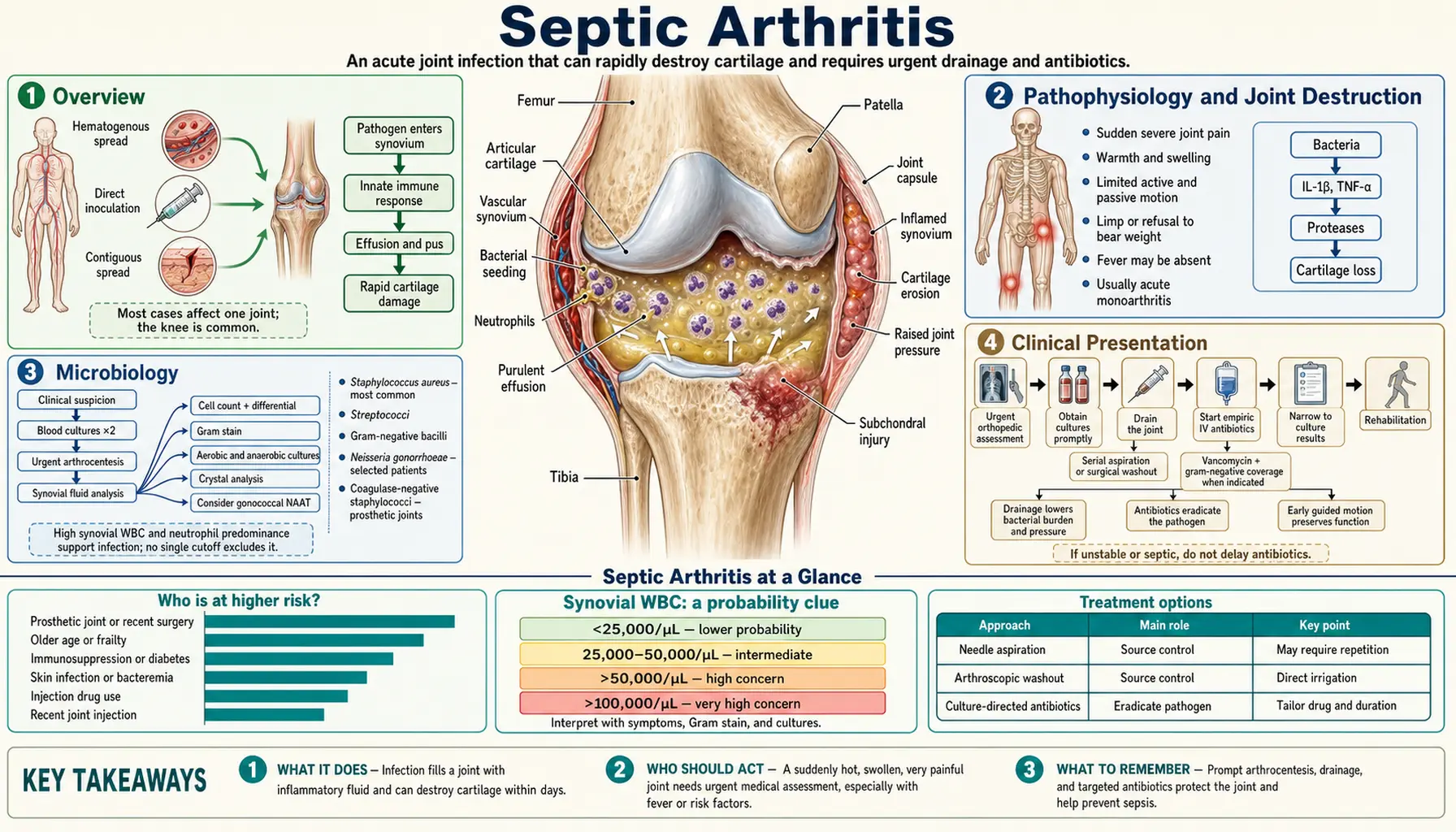
Table of Contents
1. Overview
Septic arthritis is a bacterial infection of the joint space — a true orthopedic emergency. Bacteria within a joint elicit a powerful inflammatory response that rapidly destroys articular cartilage through a combination of direct bacterial proteases, neutrophil-released enzymes, and elevated intra-articular pressure that cuts off synovial blood supply. Permanent cartilage loss can begin within 24–48 hours of infection onset, and irreversible joint damage can be established within 7–10 days. Speed of diagnosis and drainage is the single most important determinant of joint outcome.
Septic arthritis occurs at an estimated rate of 2–10 per 100,000 population per year in developed countries, with higher rates in patients over 65, in those with underlying joint disease (rheumatoid arthritis, osteoarthritis, crystal arthropathy), and in patients with conditions predisposing to bacteremia (intravenous drug use, diabetes, immunosuppression, intravenous catheters). Prosthetic joint infection, a related but distinct entity affecting implanted joint replacements, occurs in approximately 1–2% of knee and hip arthroplasties and is addressed by its own management framework.
The clinical principle that guides initial management is simple: any acute monoarthritis is treated as septic arthritis until proven otherwise. The consequences of missing septic arthritis — irreversible joint destruction, septicemia, death — are far worse than the consequences of over-treating gout or another non-infectious arthritis empirically while awaiting culture confirmation.
2. Pathophysiology and Joint Destruction
The synovial membrane is highly vascularized and lacks a basement membrane, making it uniquely accessible to bloodborne bacteria. Once bacteria enter the joint space, the following cascade unfolds:
- Bacterial adhesion and multiplication. S. aureus, the dominant pathogen, expresses multiple adhesins (fibronectin-binding protein A and B, collagen-binding adhesin, bone sialoprotein) that allow it to attach to synovial membrane and articular cartilage. Within hours, bacterial counts can reach 108–109 colony-forming units (CFU)/mL in infected synovial fluid.
- Neutrophil influx and enzymatic damage. Bacterial products activate complement and toll-like receptors, triggering a massive neutrophil recruitment into the joint space. Neutrophils release proteases (elastase, collagenase, cathepsins) and reactive oxygen species during phagocytosis. The enzymes digest the proteoglycan matrix of articular cartilage, which is not self-renewing — proteoglycan loss is irreversible within days of onset.
- Elevated intra-articular pressure. Purulent exudate accumulates under pressure in the closed joint compartment, compressing synovial vasculature and cutting off synovial fluid production. Cartilage depends on synovial fluid diffusion for nutrition; ischemia accelerates chondrocyte death. In the hip, elevated pressure can also compress the femoral head's blood supply, causing avascular necrosis — a catastrophic complication especially in children.
- Cytokine-mediated bone erosion. Interleukin-1 and tumor necrosis factor-α released by activated macrophages stimulate osteoclast-mediated bone erosion at the joint margins, producing juxta-articular osteopenia and marginal erosions visible on radiographs as early as 2–3 weeks into untreated infection.
- Biofilm and chronicity. If inadequately treated, bacteria on synovial surfaces and within articular cartilage fissures can transition to biofilm communities, making antibiotic eradication increasingly difficult and risking conversion to a chronic destructive arthritis.
3. Microbiology
The microbiology of septic arthritis is age- and risk-factor-dependent:
S. aureus — the dominant pathogen
Staphylococcus aureus is the most common cause of non-gonococcal septic arthritis across all age groups, responsible for approximately 40–50% of cases in adults. Methicillin-resistant Staphylococcus aureus (MRSA) causes 20–30% of S. aureus septic arthritis in high-risk populations (healthcare workers, people who inject drugs (intravenous drug use, IVDU), patients from long-term care facilities). S. aureus septic arthritis tends to be severe, with high rates of joint destruction and mortality.
Streptococcal species
Streptococcus pyogenes (Group A Strep), Streptococcus agalactiae (Group B, important in neonates and diabetics), and Streptococcus pneumoniae collectively account for 15–25% of non-gonococcal cases. Group B streptococcal septic arthritis in adults often occurs in patients with diabetes, cirrhosis, or malignancy.
Neisseria gonorrhoeae — in sexually active young adults
Disseminated gonococcal infection (DGI) is the most common cause of septic arthritis in sexually active adults under 40 in the United States. The clinical presentation of DGI has two phases:
- Bacteremic phase: Migratory polyarthralgia (moving from joint to joint), tenosynovitis (particularly of dorsal hand/wrist and Achilles tendon), and characteristic dermatitis (scattered painless necrotic pustules on the trunk and extremities, often fewer than 20 lesions). Blood cultures may be positive.
- Septic joint phase: Purulent monoarthritis develops, most commonly in the wrist, knee, or ankle. Cultures of urogenital, rectal, and pharyngeal sites are more sensitive than synovial fluid culture in DGI (synovial culture positive in only ~25% of cases due to fastidious growth requirements).
DGI responds rapidly to appropriate antibiotics (ceftriaxone 1 g intravenous (IV) or intramuscular every 24 hours), and joint outcome is generally favorable. The 2021 CDC treatment guidelines refer sex partners from the previous 60 days for evaluation, testing and presumptive treatment, noting that gonorrhea often causes no symptoms in the partners of people with DGI. Rising rates of cephalosporin-resistant gonorrhea are an emerging concern worldwide.
Gram-negative bacilli
Gram-negative septic arthritis occurs in neonates, elderly patients with urinary sources, IVDU (with axial joint involvement — sternoclavicular, sacroiliac), and immunocompromised patients. E. coli and Klebsiella are most common. Pseudomonas aeruginosa is specifically associated with IVDU and puncture wounds through footwear.
Kingella kingae in young children
Kingella kingae is a fastidious gram-negative coccobacillus that is the second most common cause of septic arthritis and osteomyelitis in children aged 6 months to 4 years. It colonizes the upper respiratory tract and enters the bloodstream via mucosal disruption during viral infections. Culture sensitivity is greatly improved by inoculating synovial fluid directly into blood culture bottles. Kingella infections tend to be less destructive than S. aureus and respond well to beta-lactam antibiotics.
Unusual pathogens in immunocompromised patients
Patients with human immunodeficiency virus (HIV), solid-organ transplant, or on biologic disease-modifying antirheumatic drugs (DMARDs) (tumor necrosis factor (TNF) inhibitors, JAK inhibitors) are susceptible to unusual organisms: Mycobacterium tuberculosis, atypical mycobacteria (M. marinum from aquatic exposure), Cryptococcus neoformans, Candida species, and Sporothrix schenckii. Synovial biopsy with special stains and cultures for acid-fast bacilli and fungi is the standard next step when standard cultures are negative and the host is immunocompromised.
4. Clinical Presentation
The classic presentation of non-gonococcal septic arthritis is acute onset monoarthritis with fever, pain, swelling, warmth, and severely limited range of motion in a single joint. Key features by joint:
Knee (most common, approximately 50% of cases)
Visible effusion, warmth, and exquisite pain with any attempted movement. The patient holds the knee in slight flexion (the position of maximum joint space). The inability to fully extend the knee against gravity is a helpful sign. The knee's large volume means effusions can become massive before causing the pressure complications seen in the hip.
Hip (second most common in adults; most serious in children)
The hip is a deeply situated joint, making physical examination challenging. The patient typically holds the hip in the position of comfort: flexion, abduction, and external rotation (a position that maximizes joint volume). Active and passive range of motion is severely limited and painful in all planes. The hip Patrick test (FABER — flexion, abduction, external rotation) reproduces pain. In children, hip septic arthritis is a surgical emergency due to the risk of avascular necrosis from pressure on the femoral head's blood supply; urgent ultrasound + joint aspiration is standard practice.
Shoulder, ankle, wrist
Each has characteristic effusion signs (subacromial fullness, ankle synovitis above the malleoli, dorsal wrist swelling). The shoulder is the most common site of septic arthritis in IVDU and in patients with rheumatoid arthritis. All sites present with the universal features of acute bacterial arthritis: warmth, swelling, extreme pain with movement, and erythema.
Atypical presentations
Elderly patients, patients on corticosteroids, and immunocompromised patients may present with minimal fever or only subtle joint findings. Patients on biologic DMARDs (which suppress cytokine production) may have blunted inflammatory signs even with established joint infection. In these patients clinicians keep a high index of suspicion and a low threshold for joint aspiration.
5. Diagnosis and Joint Aspiration
Joint aspiration is the standard first test whenever septic arthritis is suspected, and it is not delayed for imaging or laboratory results. Synovial fluid is sent for:
- Cell count with differential (most important test)
- Gram stain
- Culture (aerobic and anaerobic; fungal and mycobacterial cultures in appropriate hosts)
- Crystal analysis under polarized light microscopy (to assess for gout/pseudogout)
- Glucose and protein (less specific than cell count; not routinely required if WBC available)
Synovial fluid WBC count interpretation
The synovial white blood cell count is the most important diagnostic parameter:
- >100,000 cells/μL with >90% neutrophils: near-diagnostic for septic arthritis; usually treated empirically as infected pending cultures. Sensitivity ~28%, specificity ~99%.
- 50,000–100,000 cells/μL: Strongly suggestive of septic arthritis; usually treated empirically. Sensitivity ~62%, specificity ~92%.
- 20,000–50,000 cells/μL: Suspicious; requires integration with clinical context. Crystal arthropathy and inflammatory arthritis (rheumatoid arthritis (RA), reactive arthritis) can reach these levels. Treatment is usually not withheld when clinical suspicion is high.
- <20,000 cells/μL: Makes septic arthritis less likely but does not exclude it, especially in immunocompromised patients or partially treated infection.
A 2007 review of 14 studies (6,242 patients, 653 of them with septic arthritis) put numbers on this. A joint-fluid white cell count above 100,000 made infection about 28 times more likely (likelihood ratio 28.0), above 50,000 about 7.7 times, and 25,000 or more about 2.9 times, while a count below 25,000 lowered the odds (likelihood ratio 0.32). Neutrophils making up at least 90% of the cells raised the likelihood 3.4 times. Among people who did have septic arthritis, only joint pain (85%), a history of joint swelling (78%) and fever (57%) were present in more than half, so a normal temperature does not rule infection out.
Critically, crystal arthropathy and septic arthritis can coexist. Finding monosodium urate or calcium pyrophosphate crystals on polarized microscopy does not rule out concurrent bacterial infection. If Gram stain or culture suggests infection in a crystal-containing fluid, both are treated.
Gram stain and culture
Gram stain is positive in approximately 50% of non-gonococcal septic arthritis cases (higher for gram-positive cocci, lower for gram-negative rods). Culture is positive in 70–80% of non-gonococcal cases when obtained before antibiotics. Inoculating synovial fluid into blood culture bottles significantly improves yield for fastidious organisms including Kingella kingae and N. gonorrhoeae.
Blood cultures
Blood cultures are drawn before antibiotics in all patients as standard. They are positive in approximately 30–50% of hematogenous septic arthritis cases and may be the only positive culture in gonococcal bacteremic phase disease.
Imaging
Plain radiographs are obtained to assess for osteomyelitis, fracture, or pre-existing joint disease, but are normal early in septic arthritis. Ultrasound is the quickest modality for confirming joint effusion and guiding aspiration, especially in deep joints like the hip. Magnetic resonance imaging (MRI) with gadolinium can detect early synovitis, periarticular soft tissue involvement, and concurrent osteomyelitis — important when the clinical picture is complex or when adjacent bone infection is suspected.
6. Differential Diagnosis
The differential for acute monoarthritis is critically important because several conditions mimic septic arthritis:
- Crystal arthropathy: Gout (monosodium urate) and pseudogout (calcium pyrophosphate) are the most common mimics. Both can cause fever, elevated WBC, and markedly elevated synovial fluid cell counts. Definitive distinction requires crystal analysis. Key point: crystals do not exclude infection — they can coexist.
- Reactive arthritis (formerly Reiter syndrome): Sterile oligoarthritis triggered by a preceding infection (urogenital Chlamydia, enteric Salmonella, Shigella, Campylobacter). Typically occurs 1–4 weeks after the triggering infection. Synovial WBC typically 10,000–50,000 cells/μL; cultures negative.
- Hemarthrosis: Bloody joint fluid from trauma, anticoagulation, or hemophilia. WBC usually low with predominantly red cells; no organisms. Coagulation studies and trauma history guide the diagnosis.
- Lyme arthritis: In endemic areas, Borrelia burgdorferi causes a monoarthritis typically involving the knee. Synovial WBC can exceed 50,000 cells/μL. Diagnosis by serology (ELISA + Western blot). Important because it requires oral doxycycline, not surgical drainage.
- Inflammatory arthritis: Rheumatoid arthritis, psoriatic arthritis, and spondyloarthropathy can all present with acute flares mimicking infection. Characteristic extra-articular features, prior disease history, negative cultures, and appropriate serology guide the workup. Risk: patients with rheumatoid arthritis on immunosuppressive therapy can develop septic arthritis that partially mimics a disease flare.
7. Antibiotic Treatment
Antibiotics are started promptly after joint aspiration and blood cultures are obtained — ideally within the same clinical encounter. Standard practice is not to delay antibiotics while waiting for culture results when the clinical picture strongly suggests infection.
Empiric regimens
- Non-gonococcal, methicillin-susceptible Staphylococcus aureus (MSSA) risk (Gram stain shows gram-positive cocci or no organism identified, low methicillin-resistant Staphylococcus aureus risk): Nafcillin or oxacillin 2g intravenous q4h. Superior to vancomycin for confirmed MSSA based on outcomes data (lower mortality, faster bacteremia clearance).
- Non-gonococcal, methicillin-resistant Staphylococcus aureus risk (IVDU, healthcare-associated, prior methicillin-resistant Staphylococcus aureus colonization, severe illness): Vancomycin intravenous dosed by area-under-the-curve to minimum-inhibitory-concentration (AUC/MIC) monitoring (target AUC 400–600 mg·h/L). Alternative: daptomycin 6mg/kg/day intravenous.
- Gram-negative coverage required (neonates, elderly with urinary source, IVDU, immunocompromised): Ceftriaxone 2g intravenous daily (most gram-negatives including many enteric organisms) or cefepime 2g intravenous q8h if Pseudomonas risk. Ciprofloxacin has excellent oral bioavailability and bone/joint penetration for step-down therapy.
- Gonococcal arthritis: The 2021 CDC sexually transmitted infection treatment guidelines list ceftriaxone 1 g intramuscular or intravenous every 24 hours. Once there is substantial clinical improvement (24–48 hours after it), treatment can switch to an oral drug chosen by antimicrobial susceptibility testing, for a total course of at least 7 days. When chlamydia (Chlamydia trachomatis) has not been ruled out, the same guidelines add doxycycline 100 mg by mouth twice a day for 7 days.
- Children (<3 months): Antistaphylococcal beta-lactam + gram-negative coverage (e.g. nafcillin + gentamicin or cefotaxime).
Duration of therapy
Standard duration for non-gonococcal septic arthritis is 2–4 weeks, though practice varies. Vertebral and hip involvement may warrant longer courses.
- Two weeks vs four weeks: In a 2019 randomised trial of 154 adults whose infected native joint had been washed out surgically, 2 weeks of antibiotics was not inferior to 4 weeks: infection came back in 1 person in the 2-week group and 2 in the 4-week group, with no difference in side effects or lasting joint problems, and a shorter hospital stay. Most cases (99 of 154) were in the hand and wrist.
- Tablets vs drip (OVIVA trial): In the OVIVA trial (1,054 adults with bone or joint infection at 26 UK centres), antibiotics by mouth for the first 6 weeks were not inferior to intravenous antibiotics. Treatment failed within a year in 13.2% on tablets vs 14.6% on the drip, and drip-line (catheter) complications were more common with the drip (9.4% vs 1.0%).
- Children: In 2023 the Pediatric Infectious Diseases Society (PIDS) and the Infectious Diseases Society of America (IDSA) published a joint clinical practice guideline on the diagnosis and management of acute bacterial arthritis in children.
8. Joint Drainage
Antibiotics alone are insufficient — drainage of the infected joint is a fundamental component of treatment. Purulent joint fluid is evacuated to reduce intra-articular pressure, remove proteolytic enzymes, and reduce the bacterial load.
Needle aspiration
Repeated closed needle aspiration, performed daily or every other day until effusion resolves and inflammatory markers trend down, is used for most accessible joints (knee, wrist, ankle). It is less practical for deep joints (hip, shoulder) and when loculation prevents complete drainage.
Arthroscopic drainage and lavage
Arthroscopic irrigation and debridement is the preferred surgical option for most joints when:
- Needle aspiration fails (fluid re-accumulates rapidly, effusion remains thick/loculated).
- The joint is deep and poorly accessible (hip, shoulder).
- There is concurrent osteomyelitis or significant synovial debris requiring mechanical debridement.
- Clinical response is inadequate after 48–72 hours of antibiotics and aspiration.
Arthroscopy allows direct visualization of cartilage damage, thorough lavage of all joint recesses, and synovial biopsy if diagnosis is uncertain. In experienced hands it achieves superior joint clearance compared to needle aspiration alone.
Open surgical drainage
Open arthrotomy (surgical opening of the joint) is specifically indicated for the hip joint in children (to urgently decompress pressure threatening femoral head perfusion), for joints where arthroscopy is not available or technically difficult (sternoclavicular, sacroiliac), and when arthroscopic approaches have failed or there is extensive bone involvement requiring open debridement.
Post-drainage mobilization
Early mobilization and physical therapy after initial drainage are regarded as important for preserving joint function. Immobilization promotes cartilage adhesion and joint stiffness. Range-of-motion exercises usually begin as soon as pain permits, typically within 24–48 hours of initial drainage in a well-controlled infection.
9. Outcomes and Complications
The prognosis of septic arthritis depends on the causative organism, the joint involved, time to diagnosis and drainage, and the patient's baseline health:
- Cartilage destruction and chronic arthritis: Approximately 25–50% of patients with septic arthritis develop some degree of permanent joint damage. Risk factors include delay to drainage (>5 days), S. aureus infection, older age, pre-existing joint disease (especially rheumatoid arthritis), and hip or shoulder involvement.
- Avascular necrosis of the femoral head: The most feared complication of hip septic arthritis in children. Elevated intra-articular pressure cuts off blood supply to the femoral head, causing ischemic necrosis. Urgent surgical decompression (<24 hours of symptom onset when possible) is the standard approach aimed at preventing this outcome.
- Mortality: Septic arthritis still carries a mortality rate of 7–15% in adults, predominantly due to septicemia and multi-organ failure in elderly and immunocompromised patients. S. aureus bacteremia carries particularly high mortality risk (endocarditis, metastatic infection).
- Extension to bone (osteomyelitis): Adjacent bone can become secondarily infected, particularly in infants and at the hip where the metaphysis is intra-articular. This significantly complicates treatment and worsens prognosis.
- Gonococcal arthritis prognosis: Generally excellent with prompt treatment. Permanent joint damage is uncommon compared to non-gonococcal septic arthritis, reflecting the less destructive nature of N. gonorrhoeae in the joint space.
Key Research Papers
- Mathews CJ, Weston VC, Jones A, Field M, Coakley G. Bacterial septic arthritis in adults. Lancet. 2010;375(9717):846–855. — Review of the changing epidemiology, risk factors, pathogenesis and management of native-joint septic arthritis in adults. — PubMed PMID: 20206778
- Shirtliff ME, Mader JT. Acute septic arthritis. Clin Microbiol Rev. 2002;15(4):527–544. — Pathophysiology of acute septic arthritis: host-pathogen interaction, diagnosis, and the rationale for prompt antibiotics and drainage. — PubMed PMID: 12364368
- Margaretten ME, Kohlwes J, Moore D, Bent S. Does this adult patient have septic arthritis? JAMA. 2007;297(13):1478–1488. — Systematic review: likelihood ratios for synovial white blood cell (WBC) count, fever, and risk factors. — PubMed PMID: 17405973
- Goldenberg DL. Septic arthritis. Lancet. 1998;351(9097):197–202. — Septic arthritis review: clinical features, diagnosis, management, and outcomes. — PubMed PMID: 9449882
- Kaandorp CJ, Krijnen P, Moens HJ, Habbema JD, van Schaardenburg D. The outcome of bacterial arthritis: a prospective community-based study. Arthritis Rheum. 1997;40(5):884–892. — 154 patients followed a mean of 2 years: joint outcome poor in 33% of survivors; older age, pre-existing joint disease and synthetic joint material predicted a poor outcome, while treatment delay did not. — PubMed PMID: 9153550
- Li HK, Rombach I, Zambellas R, Walker AS, McNally MA, Atkins BL, Lipsky BA, et al.; OVIVA Trial Collaborators. Oral versus Intravenous Antibiotics for Bone and Joint Infection. N Engl J Med. 2019;380(5):425–436. — OVIVA trial: oral antibiotics non-inferior to IV for bone and joint infections at 1 year; supports early oral step-down. — PubMed PMID: 30699315
- Gjika E, Beaulieu JY, Vakalopoulos K, Gauthier M, Bouvet C, Gonzalez A, Morello V, Steiger C, Hirsiger S, Lipsky BA, Uçkay I. Two weeks versus four weeks of antibiotic therapy after surgical drainage for native joint bacterial arthritis: a prospective, randomised, non-inferiority trial. Ann Rheum Dis. 2019;78(8):1114–1121. — 154 adults: 2 weeks not inferior to 4 weeks after surgical lavage. — PubMed PMID: 30992295
- Kocher MS, Zurakowski D, Kasser JR. Differentiating between septic arthritis and transient synovitis of the hip in children: an evidence-based clinical prediction algorithm. J Bone Joint Surg Am. 1999;81(12):1662–1670. — The Kocher criteria: history of fever, non-weight-bearing, erythrocyte sedimentation rate (ESR) of at least 40 mm/hr, and serum WBC above 12,000/mm³. — PubMed PMID: 10608376
- Woods CR, Bradley JS, Chatterjee A, Kronman MP, Arnold SR, Robinson J, Copley LA, Arrieta AC, Fowler SL, Harrison C, Eppes SC, Creech CB, Stadler LP, Shah SS, Mazur LJ, Carrillo-Marquez MA, Allen CH, Lavergne V. Clinical Practice Guideline by the Pediatric Infectious Diseases Society (PIDS) and the Infectious Diseases Society of America (IDSA): 2023 Guideline on Diagnosis and Management of Acute Bacterial Arthritis in Pediatrics. J Pediatric Infect Dis Soc. 2024;13(1):1–59. — PubMed PMID: 37941444
PubMed Topic Searches
The links below run live searches on PubMed, the U.S. National Library of Medicine's database of biomedical literature.
- PubMed: Septic arthritis treatment outcomes
- PubMed: Septic arthritis synovial fluid diagnosis
- PubMed: S. aureus MRSA septic arthritis
- PubMed: Gonococcal arthritis DGI
- PubMed: Hip septic arthritis children avascular necrosis
- PubMed: Septic arthritis arthroscopic drainage
- PubMed: Septic arthritis vs gout differential diagnosis
- PubMed: Kingella kingae pediatric joint infection
- PubMed: Septic arthritis rheumatoid arthritis biologics
- PubMed: Septic arthritis cartilage destruction pathogenesis
- PubMed: Reactive arthritis post-infectious monoarthritis
- PubMed: Prosthetic joint infection septic arthritis
Connections
- Orthopedics
- Osteomyelitis — septic arthritis and osteomyelitis frequently coexist; infection can spread from joint to adjacent bone or from hematogenous seeding of both simultaneously.
- Gout — the most common mimic of septic arthritis; both cause acute monoarthritis with elevated synovial WBC; can coexist; differentiation by crystal analysis is critical.
- Rheumatoid Arthritis — rheumatoid arthritis patients on DMARDs and biologics have increased susceptibility to septic arthritis with blunted inflammatory signs; a flare may mask underlying infection.
- Osteoarthritis — pre-existing OA and joint arthroplasty create surfaces susceptible to hematogenous bacterial seeding.
- Spinal Stenosis — distinguishing degenerative spinal disease from infectious spondylodiscitis is a critical clinical challenge in back-pain evaluation.
- Diabetes — diabetes impairs neutrophil function and vascular supply, increasing susceptibility to both septic arthritis and osteomyelitis.
- All Conditions — complete disease index.
- Arthritis — the umbrella overview of joint inflammation, including the non-infectious forms that must be distinguished from a septic joint.