H. pylori (Helicobacter pylori)
- Overview
- Epidemiology
- Pathophysiology
- Diseases Caused
- Diagnosis
- Treatment (Eradication)
- Eradication and Cancer Prevention
- Key Research Papers
- Connections
Interactive Visualization Stomach Acid & proton pump inhibitors (PPIs) — run the proton pump Drive your stomach to pH 2, activate pepsin and free your B12 and iron — then take a proton pump inhibitor (PPI) and watch the pH climb while the nutrients stop being released. Launch →
Overview
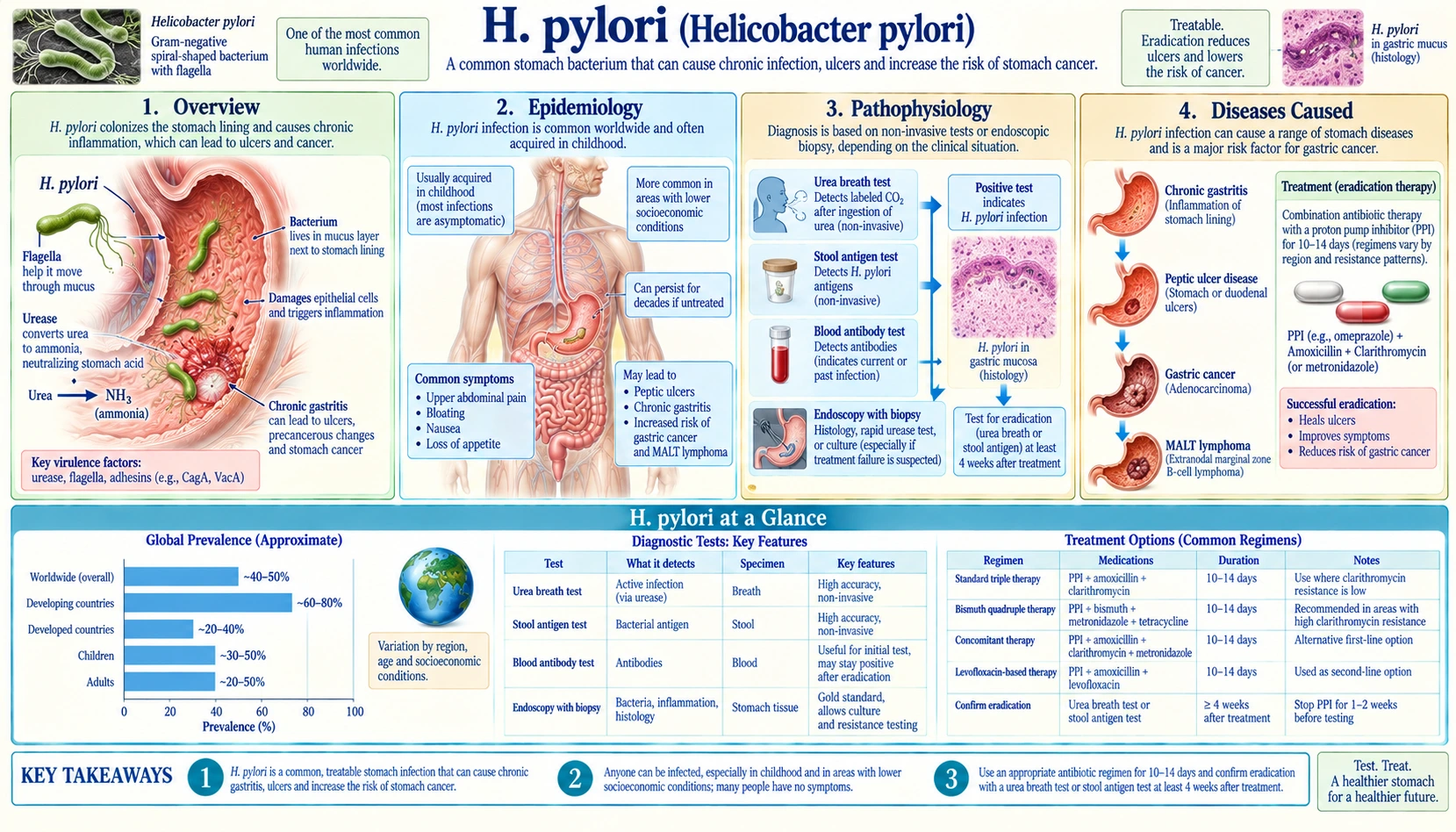
Helicobacter pylori (H. pylori) is a gram-negative, spiral-shaped bacterium that colonizes the gastric mucosa of the human stomach. It is the most common chronic bacterial infection worldwide, affecting approximately 50% of the global population — and substantially more in developing nations.
The bacterium preferentially colonizes the gastric antrum, the lower portion of the stomach closest to the pylorus. Its remarkable ability to survive in the acidic gastric environment depends on a key virulence tool: the enzyme urease. Urease splits urea into ammonia and carbon dioxide; the ammonia neutralizes stomach acid immediately surrounding the bacterium, creating a micro-environment where H. pylori can thrive.
Two additional virulence factors define its pathogenic power:
- CagA (cytotoxin-associated gene A) — present in roughly 60–70% of Western strains and nearly all East Asian strains. CagA is injected directly into gastric epithelial cells via a type IV secretion system (essentially a molecular syringe). Inside the cell, CagA disrupts tight junctions, disturbs cell polarity, and activates oncogenic signaling cascades — contributing directly to cancer risk.
- VacA (vacuolating cytotoxin A) — a pore-forming toxin that inserts into cell membranes, induces vacuolation of the cytoplasm, damages mitochondria, and triggers apoptosis (programmed cell death). VacA also suppresses local immune responses, helping the bacterium persist for decades.
H. pylori was discovered in 1982 by Barry Marshall and Robin Warren, who were awarded the Nobel Prize in Physiology or Medicine in 2005. Their discovery overturned the prevailing belief that ulcers were caused by stress and acid alone, revealing instead that a curable bacterial infection was responsible for the majority of peptic ulcers worldwide.
Epidemiology
H. pylori infects an estimated 4.4 billion people globally — roughly half of all living humans. Infection rates vary dramatically by geography and socioeconomic conditions:
- Developing countries: prevalence ranges from 70–80%, reflecting overcrowded living conditions, limited access to clean water, and inadequate sanitation.
- Developed countries: prevalence of 30–40%, with rates declining in younger generations due to improved hygiene and antibiotic use.
- Highest prevalence regions: Sub-Saharan Africa (>70%), South America, Southeast Asia, and Eastern Europe.
- Lowest prevalence regions: Western Europe, Australia, Canada, and the United States (where it has fallen to roughly 30–35%).
Transmission occurs primarily via two routes:
- Fecal-oral: contaminated water or food, particularly in areas without reliable sewage treatment.
- Oral-oral: shared utensils, kissing, and possibly through saliva — explaining intrafamilial clustering.
Infection is acquired predominantly in childhood, often before age 5. Once established, it persists for life without treatment. Spontaneous clearance is rare.
The public health stakes are enormous. H. pylori is classified as a Group 1 carcinogen by the International Agency for Research on Cancer (IARC). H. pylori-related gastric cancer kills approximately 700,000 people per year worldwide, making it one of the most deadly infectious causes of cancer. Gastric cancer remains the fifth most common cancer and fourth leading cause of cancer death globally.
Pathophysiology
H. pylori's persistence and pathogenicity stem from a sophisticated series of interactions with the gastric mucosa that, over years to decades, drive a predictable cascade of tissue damage:
Initial Colonization
After ingestion, H. pylori uses its flagella to swim through the gastric mucus layer toward the epithelial surface. Urease production begins immediately — ammonia neutralizes local pH, making the bacterium's immediate micro-environment survivable. Adhesins on the bacterial surface bind to specific receptors on gastric epithelial cells, securing attachment.
CagA Injection and Oncogenic Signaling
CagA-positive strains (cagPAI — cag pathogenicity island) assemble a type IV secretion system that functions like a molecular needle, injecting the CagA protein directly into epithelial cells. Once inside:
- CagA is phosphorylated by Src kinases, then activates the SHP-2 phosphatase (Src homology 2 domain-containing phosphatase 2) — a known proto-oncogene.
- It disrupts the E-cadherin/beta-catenin complex, impairing epithelial tight junctions and cell polarity.
- It activates nuclear factor kappa B (NF-κB), driving inflammatory cytokine production (interleukin-8 (IL-8) in particular), which recruits neutrophils and macrophages.
- Persistent CagA signaling mimics growth-factor stimulation, promoting aberrant proliferation.
VacA-Mediated Cell Injury
VacA is secreted by virtually all H. pylori strains, though toxin activity varies by allele. VacA inserts into mitochondrial and plasma membranes, forming anion-selective channels. The downstream effects include:
- Large vacuole formation in the cytoplasm (the hallmark the toxin is named for).
- Mitochondrial fragmentation and dysfunction, releasing cytochrome c and triggering apoptosis.
- Inhibition of T-cell proliferation and interleukin-2 (IL-2) secretion — local immune evasion.
The Correa Cascade (Gastric Carcinogenesis)
Decades of chronic active gastritis, driven by CagA, VacA, and the persistent inflammatory response, set in motion a stepwise progression described by Pelayo Correa:
- Normal gastric mucosa
- Chronic non-atrophic gastritis
- Atrophic gastritis (loss of parietal cells and acid-producing glands)
- Intestinal metaplasia (gastric epithelium replaced by intestinal-type cells)
- Dysplasia
- Gastric adenocarcinoma
This progression unfolds over 20–40 years. Eradication early in the cascade (before intestinal metaplasia) dramatically reduces cancer risk; eradication after intestinal metaplasia has developed still reduces risk but does not fully reverse it.
Diseases Caused
H. pylori infection causes or strongly contributes to several distinct gastrointestinal diseases:
Peptic Ulcer Disease
H. pylori is responsible for approximately 95% of duodenal ulcers and 70% of gastric ulcers in the absence of nonsteroidal anti-inflammatory drug (NSAID) use. The bacterium disrupts the balance between acid secretion and mucosal defense: in the duodenum, H. pylori-driven antral gastritis increases gastrin production → increased acid output → acid overwhelms duodenal defenses. In the stomach, direct mucosal injury and impaired prostaglandin synthesis reduce the mucus barrier. Eradication of H. pylori heals ulcers and, critically, prevents recurrence — transforming peptic ulcer disease from a chronic relapsing condition into a curable one.
Gastric Adenocarcinoma
H. pylori is the single most important risk factor for gastric cancer, accounting for roughly 75–89% of all non-cardia gastric adenocarcinomas. International Agency for Research on Cancer classifies H. pylori as a Group 1 carcinogen. The estimated lifetime risk of developing gastric cancer in an infected individual is approximately 1–3% in Western populations and up to 6% in high-incidence regions (East Asia). Gastric cancer remains a leading cancer killer globally, killing approximately 769,000 people per year.
MALT Lymphoma
Mucosa-associated lymphoid tissue (MALT) lymphoma of the stomach is directly driven by chronic H. pylori antigen stimulation of gastric B cells. In approximately 75% of low-grade cases, antibiotic eradication of H. pylori alone — without chemotherapy or radiation — causes complete lymphoma remission. This represents one of the most dramatic examples of infectious disease treatment curing a cancer. High-grade (large B-cell) gastric lymphomas also benefit from eradication as part of combined treatment.
Functional Dyspepsia
H. pylori infection is present in roughly 50% of patients with functional dyspepsia (chronic upper abdominal pain or discomfort without organic cause on endoscopy). A modest but real proportion of these patients — roughly 1 in 10 — experience long-term symptom relief with eradication. Current guidelines therefore recommend a test-and-treat strategy for uninvestigated dyspepsia under age 60 without alarm features.
Protective Associations (the H. pylori Paradox)
In an intriguing counterpoint, H. pylori infection is inversely associated with:
- Gastroesophageal reflux disease (GERD): H. pylori, particularly CagA-positive strains, appears to suppress acid output in the esophageal-junction region. Eradication can unmask or worsen gastroesophageal reflux disease in some patients — a clinical consideration before treating patients who have both H. pylori and mild reflux.
- Esophageal adenocarcinoma: paralleling the gastroesophageal reflux disease relationship, populations with lower H. pylori prevalence have higher rates of esophageal adenocarcinoma.
- Asthma and allergies: consistent with the hygiene hypothesis, early H. pylori colonization may modulate immune development in ways that reduce atopic disease risk.
Diagnosis
The appropriate diagnostic approach depends on clinical context — specifically whether the patient needs endoscopy for other reasons (alarm features) or can be managed non-invasively.
Test-and-Treat Strategy
For patients under age 60 with uninvestigated dyspepsia and no alarm features, current ACG, AGA, and ACG-CAG guidelines recommend a test-and-treat strategy: non-invasively test for H. pylori, treat if positive, and reserve endoscopy for those who don't improve. This approach is cost-effective and avoids unnecessary procedures.
Non-Invasive Tests
Urea Breath Test (UBT) — Gold Standard Non-Invasive Test
The patient swallows a capsule or solution containing 13C-labeled urea. If H. pylori is present, its urease enzyme splits the labeled urea, releasing 13CO2 that is absorbed into the bloodstream and exhaled. Breath is collected and analyzed for the isotopic signature. Sensitivity and specificity both exceed 95%. Key requirements:
- Proton pump inhibitors are stopped at least 2 weeks before testing (proton pump inhibitors suppress H. pylori activity and cause false negatives).
- Antibiotics and bismuth are stopped at least 4 weeks before testing.
Stool Antigen Test (H. pylori stool antigen, HpSA)
Detects H. pylori antigens in a stool sample. Monoclonal antibody-based assays are preferred (sensitivity and specificity >95%). Inexpensive and widely available. Subject to the same proton pump inhibitor/antibiotic restriction as UBT. Useful for both initial diagnosis and confirming eradication.
Serology (immunoglobulin G (IgG) antibody test)
Detects immunoglobulin G antibodies against H. pylori in blood. Widely available and inexpensive, but with a critical limitation: antibodies persist for months to years after successful eradication. Serology cannot distinguish active from past infection and therefore cannot be used to confirm eradication. Its use is increasingly discouraged for clinical decision-making.
Invasive Tests (Endoscopy + Biopsy)
When endoscopy is performed for clinical indications, several H. pylori tests can be performed on biopsy specimens:
- Rapid urease test (on a biopsy): biopsy placed in urea-containing medium; color change indicates urease activity. Results within minutes to hours. Sensitivity ~90–95%.
- Histology: hematoxylin and eosin (H&E) or Giemsa staining of antral/body biopsies identifies the organism and grades gastritis. Allows simultaneous assessment of intestinal metaplasia and dysplasia.
- Culture: allows antibiotic susceptibility testing — valuable when standard regimens have failed. Technically demanding and not widely available.
Alarm Features Mandating Endoscopy
Guidelines list the following features as reasons for upper endoscopy rather than empiric test-and-treat:
- Age >60 years (new dyspepsia)
- Unintentional weight loss
- Dysphagia or odynophagia
- Gastrointestinal bleeding (hematemesis, melena, hematochezia)
- Iron deficiency anemia
- Palpable abdominal mass
- Family history of gastric cancer
- Prior gastric surgery
Treatment (Eradication)
Antibiotic resistance has fundamentally changed H. pylori treatment. Clarithromycin resistance now exceeds 15–20% in many parts of the United States, Europe, and East Asia, and metronidazole resistance is even higher globally. Standard triple therapy (proton pump inhibitor + clarithromycin + amoxicillin × 7–10 days) that once achieved >90% cure rates now frequently fails below 80% in high-resistance regions. Current guidelines emphasize resistance-aware regimen selection and 14-day treatment duration.
The 2024 American College of Gastroenterology (ACG) guideline names 14 days of bismuth quadruple therapy as the preferred first treatment when the germ's antibiotic sensitivity is unknown, with 14 days of rifabutin triple therapy or vonoprazan–amoxicillin dual therapy as suitable alternatives for people without a penicillin allergy. It states that regimens containing clarithromycin or levofloxacin for people already treated should only be used when antibiotic sensitivity is confirmed, and it calls for a test of cure after treatment in everyone treated.
Bismuth Quadruple Therapy — Preferred First-Line (Most Regions)
The most reliable regimen across diverse resistance landscapes:
- Proton pump inhibitor (standard or double dose) twice daily
- Bismuth subcitrate or subsalicylate 120–300 mg four times daily
- Metronidazole 500 mg three to four times daily
- Tetracycline 500 mg four times daily
- Duration: 14 days
Bismuth quadruple therapy achieves eradication rates of approximately 85–90% even in areas with high clarithromycin and metronidazole resistance. It is also available as a three-in-one capsule of bismuth subcitrate potassium, metronidazole and tetracycline. Side effects include dark stools and tongue (harmless bismuth discoloration), nausea, and diarrhea.
Standard Triple Therapy
- Proton pump inhibitor twice daily + clarithromycin 500 mg twice daily + amoxicillin 1 g twice daily × 14 days
Older guidelines accepted it as first-line only in regions where clarithromycin resistance was confirmed to be below 15% and the person had not previously received a macrolide antibiotic; the 2016 Toronto Consensus restricted it to areas with known low clarithromycin resistance or high cure rates with it. The 2024 ACG guideline does not name it among the empiric first-line choices when antibiotic sensitivity is unknown (bismuth quadruple, rifabutin triple or vonoprazan dual therapy). Eradication rates with 14-day triple therapy are approximately 70–85% even under favorable conditions.
Concomitant (Non-Bismuth Quadruple) Therapy
- Proton pump inhibitor + clarithromycin 500 mg + amoxicillin 1 g + metronidazole 500 mg — all twice daily × 14 days
Recommended in areas with dual clarithromycin and metronidazole resistance, or where bismuth is unavailable. Eradication rates of approximately 85–90% in appropriate populations.
Vonoprazan-Based Therapy — U.S. Food and Drug Administration-Approved 2022
Vonoprazan is a potassium-competitive acid blocker (P-CAB) — a new class of acid suppressant that provides faster, more complete, and more durable acid suppression than conventional proton pump inhibitors regardless of CYP2C19 (cytochrome P450 2C19 liver enzyme) metabolism. U.S. Food and Drug Administration-approved regimens:
- Dual therapy: vonoprazan 20 mg twice daily + amoxicillin 1 g three times daily × 14 days (eradication 78.5% in strains without clarithromycin or amoxicillin resistance and 77.2% in all patients in the 2022 phase 3 trial)
- Triple therapy: vonoprazan 20 mg + amoxicillin 1 g + clarithromycin 500 mg — all twice daily × 14 days (eradication 84.7% in non-resistant strains, 80.8% in all patients and 65.8% in clarithromycin-resistant strains in the same trial)
The 2022 US and European phase 3 trial randomized 1,046 previously untreated adults. Overall, vonoprazan triple therapy cured 80.8% and vonoprazan dual therapy 77.2%, versus 68.5% for standard lansoprazole triple therapy. Against clarithromycin-resistant germs the figures were 65.8% and 69.6%, versus 31.9%. Side effects were about as frequent with vonoprazan as with lansoprazole.
Vonoprazan-based regimens consistently outperform proton pump inhibitor-based triple therapy in head-to-head trials and are particularly valuable in CYP2C19 rapid metabolizers (who clear proton pump inhibitors quickly) and in amoxicillin-susceptible strains.
Salvage (Second-Line) and Third-Line Therapy
When first-line therapy fails, culture and susceptibility testing can guide the choice of regimen. The 2024 ACG guideline prefers 14 days of "optimized" bismuth quadruple therapy for people with persistent infection who have not had it before, when susceptibility is unknown, and rifabutin triple therapy for 14 days as an alternative for those already treated with optimized bismuth quadruple therapy. Other options:
- Levofloxacin-based triple therapy: proton pump inhibitor + levofloxacin 500 mg + amoxicillin 1 g, all twice daily × 14 days — the 2024 ACG guideline states that salvage regimens containing levofloxacin or clarithromycin should only be used when antibiotic sensitivity is confirmed.
- Rifabutin-based triple therapy: proton pump inhibitor + rifabutin 150 mg + amoxicillin 1 g, twice daily × 14 days — the 2016 Toronto Consensus restricted rifabutin regimens to people who had failed at least 3 prior options, while the 2024 ACG guideline lists rifabutin triple therapy as a suitable empiric first-line alternative for people without penicillin allergy. Risk of myelotoxicity (bone-marrow suppression) has limited its use.
Confirming Eradication
Guidelines call for confirming eradication in everyone treated (the 2024 ACG guideline calls for a universal test of cure) — treatment failure is common and consequences of unrecognized failure (recurrent ulcers, ongoing cancer risk) are serious. Confirmation uses a UBT or monoclonal stool antigen test:
- At least 4 weeks after completing antibiotics
- Off proton pump inhibitors for at least 2 weeks before testing
Eradication and Cancer Prevention
One of the most consequential findings in modern gastroenterology is that eradicating H. pylori reduces the subsequent risk of gastric cancer. The evidence is now robust:
- Meta-analyses of randomized controlled trials demonstrate a 40–50% reduction in gastric cancer incidence among infected individuals who receive eradication therapy compared to those who do not.
- The benefit is greatest when eradication occurs before intestinal metaplasia develops — once the mucosa has undergone intestinal metaplasia, cancer risk is reduced but not eliminated by eradication.
- A Japanese randomized trial (Lancet, 2008) of 544 people who had an early stomach cancer removed through the endoscope found that, at 3 years, a new (metachronous) stomach cancer had developed in 9 people given eradication treatment versus 24 controls; in the analysis of the 255 versus 250 people with follow-up, the hazard ratio was 0.339 (about two-thirds lower).
- An earlier Japanese study (N Engl J Med, 2001) followed 1,526 people for a mean of 7.8 years: stomach cancer developed in 36 (2.9%) of the 1,246 infected people and in none of the 280 uninfected.
Population-Level Eradication Programs
Japan and Taiwan have run large-scale H. pylori eradication programs:
- Taiwan's mass screening and eradication program for adults aged 30 and over on the Matsu Islands (2004–2018) reached 85.5% of 7,616 residents with a breath test and treated those who tested positive. H. pylori prevalence fell from 64.2% to 15.0%, with reinfection under 1% per person-year, and atrophic gastritis and intestinal metaplasia became less common and less severe over time. Compared with 1995–2003, gastric cancer incidence fell by 53%; deaths fell by 25%, which was not yet statistically significant. Other digestive-tract cancers and the antibiotic-resistance rate did not change significantly.
- Japan's expansion of eradication therapy coverage under national health insurance for all H. pylori-positive gastritis patients (2013) has already reduced endoscopy-detected gastric cancer rates in subsequent years.
World Health Organization Recommendations
The World Health Organization and International Agency for Research on Cancer now recommend screen-and-treat strategies in intermediate- and high-incidence populations (defined as gastric cancer incidence >20 per 100,000 per year). In lower-incidence settings (most of Western Europe, North America), targeted testing of at-risk groups — first-generation immigrants from high-incidence countries, individuals with family history of gastric cancer — is emphasized alongside the standard test-and-treat approach for dyspepsia.
MALT Lymphoma Remission
For the specific case of low-grade gastric MALT lymphoma, H. pylori eradication is first-line treatment — not just an adjunct. Approximately 75% of localized low-grade gastric MALT lymphomas achieve complete remission with antibiotic eradication alone, without any need for radiation or chemotherapy. This remains one of the most compelling examples in oncology of treating an infection to cure a cancer.
Key Research Papers
- Malfertheiner P et al. Management of Helicobacter pylori infection — Maastricht IV/Florence Consensus Report. Gut. 2012;61(5):646–664. — PubMed PMID: 22491499
- Hooi JKY et al. Global Prevalence of Helicobacter pylori Infection: Systematic Review and Meta-Analysis. Gastroenterology. 2017;153(2):420–429. — PubMed PMID: 28456631
- Graham DY. Helicobacter pylori update: gastric cancer, reliable therapy, and possible benefits. Gastroenterology. 2015;148(4):719–731.e3. — PubMed PMID: 25655557
- Peek RM Jr, Blaser MJ. Helicobacter pylori and gastrointestinal tract adenocarcinomas. Nat Rev Cancer. 2002;2(1):28–37. — PubMed PMID: 11902583
- Chey WD et al. ACG Clinical Guideline: Treatment of Helicobacter pylori infection. Am J Gastroenterol. 2017;112(2):212–239. — PubMed PMID: 28071659
- Crowe SE. Helicobacter pylori infection. N Engl J Med. 2019;380(12):1158–1165. — PubMed PMID: 30893536
- Sugano K et al. Kyoto global consensus report on Helicobacter pylori gastritis. Gut. 2015;64(9):1353–1367. — PubMed PMID: 26187502
- Ford AC et al. Eradication therapy for peptic ulcer disease in Helicobacter pylori-positive people. Cochrane Database Syst Rev. 2016;4:CD003840. — PubMed PMID: 27092708
- Wroblewski LE, Peek RM Jr, Wilson KT. Helicobacter pylori and gastric cancer: factors that modulate disease risk. Clin Microbiol Rev. 2010;23(4):713–739. — PubMed PMID: 20930071
- Fallone CA et al. The Toronto Consensus for the Treatment of Helicobacter pylori Infection in Adults. Gastroenterology. 2016;151(1):51–69.e14. — PubMed PMID: 27102658
- Graham DY, Liou JM. Primer for Development of Guidelines for Helicobacter pylori Therapy Using Antimicrobial Stewardship. Clin Gastroenterol Hepatol. 2022;20(5):973–983.e1. — PubMed PMID: 33775895
- Chey WD, Mégraud F, Laine L, et al. Vonoprazan Triple and Dual Therapy for Helicobacter pylori Infection in the United States and Europe: Randomized Clinical Trial. Gastroenterology. 2022;163(3):608–619. — PubMed PMID: 35679950
- Chey WD, Howden CW, Moss SF, et al. ACG Clinical Guideline: Treatment of Helicobacter pylori Infection. Am J Gastroenterol. 2024;119(9):1730–1753. — PubMed PMID: 39626064
- Fukase K, Kato M, Kikuchi S, et al. Effect of eradication of Helicobacter pylori on incidence of metachronous gastric carcinoma after endoscopic resection of early gastric cancer: an open-label, randomised controlled trial. Lancet. 2008;372(9636):392–397. — PubMed PMID: 18675689
- Uemura N, Okamoto S, Yamamoto S, et al. Helicobacter pylori infection and the development of gastric cancer. N Engl J Med. 2001;345(11):784–789. — PubMed PMID: 11556297
- Chiang TH, Chang WJ, Chen SL, et al. Mass eradication of Helicobacter pylori to reduce gastric cancer incidence and mortality: a long-term cohort study on Matsu Islands. Gut. 2021;70(2):243–250. — PubMed PMID: 32792335
PubMed Topic Searches
Search PubMed for the latest research on these H. pylori topics:
- PubMed: H. pylori eradication therapy
- PubMed: H. pylori and gastric cancer prevention
- PubMed: H. pylori antibiotic resistance
- PubMed: Vonoprazan H. pylori treatment
- PubMed: H. pylori and MALT lymphoma
- PubMed: H. pylori urea breath test diagnosis
- PubMed: CagA virulence factor
- PubMed: H. pylori and peptic ulcer disease
- PubMed: H. pylori global prevalence
- PubMed: Bismuth quadruple therapy
- PubMed: H. pylori and functional dyspepsia
- PubMed: H. pylori screen-and-treat programs
Connections
- Gastroenterology
- Mastic Gum — the Chios resin with honest anti-H. pylori evidence
- Stomach Acid, pH & the Proton Pump Inhibitor Trade-off — interactive animation
- Peptic Ulcer Disease
- Gastritis
- Barrett's Esophagus
- Small Intestinal Bacterial Overgrowth
- Crohn's Disease
- C. diff (Clostridioides difficile)
- Mastic Gum
- Zinc
- Herbs covered on this site that discuss this condition: Kencur (Kaempferia galanga) · Galangal (Alpinia galanga) · Betel Leaf (Piper betle) · Perilla (Perilla frutescens) · Tamarind (Tamarindus indica)