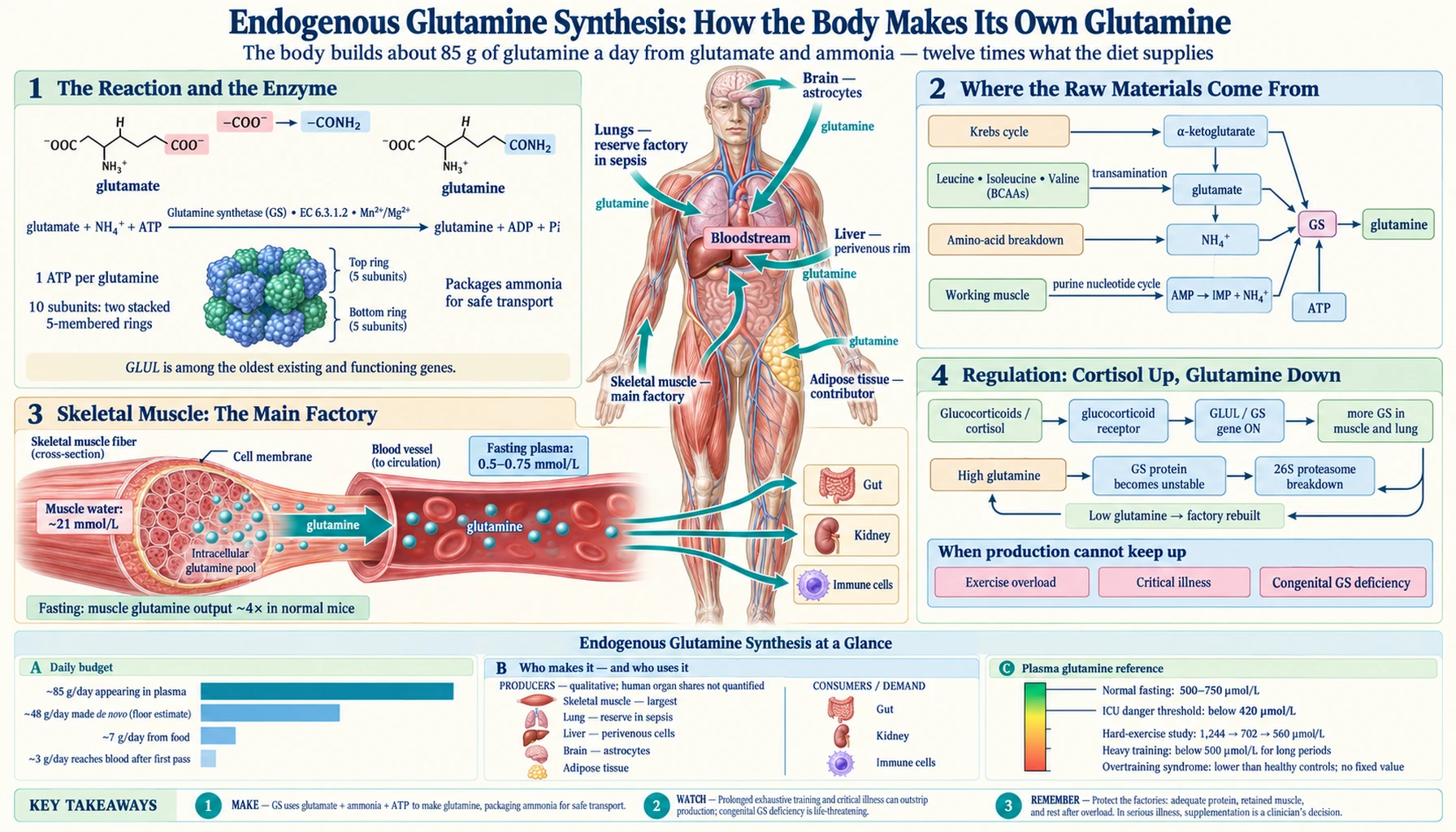
Endogenous Glutamine Synthesis: How the Body Makes Its Own Glutamine
Glutamine is called a "non-essential" amino acid, and the label is easy to misread. It does not mean the body can do without it; it means the body builds it in-house, in quantities that dwarf anything on a plate. Isotope studies in healthy adults put the amount of glutamine entering the bloodstream at roughly 85 grams a day, while the average diet supplies about 7 grams. This page follows that production line: the single enzyme that does the work, the raw materials it needs, the organs that run it — skeletal muscle above all, then lung, liver, brain and fat — the hormonal switches that turn it up and down, and what happens when demand outruns supply or when the enzyme is missing altogether.
Interactive Visualization From Sugar to Energy: Glycolysis & the Krebs Cycle The carbon skeleton of every glutamine molecule starts life as alpha-ketoglutarate in this cycle. Watch the intermediates form, then cut the oxygen and see the supply falter. Launch →
Table of Contents
- Overview: The Body's Own Glutamine Factory
- The Reaction: Glutamate + Ammonia + ATP → Glutamine
- Glutamine Synthetase: One of the Oldest Enzymes on Earth
- How Much the Body Makes Each Day: The Arithmetic
- Where the Raw Materials Come From
- Skeletal Muscle: The Main Factory
- The Lungs: A Reserve Factory That Switches On in Sepsis
- The Liver: Two Zones, Two Jobs
- The Brain: Astrocytes Make Glutamine for Neurons
- Adipose Tissue: The Quiet Contributor
- The Kidney: Mostly a Consumer, and the Acidosis Switch
- The Consumers That Set the Demand: Gut and Immune Cells
- How Production Is Regulated: Cortisol Up, Glutamine Down
- When Production Cannot Keep Up: "Conditionally Essential"
- When the Enzyme Is Missing: Congenital Glutamine Synthetase Deficiency
- How to Support Your Own Production, and What Supplements Cannot Do
- What Is Not Known
- Key Research Papers
- Connections
- Featured Videos
Overview: The Body's Own Glutamine Factory
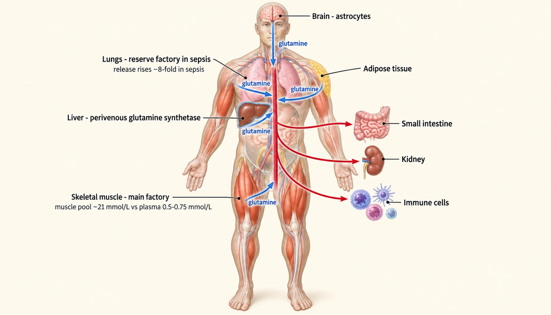
Glutamine is the most abundant free amino acid in human blood and muscle, and almost none of it was eaten as glutamine. It is assembled inside cells from two smaller pieces — the amino acid glutamate and a molecule of ammonia — by one enzyme, glutamine synthetase. Every tissue that carries that enzyme is a small glutamine factory, and the body runs several of them at once: skeletal muscle is the largest, the lungs are a reserve that opens in sepsis, a thin rim of liver cells mops up ammonia that the urea cycle missed, the brain's astrocytes make glutamine to recycle the neurotransmitter glutamate, and fat tissue contributes a share that was only measured in the 1990s.
The word "non-essential" was coined by nutritionists to mean "not required in the diet", and for glutamine it hides two facts. First, the body's own production is enormous — the arithmetic below lands near 85 grams a day appearing in plasma in a resting, fasted adult, against a dietary intake of about 7 grams. Second, that production has a ceiling. In 1990 Lacey and Wilmore proposed that glutamine becomes "conditionally essential" in the critically ill, because during severe stress the body's requirement can exceed what it is able to make. Thirty-five years of tracer studies, knockout animals and intensive-care trials have filled in the mechanism behind that sentence, and they are what this page is about.
A note on what this page is not. The companion pages under this topic cover what glutamine does once it is made — gut lining, immune fuel, exercise recovery — and where it is found in food. This one stays on the supply side: where the molecule comes from, how much, and what governs the rate.
The Reaction: Glutamate + Ammonia + ATP → Glutamine
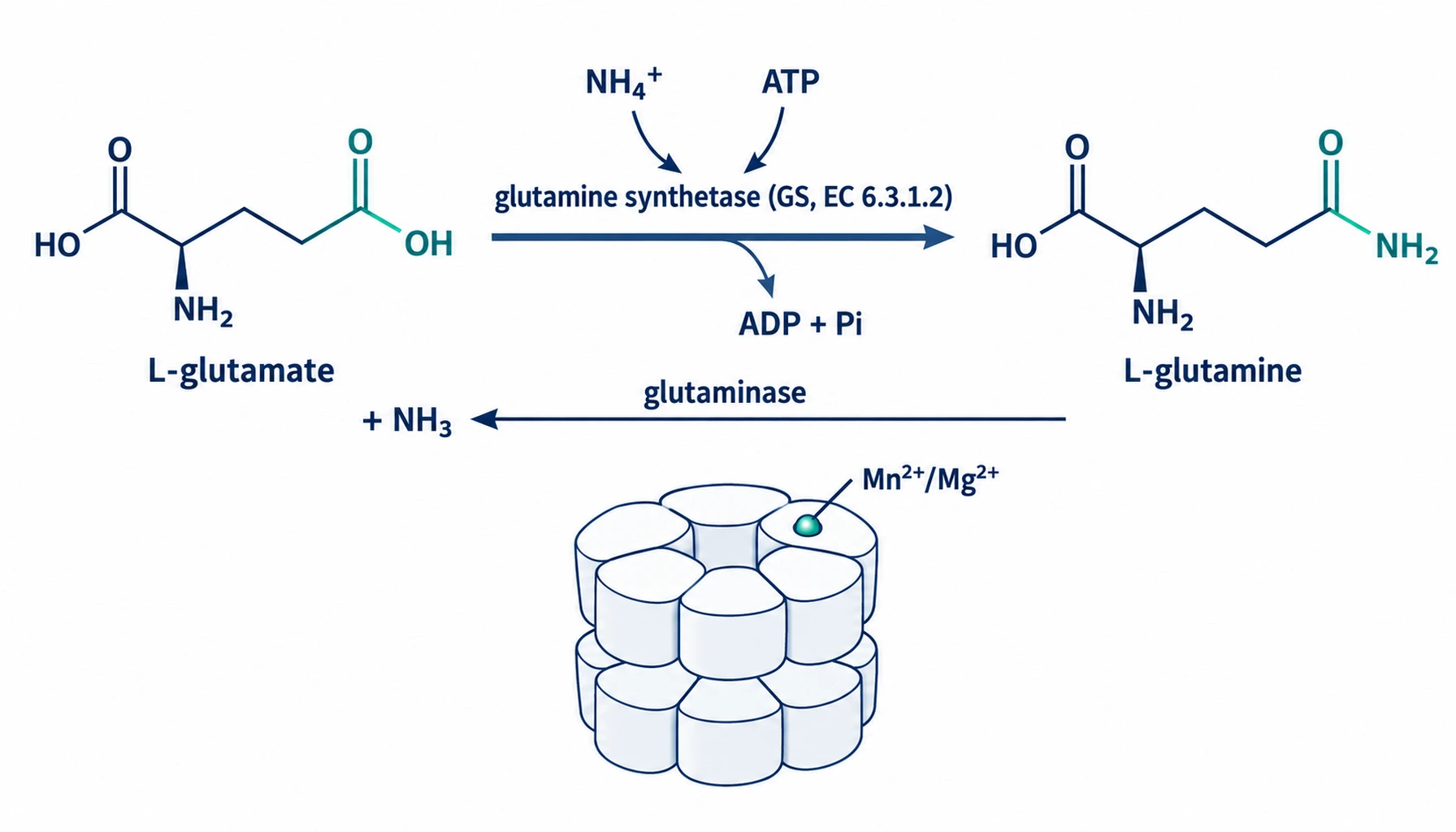
The chemistry is a single step. Glutamate carries a free carboxyl group on the end of its side chain; glutamine synthetase attaches an ammonia molecule to that carboxyl, turning it into an amide. Doing so costs one molecule of ATP: the enzyme first uses the phosphate from ATP to activate the carboxyl group (forming a short-lived γ-glutamyl phosphate), and ammonia then displaces the phosphate. The products are glutamine, ADP and inorganic phosphate. Hans Krebs described this synthesis from glutamic acid and ammonia in animal tissues in 1935 — the same year he described the urea cycle's companion chemistry — and showed in the same paper that a second enzyme, glutaminase, runs the hydrolysis in reverse.
That two-enzyme arrangement is the key to everything that follows. Glutamine synthetase (enzyme classification EC 6.3.1.2, gene GLUL, "glutamate–ammonia ligase") packages ammonia into a harmless, water-soluble amino acid that can travel in the blood. Glutaminase (EC 3.5.1.2) unpacks it again in whichever tissue needs the nitrogen, the carbon or the ammonia. Labow, Souba and Abcouwer's 2001 review puts it plainly: whether on the scale of a single cell, an organ or the whole organism, glutamine homeostasis is to a large extent set by the activities of these two enzymes.
The reaction does two useful things at once, which is why the enzyme is found in every kind of living cell. It removes free ammonia, which is toxic to the brain at concentrations not far above normal, and it converts glutamate — the brain's main excitatory neurotransmitter, and neurotoxic when it accumulates outside synapses — into glutamine, which is inert (Krajewski 2008). The enzyme needs a divalent metal ion at its active site; the human enzyme structures were solved with manganese bound, and magnesium serves the same role in most tissues.
Written out in full:
- Inputs: one glutamate, one ammonium ion (NH4+), one ATP, one Mg2+ or Mn2+ held by the enzyme.
- Intermediate: γ-glutamyl phosphate, the activated side chain, which never leaves the active site.
- Outputs: one glutamine, one ADP, one inorganic phosphate.
- Reverse route: glutaminase splits glutamine back into glutamate and ammonia without recovering the ATP — the round trip is a net energy cost, paid so that ammonia can be shipped safely between organs.
Glutamine Synthetase: One of the Oldest Enzymes on Earth
Glutamine synthetase is not a human invention or even an animal one. A 1993 phylogenetic analysis of thirty glutamine synthetase gene sequences from bacteria and eukaryotes concluded that the two families of the gene arose by a duplication that preceded the split between prokaryotes and eukaryotes, perhaps by more than a thousand million years, and that the enzyme is present in every extant life form. The authors placed it among the oldest existing and still-functioning genes, consistent with a role in nitrogen metabolism dating back toward the earliest fossils at 3,800 million years (Kumada 1993). Life learned to make glutamine very early, and never stopped.
The human enzyme is a large assembly: ten identical subunits, arranged as two stacked five-membered rings, with an active site formed between neighbouring subunits. The first structures of mammalian glutamine synthetase, published in 2008, captured the human enzyme with ADP, phosphate and manganese bound, and a second time with the classic inhibitor methionine sulfoximine locked into the active site in its phosphorylated form. Loops near the active site close down over the substrates as they bind, with the largest movement triggered by the nucleotide (Krajewski 2008). The amino-acid binding site is almost identical between bacteria and mammals; the nucleotide site is not, which is why drugs aimed at bacterial glutamine synthetase can in principle spare the human one.
Two properties of the enzyme matter for the rest of this page. It is highly regulated — Eisenberg's 2000 review calls it a highly regulated enzyme at the core of nitrogen metabolism — with control exerted at the level of gene transcription, protein stability and direct feedback by its own product. And it is unusually vulnerable to oxidative damage. In post-mortem human brain, glutamine synthetase activity falls with normal ageing in step with rising protein-oxidation products, more in the frontal lobe than the occipital, and it is the one enzyme whose loss distinguished Alzheimer brains from age-matched controls (Smith 1991). An old enzyme, but not an indestructible one.
How Much the Body Makes Each Day: The Arithmetic
Nobody can weigh the glutamine a person makes; it is synthesised, released and consumed continuously, and most of it never leaves the cell that made it. What can be measured is the rate at which glutamine appears in the blood, by infusing a trace of glutamine labelled with a heavy nitrogen isotope and watching how quickly the body dilutes it. Two classic human studies, done a decade apart with different tracers, agree closely.
- Darmaun, Matthews and Bier (1986) infused 15N-labelled glutamine and glutamate into healthy young men after an overnight fast and measured a plasma glutamine appearance rate of 348 ± 33 µmol per kg per hour.
- Nurjhan and colleagues (1995) infused 14C-glutamine alongside labelled alanine, phenylalanine and glucose in post-absorptive volunteers and measured 5.76 ± 0.26 µmol per kg per minute — which is 346 µmol per kg per hour, within one per cent of the 1986 figure.
Turning that into grams requires two stated assumptions: a 70-kg adult, and a day spent in the resting, post-absorptive state the volunteers were measured in. Then 348 µmol × 70 kg × 24 h = 585 mmol per day, and at glutamine's molecular weight of 146 g/mol that is about 85 grams of glutamine entering the plasma every day. Not all of it is newly built. Nurjhan's group traced the fraction that came straight from protein breakdown — glutamine released as existing proteins were dismantled — at 2.45 of the 5.76 µmol per kg per minute, or 43 per cent. The remaining 57 per cent, about 3.3 µmol per kg per minute or roughly 48 grams a day, had to be made de novo by glutamine synthetase. Review articles commonly quote a range of 40 to 80 grams a day for total production; this arithmetic lands inside it.
Even that is an under-count, and the 1986 authors said so. Only about 5 per cent of the glutamine synthesised inside cells and released into plasma had been built from glutamate that passed through the plasma first; the tracers did not mix thoroughly with the intracellular pools, so what they measured was transport through the blood, not the whole-body flux. Glutamine made and consumed inside a muscle fibre, or handed from an astrocyte to the neuron beside it, is invisible to a plasma tracer. The true figure for synthesis is therefore higher than 48 grams, not lower.
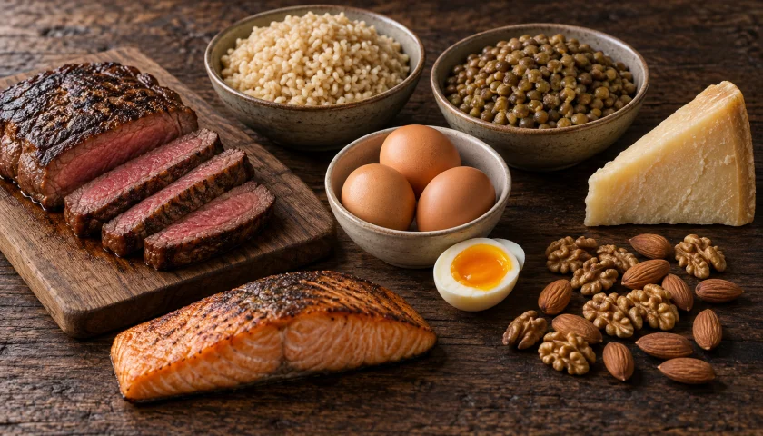
Against this, the diet. Using a food-composition database built from gene-sequence data, Lenders and colleagues estimated the mean glutamine intake of 70,356 women in the Nurses' Health Study at 6.84 grams a day (standard deviation 2.19). Glutamine made up anywhere from 1 to 33 per cent of the protein in individual foods and 4.8 per cent of the protein in meat (Lenders 2009). And of what is eaten, about half never reaches the general circulation: when glutamine was delivered by nasogastric tube, 54 per cent was captured by the gut and liver on the first pass, and 88 per cent of glutamate (Matthews 1993). So the plate supplies about 7 grams, of which perhaps 3 grams reach the bloodstream, against 85 grams appearing there from inside the body. On those numbers the body's plasma glutamine supply is about twelve times its dietary intake, and de novo synthesis alone is about seven times it.
One comparison makes the scale vivid. Alanine has long been taught as the main carrier of nitrogen and carbon from muscle to liver, yet in the same volunteers glutamine beat it on every measure: more of it appeared in plasma (5.76 versus 4.40 µmol per kg per minute), more of it came directly from protein, forearm muscle released nearly twice as much of it (0.88 versus 0.48 µmol per 100 ml per minute), and nearly twice as much new glucose carbon came from protein-derived glutamine as from alanine (Nurjhan 1995). Glutamine, Stumvoll's group concluded in 1999, is quantitatively the more important vehicle for interorgan carbon transport, and the kidney — which prefers glutamine, where the liver prefers alanine — makes 20 to 25 per cent of the body's glucose from it.
Where the Raw Materials Come From
A factory is only as productive as its supply lines, and glutamine synthetase needs three: a carbon skeleton, a nitrogen atom, and energy.
The carbon skeleton: glutamate from the Krebs cycle
The five-carbon backbone of glutamine is the five-carbon backbone of glutamate, and glutamate is made from α-ketoglutarate, an intermediate of the Krebs (tricarboxylic acid) cycle inside mitochondria. Attaching an amino group to α-ketoglutarate produces glutamate; that transfer is done by transaminase enzymes, which take the amino group from another amino acid and leave behind that amino acid's keto-acid skeleton. In skeletal muscle the main donors are the three branched-chain amino acids — leucine, isoleucine and valine. Muscle is the body's initial site of branched-chain amino acid breakdown, and the first step of that breakdown hands their nitrogen to α-ketoglutarate, with alanine and glutamine released into the blood as the products (Holeček 2018). This is also why branched-chain amino acid levels fall in liver cirrhosis and urea-cycle disorders: muscle burns through them to trap ammonia as glutamine.
Rennie's group showed the traffic runs both ways. Intramuscular glutamate normally supplies α-ketoglutarate to the mitochondrion to keep the cycle topped up, and glutamine given before exercise expanded the pool of Krebs-cycle intermediates in muscle more than placebo did (Rennie 2001). Glutamate and glutamine are not merely products of the cycle; they are its buffer stock. Other amino acids can feed the same funnel through glutamate: isolated mouse kidney tubules built glutamine at high rates from glutamate and proline and, at lower rates, from ornithine, alanine and aspartate (Conjard 2003, mouse).
The nitrogen: ammonia from protein turnover and working muscle
The second input is ammonia, and the body has more of it than it wants. Every amino acid that is oxidised for fuel gives up its nitrogen as ammonia. Working muscle adds a second source: the purine nucleotide cycle, described by Lowenstein in 1972, in which AMP is deaminated to inosine monophosphate during heavy contraction, releasing ammonia in proportion to how hard the muscle is working. Glutamine synthetase is the trap that catches it. Mouse muscle can detoxify about 2.5 µmol of ammonia per gram of muscle per hour through the enzyme, and mice engineered without muscle glutamine synthetase leak roughly three times as much ammonia from their hindquarters even at rest (He 2010, mouse). The same enzyme in the liver's outflow zone catches what the urea cycle missed (see the liver below).
The energy: one ATP per molecule
Each glutamine costs one ATP to make, and glutaminase does not give it back. At 48 grams of de novo synthesis a day — about 330 mmol — the bill is roughly 330 mmol of ATP a day, a small fraction of the body's turnover but not nothing. It is paid because the alternative, free ammonia in the blood, is worse. This is also why a cell short of energy makes less glutamine: the enzyme is fed by the same mitochondria that make the ATP and the α-ketoglutarate, so anything that stalls oxidative metabolism stalls the factory upstream.
Skeletal Muscle: The Main Factory
Ask where the body's glutamine comes from and the honest short answer is muscle. Wernerman's 2008 review states it as settled: the major part of endogenously produced glutamine comes from skeletal muscle. Biolo puts the corollary just as plainly: glutamine is primarily synthesised in skeletal muscle and enables the transfer of nitrogen to the gut, the kidneys and the immune system.
Muscle also stores it. When Bergström, Fürst and Vinnars took needle biopsies from healthy volunteers in 1974 and measured the free amino acids inside the fibres, glutamine dominated the pool. Modern measurements put the free glutamine concentration in human muscle water at about 21 mmol per litre (Mittendorfer 2001), against a fasting plasma concentration of 0.5 to 0.75 mmol per litre (Walsh 1998) — a gradient of thirty- to forty-fold, held in place by active transport across the muscle membrane. Because muscle is roughly 40 per cent of body weight, that intracellular pool is the body's glutamine reserve, and it is the reserve that stress draws down.
Human muscle exports glutamine continuously. In post-absorptive volunteers the forearm released 0.88 µmol of glutamine per 100 ml of tissue per minute, nearly double its release of alanine (Nurjhan 1995), and Felig's 1975 review had already established alanine and glutamine as the two amino acids that dominate the outflow from muscle. Mittendorfer's group measured the synthesis rate directly in the leg: 0.82 µmol per 100 ml of leg per minute in the fasted state. Feeding an amino-acid mixture raised arterial glutamine by about 20 per cent and increased muscle uptake, but did not enlarge the intramuscular pool; adding glucose to the mixture actually lowered muscle glutamine, from 21.0 to 16.4 mmol per litre, because the insulin response reduced both protein breakdown and glutamine synthesis (Mittendorfer 2001). The factory slows when the body is fed and well supplied, and speeds up when it is not.
The knockout experiment shows how much the switch matters. Mice whose muscle glutamine synthetase was deleted were healthy and fertile when fed, with normal circulating glutamine — other organs covered for the loss. After 20 hours of fasting, however, normal mice roughly quadrupled glutamine production across the hindquarter, and the knockouts could not; instead their muscle ammonia output rose about fivefold (He 2010, mouse). Muscle glutamine synthetase, the authors concluded, is dispensable in the fed state but plays the key role in mounting the adaptive response to fasting. The same is presumably true of every state that resembles fasting to the body: injury, infection, and the days after surgery.
Those days are when the reserve empties. Vinnars measured muscle free amino acids two to three days after major uncomplicated abdominal surgery and found that glutamine showed the largest fall of any amino acid in the fibres — a change that plasma levels did not reflect (Vinnars 1975). In intensive care the picture is worse still, because three things go wrong at once: whole-body consumption rises, de novo synthesis in muscle becomes relatively impaired, and the outward transporter that exports glutamine from the fibre is upregulated without any matching increase in production (Biolo 2005). Giving glutamine intravenously refills the plasma but not the muscle, which Biolo attributes to muscle's relative inability to seize glutamine from the bloodstream; the proposal that follows — support de novo synthesis rather than pour in the product — has yet to be tested in a trial. And in long intensive-care stays muscle mass itself wastes rapidly, leaving, in Wernerman's words, a tissue of diminishing size to maintain the export of glutamine.
The Lungs: A Reserve Factory That Switches On in Sepsis
In a healthy person the lungs neither add nor remove much glutamine; they are, in Plumley's phrase, an organ of amino-acid balance. In 1990 his group measured amino-acid flux across the lungs of three groups of surgical patients through pulmonary-artery catheters. Pre-operative controls released 0.80 µmol of glutamine per kg per minute, elective post-operative patients about the same, and hyperdynamic septic patients 6.80 µmol per kg per minute — more than eight times the control rate. The rise came from both increased blood flow through the lungs and a larger glutamine concentration difference across them, and the septic lung began releasing alanine as well (Plumley 1990). The lung, the authors argued, may be a key regulator of interorgan nitrogen flux after major injury and infection.
What throws the switch is not the infection itself but the hormone it provokes. In rat lung epithelial cells, glutamine synthetase messenger RNA and protein rose severalfold within hours of exposure to physiologically relevant levels of the glucocorticoid dexamethasone, and the rise was blocked by the glucocorticoid-receptor antagonist RU-486. Bacterial lipopolysaccharide, cytokines, activated complement and prostaglandins — the inflammatory mediators of septic shock — did not induce the enzyme (Abcouwer 1995, rat cells). The lung reads the cortisol surge of sepsis as an order to make glutamine, and the surge is the same signal that turns muscle production up (see regulation).
The Liver: Two Zones, Two Jobs
The liver is usually described as a glutamine consumer: its glutaminase releases ammonia from arriving glutamine to feed the urea cycle, and the urea cycle is the body's main route for disposing of nitrogen. That is true of most of the organ. But Häussinger's perfusion experiments in the early 1980s showed the liver is built in two zones with opposite jobs. Hepatocytes around the portal inflow (the periportal zone) carry glutaminase and the urea-cycle enzymes; a thin rim of cells around the venous outflow (the perivenous zone) carries glutamine synthetase instead. Blood flows past the urea-makers first and the glutamine-makers last. When ammonia was added to a rat liver perfused in the normal direction it was converted mainly to urea, and what escaped the urea cycle was captured as glutamine by the last cells before the exit; reverse the flow, and the same ammonia came out as glutamine instead (Häussinger 1983, rat). Häussinger called the arrangement an intercellular glutamine cycle: periportal cells break glutamine down to release ammonia for urea, and perivenous cells rebuild glutamine from whatever ammonia slips through — a high-affinity scavenger placed downstream of a high-capacity one.
A 2017 study in mice quantified the division of labour. Of ammonia arriving from the gut by the portal vein, about 35 per cent was detoxified by hepatic glutamine synthetase, about 35 per cent by the urea-cycle enzymes, and about 30 per cent was not cleared by the liver at all and passed into the systemic circulation, where the peripheral factories — muscle above all — had to deal with it. Ammonia given directly into a systemic vein was cleared almost entirely by glutamine synthetase activity somewhere in the body. Mice lacking liver glutamine synthetase had only mild hyperammonaemia and low glutamine, but a marked loss of muscle relative to fat, with more of the muscle-wasting protein myostatin. And the rate at which glutamine-bound ammonia was eventually converted to urea tracked the rate of glutamine synthesis, ranging from 7 per cent of normal when the enzyme was inhibited to 500 per cent when it was induced with dexamethasone (Hakvoort 2017, mouse). The authors' conclusion — that peripheral glutamine synthesis plus periportal glutamine breakdown is the body's high-affinity ammonia-detoxifying system, and that enhancing peripheral synthesis is a promising strategy for hyperammonaemia — is the strongest modern statement of why the body makes glutamine at all.
The clinical shadow of this system is hepatic encephalopathy: when a cirrhotic liver can neither make urea nor scavenge ammonia, the blood ammonia rises and the brain's own glutamine synthetase, in astrocytes, is left to absorb it.
The Brain: Astrocytes Make Glutamine for Neurons
In the brain glutamine synthetase belongs to a single cell type. Norenberg and Martinez-Hernandez mapped the enzyme in rat brain by electron-microscope immunocytochemistry in 1979 and found it in astrocytes and nowhere else — not in neurons, not in synaptic endings, not in oligodendrocytes, microglia, pericytes or blood-vessel cells (Norenberg 1979, rat). That single fact organises the brain's nitrogen economy.
Neurons, as Bak, Schousboe and Waagepetersen put it, are metabolically handicapped: they cannot synthesise their neurotransmitters glutamate and GABA from glucose on their own. So the brain runs a loop. A neuron releases glutamate at a synapse; the neighbouring astrocyte takes it up and, using glutamine synthetase, converts it to glutamine, which is inert and cannot over-excite anything; the astrocyte hands the glutamine back to the neuron, which strips off the ammonia with glutaminase to regenerate glutamate (or, in inhibitory neurons, goes one step further to GABA). This is the glutamate/GABA–glutamine cycle, and Bak's review insists it be seen as a two-way transfer of nitrogen as well as carbon, because the ammonia released in the neuron must travel back to the astrocyte to be re-fixed (Bak 2006). Hertz and colleagues make the same point from the other side: astrocytes are the glutamate producers for neurons (Hertz 1999). Glutamine synthesis in the brain is not about export; it is about recycling the most abundant neurotransmitter safely, thousands of times a second.
The astrocyte enzyme is also the brain's only defence against ammonia, since the brain has no urea cycle (Cooper 1987). When blood ammonia rises in liver failure, astrocytes fix it as glutamine faster than they can dispose of it, glutamine accumulates osmotically, and the cells swell — one of the leading explanations for the brain swelling of acute liver failure. And the enzyme ages badly. In post-mortem human frontal cortex, glutamine synthetase activity was lower in every aged brain than in young controls, tracking an exponential rise in protein oxidation with age, and lower again in Alzheimer disease (Smith 1991). Brain glutamine synthetase is a manganese enzyme, which is why manganese is concentrated in astrocytes and why manganese excess, as much as deficiency, disturbs it.
Adipose Tissue: The Quiet Contributor
Fat was left off the list of glutamine producers for most of the twentieth century because nobody had measured it in a living animal. Kowalski and Watford did, in 1994, by threading a microdialysis probe into the subcutaneous fat pad of rats and comparing the fluid between the fat cells with arterial blood. Glutamine was 122 µmol per litre higher in the tissue than in the artery, and glutamate 61 µmol per litre lower: adipose tissue was taking up glutamate, fixing ammonia onto it, and releasing glutamine. The magnitude, they wrote, suggests adipose tissue may play a significant role in whole-body glutamine homeostasis (Kowalski 1994, rat). Given how much adipose tissue an adult carries, even a modest rate per gram adds up.
The human relevance arrived in 2020. Comparing metabolites released from the white adipose tissue of 81 obese and non-obese women, Petrus and colleagues found glutamine was the metabolite most consistently reduced in obesity, and its level was inversely related to a pro-inflammatory tissue phenotype. Restoring glutamine, in cultured human fat cells and in mice, damped inflammatory gene expression and macrophage infiltration through a specific mechanism: glutamine lowered glycolysis and the supply of UDP-GlcNAc, the sugar that is attached to nuclear proteins in a modification (O-GlcNAcylation) that drives inflammatory transcription (Petrus 2020). A fat depot that makes less of its own glutamine, on this evidence, is a fat depot that inflames.
The Kidney: Mostly a Consumer, and the Acidosis Switch
The kidney is the one major organ that is, on balance, a glutamine user, and understanding why explains a large part of interorgan glutamine traffic. Renal tubule cells take up glutamine and split it with glutaminase, releasing ammonia into the urine. Every ammonium ion excreted carries away a hydrogen ion, and the bicarbonate generated in the process is returned to the blood. This is how the body defends itself against an acid load. Welbourne's 1987 review frames acid–base balance as depending on the flow of glutamine from producer organs to the kidney: normally glutamine flows from muscle to the splanchnic bed, where it fuels the gut and supports urea synthesis, but in chronic metabolic acidosis the flow is rerouted to the kidneys, arterial glutamine falls, and the whole system — increased glutamine synthetase in muscle, increased glutaminase in the kidney — is retuned to turn muscle nitrogen into urinary ammonium (Welbourne 1987). Glutamine homeostasis, in his phrase, is sacrificed to impart direction to interorgan glutamine flow.
The kidney can make glutamine too; it simply chooses not to when acid is the problem. Isolated mouse proximal tubules synthesised glutamine at high rates from glutamate and proline, but 48 hours of metabolic acidosis markedly reduced renal glutamine synthetase protein and activity — without touching its messenger RNA — while stimulating glutamine breakdown and ammonia production. Shutting down the kidney's own synthesis, the authors argued, keeps more ammonium available for excretion. The regulation is species-specific, since rat kidney behaves differently, so its human form is unknown (Conjard 2003, mouse).
The kidney also turns glutamine into glucose. Stumvoll's group showed that in fasting humans glutamine is predominantly a renal gluconeogenic substrate while alanine is essentially a hepatic one, that renal gluconeogenesis supplies 20 to 25 per cent of whole-body glucose production, and that in type 2 diabetes the conversion of glutamine to glucose is increased (Stumvoll 1999). The carbon that muscle exports as glutamine comes back, in part, as blood sugar.
The Consumers That Set the Demand: Gut and Immune Cells
Production is only half of a supply chain. The reason the body makes 50-plus grams of glutamine a day is that a few tissues burn it as their preferred fuel, and their appetite sets the pace.
The gut. Windmueller and Spaeth showed in 1974 that the small intestine takes up glutamine from arterial blood and metabolises it, establishing glutamine as the gut's major fuel from the blood side (Windmueller 1974, rat). In humans, the gut and liver together captured 54 per cent of nasogastric glutamine on first pass and 88 per cent of glutamate, and the splanchnic bed was found to be making glutamine from glutamate and glutamate from glutamine at the same time (Matthews 1993). Reeds and colleagues refined the picture in fed piglets: 95 per cent of the glutamate in the diet was consumed by the gut wall before it could reach the portal blood, glutamate was the single largest contributor to the gut's energy generation, and arterial glutamine supplied no more than 15 per cent of the gut's CO2 output in the fed state (Reeds 2000, piglet). The enterocyte, in other words, eats dietary glutamate when there is a meal and draws on blood glutamine between meals — and the latter is glutamine the body made. Souba's 1991 review named glutamine the key substrate for the splanchnic bed.
Immune cells. Ardawi and Newsholme's 1983 study of rat lymphocytes found glutamine metabolised at high rates to glutamate, aspartate and ammonia, contributing about 30 per cent of the cells' respiration; a mitogen raised glutamine use by 51 per cent, and DNA synthesis in stimulated lymphocytes was almost absent without glutamine and maximal at 0.3 mmol per litre, a concentration no other amino acid or ammonia could substitute for (Ardawi 1983, rat). Cruzat's 2018 review summarises the human evidence since: immune cells consume glutamine at rates similar to or greater than glucose, and lymphocyte proliferation, macrophage phagocytosis and neutrophil bacterial killing all depend on it. An infection is therefore a sudden new customer for muscle's output — which is the logic behind the glucocorticoid switch described next.
Newsholme's 2003 list of what the molecule is used for explains why the demand never stops: substrate for protein synthesis, precursor for muscle growth, acid–base regulation in the kidney, urea synthesis in the liver, gluconeogenesis in liver and kidney, oxidative fuel for gut and immune cells, interorgan nitrogen transport, neurotransmitter precursor, nucleotide precursor, and precursor of glutathione. Most of those uses begin by converting glutamine back to glutamate.
How Production Is Regulated: Cortisol Up, Glutamine Down
Two controls govern how much glutamine synthetase a tissue carries, and they pull in opposite directions. The first is hormonal and raises the enzyme; the second is the product itself, and lowers it.
Glucocorticoids raise the enzyme
Give a rat daily dexamethasone at 0.5 mg per kg — a dose the authors chose because it approximates the corticosteroid levels of an animal under severe stress — and glutamine synthetase activity and messenger RNA rise strikingly in plantaris, soleus and diaphragm muscle. The heart barely responds (about 15 per cent, with no change in mRNA), and an unrelated gene is unaffected, so this is a targeted induction rather than a general drug effect (Max 1988, rat). Lung epithelial cells respond the same way, through the glucocorticoid receptor (Abcouwer 1995). Labow's review generalises: the glutamine synthetase gene is transcriptionally activated by glucocorticoid hormones in a tissue-specific fashion, which lets messenger RNA rise in selected organs during catabolic states (Labow 2001). Cortisol, the hormone of injury, infection, starvation and hard training, is the order to build more of the enzyme where it is needed. In Hakvoort's mice dexamethasone pushed the disposal of glutamine-bound ammonia to five times normal (Hakvoort 2017).
Glutamine itself lowers the enzyme
The second control is a thermostat. In mouse skeletal-muscle cells, glutamine in the culture medium at concentrations as low as 0.25 mmol per litre — below normal plasma — reduced glutamine synthetase protein, with the maximal effect at 2 mmol per litre. Glutamine did not switch the gene off; it made the existing enzyme protein unstable, and a proteasome inhibitor partly blocked the loss. Only glutamine and the amino acids that can be converted into it (glutamate, alanine, ornithine) had the effect; methionine sulfoximine, which locks the enzyme, stabilised it (Huang 2007, mouse cells). Labow describes the same mechanism in vivo as a unique form of product feedback: glutamine increases glutamine synthetase protein turnover through the 26S proteasome, which appears to index the production of glutamine to its intracellular concentration and therefore to systemic demand (Labow 2001). When the cell is full of glutamine, it dismantles the factory; when the level falls, the factory is rebuilt. This is also the mechanism by which supplemental glutamine, in cell culture at least, dampens the body's own enzyme (see below).
Cancer cells rewrite the rule
Tumours are famous for being glutamine-hungry, importing it through the same glutaminase route that immune cells use. Bott and colleagues found that the oncogene Myc also does the opposite in a range of human and mouse cancers: it induces glutamine synthetase by activating an enzyme that strips the methyl groups silencing the GLUL promoter. Cells that make their own glutamine survive when the supply runs out, and silencing the enzyme slowed their growth as tumour grafts (Bott 2015). The body's oldest nitrogen enzyme, in other words, is one that some cancers switch back on to feed themselves.
When Production Cannot Keep Up: "Conditionally Essential"
Lacey and Wilmore's 1990 proposal was that during stress the body's requirement for glutamine appears to exceed its ability to produce it. The evidence since then runs from the athletics track to the intensive-care unit, and it is an argument about rates: consumption by gut, kidney, liver and immune cells goes up faster than the factories can be retooled.
Exercise. Normal fasting plasma glutamine runs 500 to 750 µmol per litre. It is unchanged or briefly raised after short intense exercise, but it falls after endurance events, after prolonged exercise, in untreated diabetes and in diet-induced metabolic acidosis — states that share a rise in cortisol and glucagon and an increased uptake of glutamine by liver, gut and kidney for gluconeogenesis (Walsh 1998). Keast's group measured the fall in men run to 90 per cent of maximal oxygen uptake: plasma glutamine dropped from 1,244 to 702 µmol per litre, and to 560 at 120 per cent. Ten days of twice-daily interval training lowered resting glutamine in every athlete by day 11, and two of five had not recovered six days later (Keast 1995). Heavy training, Walsh notes, can leave plasma glutamine below 500 µmol per litre for long periods, and athletes with overtraining syndrome sit lower at rest than healthy controls. The factories are not broken; the customers have simply multiplied.
Critical illness. Among 80 patients admitted acutely to a Dutch intensive-care unit, the 25 with a plasma glutamine below 0.420 mmol per litre on arrival had a hospital mortality of 60 per cent against 29 per cent in the rest, and adding the low level to the standard severity score significantly improved its prediction of death (Oudemans-van Straaten 2001). Biolo's account of why — rising use, relatively impaired muscle synthesis, and an export transporter running faster than the enzyme behind it — is given in the muscle section. Wernerman reports that 20 to 25 grams of glutamine a day normalises plasma glutamine in most such patients, with strong evidence for intravenous supplementation in patients on parenteral nutrition, whose feeding formulas usually contain no glutamine at all (Wernerman 2008).
The harm signal. More is not automatically better, and the largest trial says so. REDOXS randomised 1,223 mechanically ventilated adults with multi-organ failure in 40 intensive-care units to glutamine, antioxidants, both or placebo, given both intravenously and enterally from the first day. Glutamine did not reduce organ failure or infections; 28-day mortality was 32.4 per cent with glutamine against 27.2 per cent without (adjusted odds ratio 1.28, 95 per cent confidence interval 1.00 to 1.64), and in-hospital and six-month mortality were significantly higher in the glutamine groups (Heyland 2013). A low glutamine level in a failing body marks a system whose factories and customers are both in trouble, and flooding the plasma does not repair either. That is the distinction this page turns on: the body's own synthesis is regulated, local and paid for in ATP; a bolus from outside is none of those things.
When the Enzyme Is Missing: Congenital Glutamine Synthetase Deficiency
The clearest proof that the body must make its own glutamine is what happens to a child who cannot. In 2005 Häberle and colleagues described two unrelated newborns with congenital glutamine synthetase deficiency. Each carried a homozygous mutation in the gene (R324C in one, R341C in the other), each had glutamine largely absent from serum, urine and cerebrospinal fluid, and each had severe brain malformations leading to multi-organ failure and death in the neonatal period. Cells engineered to express the mutant enzymes showed reduced activity (Häberle 2005). The brain damage was present at birth: the deficit acted throughout fetal development, before any diet could have been involved, and the mother's glutamine, delivered across the placenta, was not enough to compensate.
Set that against the animal work and the redundancy of the system becomes visible. Delete the enzyme from muscle alone and mice are healthy when fed, failing only when fasted (He 2010). Delete it from the liver alone and they show mild hyperammonaemia and lose muscle relative to fat (Hakvoort 2017). Delete it everywhere, as the human mutations effectively did, and the organism does not survive. Every factory can cover for another; none can be replaced by food.
How to Support Your Own Production, and What Supplements Cannot Do
The evidence above is mostly about mechanism and about sick people; nobody has run a trial of "how to make more glutamine" in healthy adults. What follows is therefore labelled by the kind of evidence behind it.
- Eat enough protein, including the branched-chain amino acids. Human physiology, no outcome trial. Glutamine synthesis in muscle runs on glutamate made from α-ketoglutarate and the nitrogen of leucine, isoleucine and valine (Holeček 2018). Ingesting an amino-acid mixture raised arterial glutamine by about 20 per cent and increased muscle uptake in healthy volunteers (Mittendorfer 2001). Complete proteins supply both the carbon and the nitrogen; the Sources page lists foods by glutamine and protein content, and bone broth is discussed separately.
- Keep your muscle. Strong for the premise, inferred for the advice. Muscle is the main factory and the main reserve; when it wastes, in Wernerman's phrase, a tissue of diminishing size is left to maintain the export. Resistance exercise preserves muscle mass. That it thereby preserves glutamine output is a reasonable inference, not a measured result.
- Do not train yourself into chronic depletion. Observational, moderate. Plasma glutamine falls with prolonged exhaustive exercise and with overload training, and recovery can take more than a week (Keast 1995; Walsh 1998). Rest days are part of the production schedule.
- Mind the acid load. Mechanism only. Chronic metabolic acidosis reroutes glutamine from gut and liver to the kidney and lowers arterial glutamine (Welbourne 1987). This is a statement about frank acidosis, not about ordinary dietary variation, and no study has shown that adjusting diet acidity changes glutamine status in healthy people.
- Cofactors. Mechanism only. The enzyme needs magnesium or manganese at its active site (Krajewski 2008). Deficiency of either is uncommon; there is no evidence that extra improves glutamine synthesis, and manganese excess damages the very astrocytes that depend on it.
- What an oral supplement does, by the numbers. Arithmetic from human data. A 5-gram dose loses about half to the gut and liver on the first pass (Matthews 1993), so roughly 2 to 3 grams reach the circulation, against about 85 grams appearing there each day from inside the body — an addition of around 3 per cent. In cell culture, raising glutamine lowers glutamine synthetase protein within hours (Huang 2007), so the immediate effect of a supplement is, if anything, to tell your own enzyme to stand down. None of this means supplements have no uses — the Benefits pages weigh the trials for gut, immune and exercise outcomes — but it does mean a healthy person cannot meaningfully out-supply their own production.
- In illness, this is a clinician's decision. Trial evidence, both directions. Twenty to 25 grams a day normalises plasma glutamine in intensive care (Wernerman 2008), and the evidence favours intravenous glutamine for patients on glutamine-free parenteral feeding; but in ventilated patients with multi-organ failure the REDOXS trial found higher mortality with glutamine (Heyland 2013). The two findings are not contradictory — they describe different patients — but they are why glutamine is not something to self-prescribe for a serious illness.
The theme running through the list is that the body's glutamine supply is best protected by protecting the factories — adequate protein, retained muscle, avoided exhaustion — rather than by trying to replace their output.
What Is Not Known
- The true whole-body synthesis rate in humans. Every figure on this page comes from plasma tracers, which Darmaun's group showed under-measure intracellular fluxes. The 48-gram de novo estimate is a floor.
- Each organ's share in humans. Muscle and lung have been measured by limb and pulmonary balance; the liver's zonation, the astrocyte enzyme, adipose production and renal regulation rest largely on rat, mouse and piglet data. Adipose glutamine output has never been quantified in a living person.
- Whether promoting synthesis beats supplying the product. Biolo proposed, and Hakvoort's group echoed for hyperammonaemia, that stimulating peripheral glutamine synthesis could succeed where supplementation fails. No trial has tried it.
- The human kidney in acidosis. Conjard's finding that acidosis suppresses renal glutamine synthetase is explicitly species-specific; whether human kidney does the same is unknown.
- How much the enzyme declines with age outside the brain. The post-mortem data are for brain only. Whether muscle and liver glutamine synthetase fall with age, and whether that contributes to sarcopenia or to the higher mortality of glutamine-depleted older intensive-care patients, has not been measured.
- Whether dietary glutamine matters at all in health. Lenders found a 16 per cent between-person variation in energy-adjusted intake and proposed studying it against disease; on the arithmetic above, a few grams either way is a rounding error against endogenous supply, but that has not been tested.
Key Research Papers
Every citation below was checked against the Crossref record for its DOI and against the PubMed record for its PMID before this page was written, and the abstracts of the papers whose numbers are quoted above were read for support. Species are noted in the text where a finding comes from animals or cultured cells. Reviews are marked by their journals; the primary human tracer studies are Darmaun 1986, Nurjhan 1995, Matthews 1993, Mittendorfer 2001 and Stumvoll 1999.
The enzyme and the reaction
- Krebs HA. Metabolism of amino-acids: IV. The synthesis of glutamine from glutamic acid and ammonia, and the enzymic hydrolysis of glutamine in animal tissues. Biochemical Journal. 1935;29(8):1951-1969. — doi:10.1042/bj0291951 (PubMed PMID: 16745865)
- Eisenberg D, Gill HS, Pfluegl GMU, et al. Structure-function relationships of glutamine synthetases. Biochimica et Biophysica Acta (BBA) - Protein Structure and Molecular Enzymology. 2000;1477(1-2):122-145. — doi:10.1016/s0167-4838(99)00270-8 (PubMed PMID: 10708854)
- Krajewski WW, Collins R, Holmberg-Schiavone L, et al. Crystal structures of mammalian glutamine synthetases illustrate substrate-induced conformational changes and provide opportunities for drug and herbicide design. Journal of Molecular Biology. 2008;375(1):217-228. — doi:10.1016/j.jmb.2007.10.029 (PubMed PMID: 18005987)
- Kumada Y, Benson DR, Hillemann D, et al. Evolution of the glutamine synthetase gene, one of the oldest existing and functioning genes. Proceedings of the National Academy of Sciences. 1993;90(7):3009-3013. — doi:10.1073/pnas.90.7.3009 (PubMed PMID: 8096645)
- Häberle J, Görg B, Rutsch F, et al. Congenital glutamine deficiency with glutamine synthetase mutations. New England Journal of Medicine. 2005;353(18):1926-1933. — doi:10.1056/nejmoa050456 (PubMed PMID: 16267323)
How much the body makes, and where it goes
- Darmaun D, Matthews DE, Bier DM. Glutamine and glutamate kinetics in humans. American Journal of Physiology-Endocrinology and Metabolism. 1986;251(1):E117-E126. — doi:10.1152/ajpendo.1986.251.1.e117 (PubMed PMID: 2873746)
- Nurjhan N, Bucci A, Perriello G, et al. Glutamine: a major gluconeogenic precursor and vehicle for interorgan carbon transport in man. Journal of Clinical Investigation. 1995;95(1):272-277. — doi:10.1172/jci117651 (PubMed PMID: 7814625)
- Matthews DE, Marano MA, Campbell RG. Splanchnic bed utilization of glutamine and glutamic acid in humans. American Journal of Physiology-Endocrinology and Metabolism. 1993;264(6):E848-E854. — doi:10.1152/ajpendo.1993.264.6.e848 (PubMed PMID: 8101428)
- Lenders CM, Liu S, Wilmore DW, et al. Evaluation of a novel food composition database that includes glutamine and other amino acids derived from gene sequencing data. European Journal of Clinical Nutrition. 2009;63(12):1433-1439. — doi:10.1038/ejcn.2009.110 (PubMed PMID: 19756030)
- Stumvoll M, Perriello G, Meyer C, et al. Role of glutamine in human carbohydrate metabolism in kidney and other tissues. Kidney International. 1999;55(3):778-792. — doi:10.1046/j.1523-1755.1999.055003778.x (PubMed PMID: 10027916)
- Newsholme P, Procopio J, Lima MMR, et al. Glutamine and glutamate--their central role in cell metabolism and function. Cell Biochemistry and Function. 2002;21(1):1-9. — doi:10.1002/cbf.1003 (PubMed PMID: 12579515)
- Watford M. Glutamine and glutamate: Nonessential or essential amino acids?. Animal Nutrition. 2015;1(3):119-122. — doi:10.1016/j.aninu.2015.08.008 (PubMed PMID: 29767158)
- Cruzat V, Macedo Rogero M, Noel Keane K, et al. Glutamine: Metabolism and Immune Function, Supplementation and Clinical Translation. Nutrients. 2018;10(11):1564. — doi:10.3390/nu10111564 (PubMed PMID: 30360490)
Skeletal muscle, the main factory
- Bergström J, Fürst P, Norée LO, et al. Intracellular free amino acid concentration in human muscle tissue. Journal of Applied Physiology. 1974;36(6):693-697. — doi:10.1152/jappl.1974.36.6.693 (PubMed PMID: 4829908)
- Mittendorfer B, Volpi E, Wolfe RR. Whole body and skeletal muscle glutamine metabolism in healthy subjects. American Journal of Physiology-Endocrinology and Metabolism. 2001;280(2):E323-E333. — doi:10.1152/ajpendo.2001.280.2.e323 (PubMed PMID: 11158937)
- He Y, Hakvoort TBM, Köhler SE, et al. Glutamine synthetase in muscle is required for glutamine production during fasting and extrahepatic ammonia detoxification. Journal of Biological Chemistry. 2010;285(13):9516-9524. — doi:10.1074/jbc.m109.092429 (PubMed PMID: 20064933)
- Felig P. Amino acid metabolism in man. Annual Review of Biochemistry. 1975;44(1):933-955. — doi:10.1146/annurev.bi.44.070175.004441 (PubMed PMID: 1094924)
- Holeček M. Branched-chain amino acids in health and disease: metabolism, alterations in blood plasma, and as supplements. Nutrition & Metabolism. 2018;15(1). — doi:10.1186/s12986-018-0271-1 (PubMed PMID: 29755574)
- Lowenstein JM. Ammonia production in muscle and other tissues: the purine nucleotide cycle. Physiological Reviews. 1972;52(2):382-414. — doi:10.1152/physrev.1972.52.2.382 (PubMed PMID: 4260884)
- Rennie MJ, Khogali SEO, Bowtell JL, et al. Interaction between glutamine availability and metabolism of glycogen, tricarboxylic acid cycle intermediates and glutathione. The Journal of Nutrition. 2001;131(9):2488S-2490S. — doi:10.1093/jn/131.9.2488s (PubMed PMID: 11533298)
- VINNARS E, BERGSTÖM J, FÜRST P. Influence of the postoperative state on the intracellular free amino acids in human muscle tissue. Annals of Surgery. 1975;182(6):665-671. — doi:10.1097/00000658-197512000-00001 (PubMed PMID: 1190870)
- Biolo G, Zorat F, Antonione R, et al. Muscle glutamine depletion in the intensive care unit. The International Journal of Biochemistry & Cell Biology. 2005;37(10):2169-2179. — doi:10.1016/j.biocel.2005.05.001 (PubMed PMID: 16084750)
Lung, liver, brain, fat and kidney
- Plumley DA. Accelerated lung amino acid release in hyperdynamic septic surgical patients. Archives of Surgery. 1990;125(1):57. — doi:10.1001/archsurg.1990.01410130063008 (PubMed PMID: 1967211)
- Abcouwer SF, Lukaszewicz GC, Souba WW. Glucocorticoids regulate glutamine synthetase expression in lung epithelial cells. American Journal of Physiology-Lung Cellular and Molecular Physiology. 1996;270(1):L141-L151. — doi:10.1152/ajplung.1996.270.1.l141 (PubMed PMID: 8772537)
- HÄUSSINGER D. Hepatocyte heterogeneity in glutamine and ammonia metabolism and the role of an intercellular glutamine cycle during ureogenesis in perfused rat liver. European Journal of Biochemistry. 1983;133(2):269-275. — doi:10.1111/j.1432-1033.1983.tb07458.x (PubMed PMID: 6852039)
- Haüssinger D. Nitrogen metabolism in liver: structural and functional organization and physiological relevance. Biochemical Journal. 1990;267(2):281-290. — doi:10.1042/bj2670281 (PubMed PMID: 2185740)
- Hakvoort TBM, He Y, Kulik W, et al. Pivotal role of glutamine synthetase in ammonia detoxification. Hepatology. 2017;65(1):281-293. — doi:10.1002/hep.28852 (PubMed PMID: 27641632)
- Norenberg MD, Martinez-Hernandez A. Fine structural localization of glutamine synthetase in astrocytes of rat brain. Brain Research. 1979;161(2):303-310. — doi:10.1016/0006-8993(79)90071-4 (PubMed PMID: 31966)
- Bak LK, Schousboe A, Waagepetersen HS. The glutamate/GABA-glutamine cycle: aspects of transport, neurotransmitter homeostasis and ammonia transfer. Journal of Neurochemistry. 2006;98(3):641-653. — doi:10.1111/j.1471-4159.2006.03913.x (PubMed PMID: 16787421)
- Hertz L, Dringen R, Schousboe A, et al. Toxoplasma gondii infection of neurons alters the production and content of extracellular vesicles directing astrocyte phenotype and contributing to the loss of GLT-1 in the infected brain. Journal of Neuroscience Research. 1999;57(4):417-428. — doi:10.1002/(sici)1097-4547(19990815)57:4<417::aid-jnr1>3.0.co;2-n (PubMed PMID: 10440891)
- Cooper AJ, Plum F. Biochemistry and physiology of brain ammonia. Physiological Reviews. 1987;67(2):440-519. — doi:10.1152/physrev.1987.67.2.440 (PubMed PMID: 2882529)
- Smith CD, Carney JM, Starke-Reed PE, et al. Excess brain protein oxidation and enzyme dysfunction in normal aging and in Alzheimer disease. Proceedings of the National Academy of Sciences. 1991;88(23):10540-10543. — doi:10.1073/pnas.88.23.10540 (PubMed PMID: 1683703)
- Kowalski TJ, Watford M. Production of glutamine and utilization of glutamate by rat subcutaneous adipose tissue in vivo. American Journal of Physiology-Endocrinology and Metabolism. 1994;266(1):E151-E154. — doi:10.1152/ajpendo.1994.266.1.e151 (PubMed PMID: 7905708)
- Petrus P, Lecoutre S, Dollet L, et al. Glutamine Links Obesity to Inflammation in Human White Adipose Tissue. Cell Metabolism. 2020;31(2):375-390.e11. — doi:10.1016/j.cmet.2019.11.019 (PubMed PMID: 31866443)
- Welbourne TC. Interorgan glutamine flow in metabolic acidosis. American Journal of Physiology-Renal Physiology. 1987;253(6):F1069-F1076. — doi:10.1152/ajprenal.1987.253.6.f1069 (PubMed PMID: 3322041)
- Conjard A, Komaty O, Delage H, et al. Inhibition of glutamine synthetase in the mouse kidney: a novel mechanism of adaptation to metabolic acidosis. Journal of Biological Chemistry. 2003;278(40):38159-38166. — doi:10.1074/jbc.m302885200 (PubMed PMID: 12871952)
Who consumes it, and how production is regulated
- Windmueller HG, Spaeth AE. The uptake of glutamine and release of arginine, citrulline and proline by the small intestine of developing pigs. Journal of Biological Chemistry. 1974;249(16):5070-5079. — doi:10.1016/s0021-9258(19)42329-6 (PubMed PMID: 4605420)
- Reeds PJ, Burrin DG, Stoll B, et al. Intestinal glutamate metabolism. The Journal of Nutrition. 2000;130(4):978S-982S. — doi:10.1093/jn/130.4.978s (PubMed PMID: 10736365)
- Souba WW. Glutamine: a key substrate for the splanchnic bed. Annual Review of Nutrition. 1991;11(1):285-308. — doi:10.1146/annurev.nu.11.070191.001441 (PubMed PMID: 1892702)
- Ardawi MSM, Newsholme EA. Glutamine metabolism in lymphocytes of the rat. Biochemical Journal. 1983;212(3):835-842. — doi:10.1042/bj2120835 (PubMed PMID: 6882397)
- Max SR, Mill J, Mearow K, et al. Dexamethasone regulates glutamine synthetase expression in rat skeletal muscles. American Journal of Physiology-Endocrinology and Metabolism. 1988;255(3):E397-E402. — doi:10.1152/ajpendo.1988.255.3.e397 (PubMed PMID: 2901814)
- Labow BI, Souba WW, Abcouwer SF. Mechanisms governing the expression of the enzymes of glutamine metabolism--glutaminase and glutamine synthetase. The Journal of Nutrition. 2001;131(9):2467S-2474S. — doi:10.1093/jn/131.9.2467s (PubMed PMID: 11533295)
- Huang YF, Wang Y, Watford M. Glutamine directly downregulates glutamine synthetase protein levels in mouse C2C12 skeletal muscle myotubes. The Journal of Nutrition. 2007;137(6):1357-1362. — doi:10.1093/jn/137.6.1357 (PubMed PMID: 17513391)
- Bott AJ, Peng IC, Fan Y, et al. Oncogenic Myc Induces Expression of Glutamine Synthetase through Promoter Demethylation. Cell Metabolism. 2015;22(6):1068-1077. — doi:10.1016/j.cmet.2015.09.025 (PubMed PMID: 26603296)
When production cannot keep up
- Lacey JM, Wilmore DW. Is glutamine a conditionally essential amino acid?. Nutrition Reviews. 2009;48(8):297-309. — doi:10.1111/j.1753-4887.1990.tb02967.x (PubMed PMID: 2080048)
- Walsh NP, Blannin AK, Robson PJ, et al. Glutamine, exercise and immune function. Links and possible mechanisms. Sports Medicine. 1998;26(3):177-191. — doi:10.2165/00007256-199826030-00004 (PubMed PMID: 9802174)
- Keast D, Arstein D, Harper W, et al. Depression of plasma glutamine concentration after exercise stress and its possible influence on the immune system. Medical Journal of Australia. 1995;162(1):15-18. — doi:10.5694/j.1326-5377.1995.tb138403.x (PubMed PMID: 7845291)
- Oudemans-van Straaten HM, Bosman RJ, Treskes M, et al. Plasma glutamine depletion and patient outcome in acute ICU admissions. Intensive Care Medicine. 2000;27(1):84-90. — doi:10.1007/s001340000703 (PubMed PMID: 11280678)
- Wernerman J. Clinical use of glutamine supplementation. The Journal of Nutrition. 2008;138(10):2040S-2044S. — doi:10.1093/jn/138.10.2040s (PubMed PMID: 18806121)
- Heyland D, Muscedere J, Wischmeyer PE, et al. A randomized trial of glutamine and antioxidants in critically ill patients. New England Journal of Medicine. 2013;368(16):1489-1497. — doi:10.1056/nejmoa1212722 (PubMed PMID: 23594003)
PubMed Topic Searches
- PubMed: Glutamine synthetase and muscle glutamine production
- PubMed: De novo glutamine synthesis in humans by stable isotope
- PubMed: Hepatic glutamine synthetase and ammonia detoxification
- PubMed: The astrocyte glutamate–glutamine cycle
- PubMed: Glucocorticoid regulation of glutamine synthetase
- PubMed: Congenital glutamine synthetase deficiency
- PubMed: Plasma glutamine and outcome in critical illness
This content is provided for informational purposes only and does not constitute medical advice. Consult a qualified healthcare provider before beginning any supplementation regimen.
Connections
- All Amino Acids
- Glutamine
- Glutamine Benefits
- Glutamine Sources
- Glutamine History
- Glutamine for Immune Function
- Glutamine for Exercise Recovery
- Glutamic Acid
- Glutamic Acid: Nitrogen Metabolism
- Asparagine: Ammonia Detoxification
- Aspartic Acid: The Urea Cycle
- Alanine: The Glutamine–Alanine Relay
- Valine: Nitrogen Balance
- Leucine (BCAA)
- GABA
- Ammonia (Lab Test)
- Hepatic Encephalopathy
- Hans Krebs
- Manganese
- Magnesium
- Bone Broth and Glutamine
- Fasting
- Glycolysis & the Krebs Cycle — interactive animation
- Mitochondria & ATP — interactive animation
Featured Videos