Carnitine: History and Discovery

Carnitine has one of the stranger biographies in nutrition. It was pulled out of a jar of commercial beef extract in 1905, named after the Latin word for flesh, and then more or less forgotten for half a century. In the late 1940s it briefly became a "vitamin" — but only for a beetle larva. In the 1950s and 1960s biochemists discovered that it is the gate through which fat enters the cell's furnaces, and in the 1970s, 1980s and 1990s physicians found the children whose bodies could not hold on to it, traced the fault to a single transporter gene, and turned carnitine into a prescription medicine. Today it is sold in gyms and pharmacies alike, argued over in cardiology journals, and implicated — perhaps — in how red meat and gut bacteria interact. This article follows that documented arc from Moscow to the modern supplement aisle. Where the record is firm we say so; where a claim is contested, we say that too.
Table of Contents
- A Compound Named for Flesh: Moscow, 1905
- Two Decades to a Structure (1905–1927)
- The Mealworm and "Vitamin BT" (1948–1952)
- Irving Fritz and the Burning of Fat (1955–1959)
- The Carnitine Shuttle Takes Shape (1962–1975)
- The Children Who Could Not Keep Carnitine (1973–1999)
- From Laboratory to Pharmacy: Levocarnitine, Dialysis and Valproate
- New Forms: Acetyl-L-Carnitine and Propionyl-L-Carnitine
- Heart Trials and the TMAO Debate (2013 Onward)
- Carnitine Today
- Key Research Papers
- Connections
- Featured Videos
A Compound Named for Flesh: Moscow, 1905
The story begins not in a hospital or a kitchen but in a laboratory bench-full of commercial meat extract. In the decades around 1900, physiological chemists in Germany and Russia were systematically taking apart the "extractives" of muscle — the small nitrogen-containing molecules left behind when meat is boiled and the broth concentrated. The convenient raw material was the concentrated beef extract sold under Justus von Liebig's name, a pantry staple of the era that happened to be a rich, standardised source of exactly these compounds. Creatine, carnosine and several other muscle molecules were characterised from it.
In 1905 two Russian chemists working in Moscow, Vladimir Gulewitsch (often transliterated Gulevich) and Robert Krimberg, reported a new base from this extract in the German journal Hoppe-Seyler's Zeitschrift für physiologische Chemie, in a paper titled, in English, "On the extractive substances of muscle. Part II. On carnitine." They gave it the empirical formula C₇H₁₅NO₃ and, because it came from meat, coined its name from the Latin caro, genitive carnis, "flesh." The name is the same root as carnivore and carnal, and it was well chosen: more than a century later, red meat remains by far the richest food source of carnitine, and roughly 95 percent of the body's own supply sits in skeletal and heart muscle.
Almost simultaneously, the Marburg physiologist Friedrich Kutscher was picking through the same commercial extract. In papers on Liebig's meat extract published in 1905 and 1906 he described a base he called novaine. For a few years the literature carried two names for what turned out to be one molecule. Krimberg settled the matter himself in 1908 with a paper whose title says it all: "On the identity of novaine with carnitine." A 1932 review of muscle extractives, looking back on the episode, credits Gulewitsch and Krimberg with the discovery, notes that Kutscher's novaine "was apparently identical with carnitine," and records that carnitine had by then been found in the muscles of many animals at 0.02 to 0.05 percent by weight, but not in other tissues — a first hint that it had something to do with muscle's particular way of making energy.
Nobody in 1905 had any idea what carnitine did. It was a curiosity: a small, strongly alkaline, very water-soluble base that formed neat crystalline salts with gold, platinum and mercury chlorides, and that rotated polarised light to the left. That last detail — carnitine is levorotatory — would matter a great deal later, because it is the origin of the "L" in L-carnitine.
Two Decades to a Structure (1905–1927)
Knowing a molecule's formula is not the same as knowing its shape. Carnitine's structure resisted the chemists of the early twentieth century for more than twenty years, and the 1932 reviewer remarked drily that "considerable difficulty was experienced" in working it out.
The clues came from breaking the molecule apart. Kutscher showed that heating novaine with barium hydroxide released trimethylamine (the fishy-smelling gas that also forms when seafood spoils) together with crotonic and succinic acids; Krimberg found that carnitine behaved the same way. From this, Krimberg proposed a structure — a betaine (a molecule carrying a positively charged nitrogen with three methyl groups) built on a four-carbon acid with a hydroxyl group on the middle carbon — which we now write as 3-hydroxy-4-(trimethylammonio)butanoate, or β-hydroxy-γ-butyrobetaine. It is worth pausing on that trimethylamine fragment: a century later, the same three-methyl nitrogen would be at the centre of the TMAO controversy described below. The chemistry that would make carnitine controversial in 2013 was, in a sense, visible on the lab bench in 1908.
Krimberg's proposal was correct, but it was contested. Other chemists argued the hydroxyl group sat on a different carbon, and a rival structure was synthesised and shown not to match. The definitive proof came from Japan. In 1927 Masaji Tomita and Y. Sendju, publishing in the same German journal where carnitine had been announced, synthesised γ-amino-β-hydroxybutyric acid, painstakingly separated it into its optical isomers, converted each to its betaine, and showed that one of the left-rotating isomers was identical in every property to natural carnitine. That settled the constitution of the molecule, and it also established a fact that would take on practical importance once carnitine became a supplement: carnitine exists as two mirror-image forms, and the body makes and uses only one of them. The natural form is L-carnitine (levocarnitine); its mirror image, D-carnitine, is biologically inactive and can interfere with the natural form. A 1983 review by the Norwegian biochemist Jon Bremer, who did much of the later mechanistic work, still made a point of distinguishing "the natural isomer" from "the unphysiological isomer," which mammals handle quite differently.
After 1927, then, carnitine was a solved chemical problem and an unsolved biological one. Bremer's later verdict on this period was blunt: carnitine "was detected at the beginning of this century, but it was nearly forgotten among biochemists until its importance in fatty acid metabolism was established 50 years later."
The Mealworm and "Vitamin BT" (1948–1952)
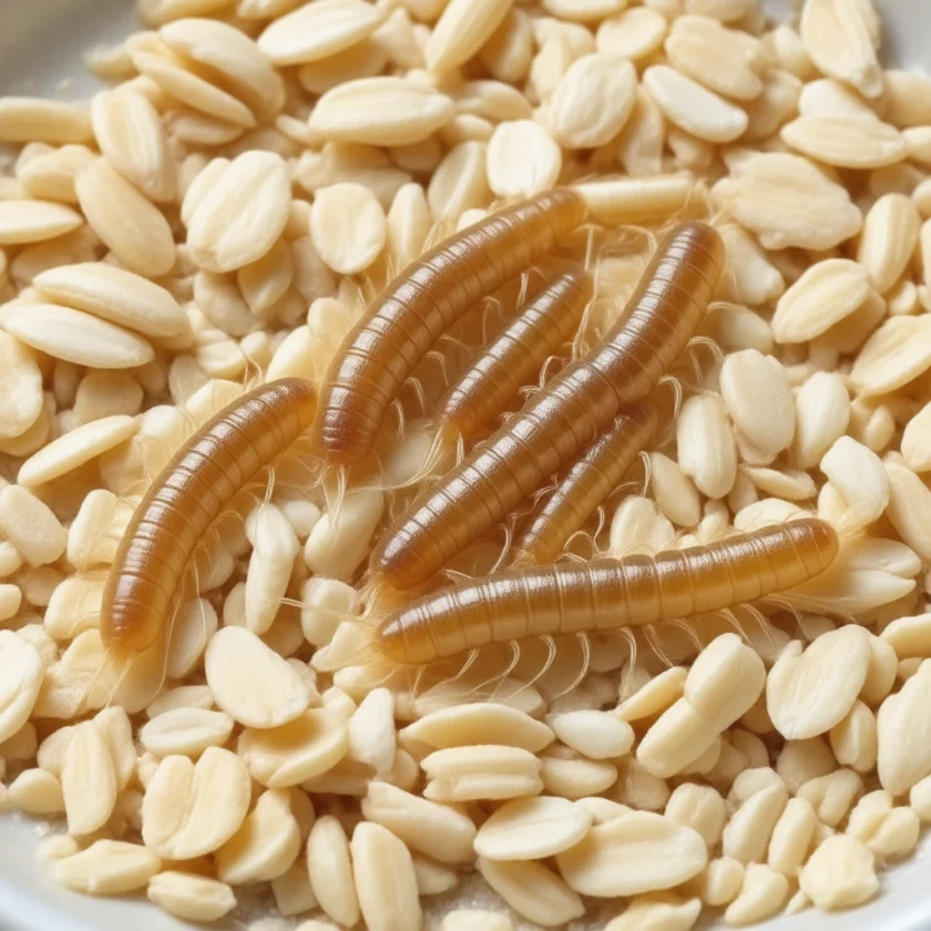
The rediscovery of carnitine came from an unlikely direction: the diet of a beetle. In the 1940s the insect physiologist Gottfried Fraenkel was working out the nutritional requirements of the yellow mealworm, Tenebrio molitor, the larva of a darkling beetle that is a familiar pest of stored grain and a familiar food for pet reptiles. Fed a purified diet containing all the then-known B vitamins, the larvae still failed to grow and could not complete their development into pupae. Something else in natural foods was required. In June 1948 Fraenkel and his colleagues announced in Nature "BT, a new vitamin of the B-group," the subscript T standing for Tenebrio. The factor was present in a range of natural foods.
Isolating it took four more years and a collaboration with organic chemists. In 1952 two short papers in Archives of Biochemistry and Biophysics — the first titled simply "The identity of vitamin BT with carnitine," the second the fuller "Chemical studies on vitamin BT: isolation and characterization as carnitine" — by Herbert Carter, P. K. Bhattacharyya, K. R. Weidman and Fraenkel showed that the mealworm's mystery vitamin was the forty-seven-year-old curiosity from Liebig's extract. The name stuck for a while, and older textbooks and some modern reviews still mention "vitamin BT" as a historical synonym. Fraenkel went on to map the distribution of carnitine across the animal kingdom, and in 1957 he and S. Friedman wrote the first major review of the compound, published in the series Vitamins and Hormones.
Why it stopped being a vitamin. A vitamin, by definition, is a substance the body needs but cannot make. The mealworm genuinely cannot make carnitine, so for Tenebrio the label is accurate. Humans and other mammals can. Over the 1960s and early 1970s the biosynthetic pathway was worked out: the amino acid lysine, once built into proteins, is methylated to trimethyllysine; that is converted step by step to γ-butyrobetaine in most tissues; and the final hydroxylation to carnitine takes place in the liver and, in some species, the kidney. The methyl groups come from methionine, and the enzymes need vitamin C, iron and vitamin B₆ as helpers. Work in the rat by Vichai Tanphaichitr and Harry Broquist in 1973 showed that lysine-deficient animals made less carnitine, nailing down the amino-acid origin. Bremer's 1983 review summarised the situation that had emerged: carnitine is synthesised "in most eucaryotic organisms, although a few insects (and most likely some newborn animals) require it as a nutritional factor (vitamin BT)." Because a healthy adult makes what he or she needs and the kidney reabsorbs almost all of it, carnitine failed the essentiality test and was reclassified. The modern term is conditionally essential: usually made in sufficient quantity, but sometimes not — in premature infants, in certain inherited disorders, on dialysis, and with certain drugs, as later sections describe.
Irving Fritz and the Burning of Fat (1955–1959)
The person who finally asked what carnitine does in a mammal was a young American physiologist, Irving B. Fritz. In a 1955 paper in Acta Physiologica Scandinavica, "The effect of muscle extracts on the oxidation of palmitic acid by liver slices and homogenates," Fritz reported a simple but startling observation: adding an extract of muscle to liver tissue made the liver burn a long-chain fatty acid (palmitic acid, the commonest saturated fat in the body) much faster. The stimulating factor in the extract turned out to be carnitine. Through the rest of the decade he pinned the effect down. In 1959 he showed in the American Journal of Physiology that carnitine acts specifically on the oxidation of long-chain fatty acids by liver, and with B. McEwen he reported in Science the same year that it does the same in muscle. Fritz was already speculating in print, in an essay titled "Facts and speculations about the function of carnitine," that carnitine might be helping fatty acids get to the site where they are burned.
To appreciate why this mattered, it helps to know what biochemists had learned in the previous decade. The mitochondrion had been identified as the cell's power plant; the enzymes of β-oxidation, which chop fatty acids into two-carbon units for the citric-acid cycle, had been shown to sit inside it; and it was known that fatty acids must first be "activated" by attaching them to coenzyme A. What nobody could explain was how a long, activated fatty acid, which cannot pass through the mitochondrion's inner membrane, ever reached the enzymes on the far side. Fritz's muscle-extract factor was the missing piece. Carnitine, the forgotten base from Liebig's extract, was the key to the door.
The Carnitine Shuttle Takes Shape (1962–1975)
The mechanism was assembled within a few years by two groups working in parallel: Fritz, now with K. T. Yue, and Jon Bremer in Oslo. In 1963 Bremer published "Carnitine in intermediary metabolism: the biosynthesis of palmitylcarnitine by cell subfractions" in the Journal of Biological Chemistry, showing that mitochondrial fractions could transfer the long-chain fatty acid from coenzyme A onto carnitine, making palmitoylcarnitine. The same year, in the Journal of Lipid Research, Fritz and Yue described the enzyme responsible — a "long-chain carnitine acyltransferase" — and showed that acylcarnitine derivatives were the actual intermediates through which carnitine increased fatty-acid oxidation. The picture that emerged, and that every biochemistry student now learns as the carnitine shuttle, has three parts:
- Carnitine palmitoyltransferase I (CPT I), on the outer face of the inner membrane, swaps the fatty acid from coenzyme A onto carnitine, making a fatty acyl-carnitine.
- A translocase in the inner membrane carries the acyl-carnitine inside in exchange for a free carnitine coming out — a one-for-one swap, so the mitochondrion's carnitine pool is neither drained nor flooded.
- Carnitine palmitoyltransferase II (CPT II), on the inner face, hands the fatty acid back to a fresh coenzyme A inside the matrix, where β-oxidation can begin. The freed carnitine returns through the translocase for the next passenger.
The two transferases were separated and characterised through the 1960s; Bremer's 1983 review counts "two acyl-CoA:carnitine acyltransferases with overlapping chain-length specificities" plus a third, octanoyltransferase, in the peroxisomes. The exchange carrier — the middle step — was the last to be pinned down. In 1975 Shri Pande, in the Proceedings of the National Academy of Sciences, described "a mitochondrial carnitine acylcarnitine translocase system," and the same year Rona Ramsay and Philip Tubbs in Cambridge reported in FEBS Letters that fatty-acid uptake by heart mitochondria worked as "an acylcarnitine-carnitine exchange." Even then the carrier was, in Bremer's words, "deduced from functional studies" rather than isolated as a protein; its gene was cloned only in the 1990s.
Two further discoveries rounded out the classical picture. In 1980 Denis McGarry and Daniel Foster in Dallas showed that CPT I is inhibited by malonyl-CoA, the first building block of fat synthesis — an elegant switch that stops a cell from making fat and burning it at the same time, and the molecular reason a high-carbohydrate meal turns off fat burning. And the shuttle turned out to run in reverse for short molecules: carnitine acetyltransferase moves two-carbon acetyl groups out of the mitochondrion, buffering the cell's supply of free coenzyme A. That second, less famous job is why acetyl-L-carnitine exists as a natural metabolite, and it explains several of the clinical findings described later. By the mid-1970s carnitine had gone from a forgotten curiosity to one of the best-understood small molecules in energy metabolism.
The Children Who Could Not Keep Carnitine (1973–1999)
If carnitine is the gate for fat, then a person without it should be unable to burn fat — and should get into trouble precisely when the body most needs fat for fuel: during fasting, illness and prolonged exercise. In March 1973 Andrew Engel and Corrado Angelini at the Mayo Clinic reported exactly such a patient in Science, in a paper titled "Carnitine deficiency of human skeletal muscle with associated lipid storage myopathy: a new syndrome." The patient had progressive muscle weakness; under the microscope her muscle fibres were stuffed with "myriad lipid-filled vacuoles," fat that had been delivered to the muscle but could not be burned. Homogenates of her muscle oxidised fatty acids more slowly than those of eleven controls; adding carnitine restored the rate to normal; and her muscle carnitine measured less than 20 percent of the level in 42 controls. The authors wrote that this was "the first recognized instance of carnitine deficiency in human skeletal muscle." Two years later Karpati, Carpenter, Engel and colleagues described a more severe "systemic" form in Neurology, with low carnitine in blood and liver as well as muscle, episodes of encephalopathy resembling Reye's syndrome, and cardiomyopathy.
Why would a body run short of something it can make? The answer was not a failure of synthesis but of retention. In 1988 William Treem, Charles Stanley and colleagues showed in the New England Journal of Medicine that in primary carnitine deficiency the fault lay in a failure of carnitine transport in kidney, muscle and cultured skin cells: these patients' kidneys could not reabsorb carnitine, so it poured out in the urine, and their tissues could not concentrate what little remained. Treatment was, and is, remarkably simple — large oral doses of L-carnitine, which push enough into cells by mass action to keep the shuttle running and can reverse the cardiomyopathy.
The gene followed a decade later, from Japan. In 1998 Ikumi Tamai, Akira Tsuji and colleagues at Kanazawa University cloned a "sodium ion-dependent, high affinity human carnitine transporter" they named OCTN2. In January 1999, in Nature Genetics, Jun-ichi Nezu, Tamai and a large collaboration showed that mutations in the gene encoding OCTN2, SLC22A5, cause primary systemic carnitine deficiency — first in a mouse strain with fatty liver and cardiomyopathy, then in three human families with deletions, frameshifts and a splice-site mutation. The paper describes the disorder as an autosomal recessive condition "characterized by progressive cardiomyopathy, skeletal myopathy, hypoglycaemia and hyperammonaemia" that "has also been linked to sudden infant death syndrome." The condition is rare — the U.S. National Library of Medicine puts it at roughly 1 in 100,000 newborns worldwide, and about 1 in 40,000 in Japan — and it is now picked up by newborn-screening programmes in many countries, which is a fitting end to a story that began with a beetle that could not grow. The site's Carnitine Deficiency page covers the modern clinical picture.
From Laboratory to Pharmacy: Levocarnitine, Dialysis and Valproate
Once a genuine deficiency disease existed, carnitine became a drug. The U.S. Food and Drug Administration approved levocarnitine tablets as a new molecular entity on 27 December 1985, an oral solution in April 1986 and an intravenous form in December 1992. The prescribing information is narrow and precise: oral levocarnitine is indicated "in the treatment of primary systemic carnitine deficiency," whose reported presentation "consisted of recurrent episodes of Reye-like encephalopathy, hypoketotic hypoglycemia, and/or cardiomyopathy," and for certain secondary deficiencies caused by inborn errors of metabolism; the injectable form is indicated "for the prevention and treatment of carnitine deficiency in patients with end stage renal disease who are undergoing dialysis." Those two indications mark the two kinds of deficiency medicine had come to recognise: primary (the transporter is broken) and secondary (the transporter works but something else drains the pool).
Dialysis. Carnitine is a small, water-soluble molecule, which is exactly what a dialysis membrane is designed to remove. Patients on long-term haemodialysis lose carnitine into the dialysate at every session, and by the 1980s low plasma and muscle carnitine were well documented in this group, alongside the muscle weakness, cramps, cardiomyopathy and hard-to-treat anaemia that plague it. In 2003 the U.S. National Kidney Foundation convened a Carnitine Consensus Conference, chaired by Garabed Eknoyan, whose practice recommendations in the American Journal of Kidney Diseases defined a "dialysis-related carnitine disorder" and set out when a trial of levocarnitine was reasonable. The evidence for hard outcomes remains debated, but the biology is not: this is the one large adult population in which a real, measurable carnitine deficit is routine.
Valproate. The second well-established secondary deficiency came from the epilepsy clinic. Valproic acid, introduced as an anticonvulsant in the 1960s and 1970s, is itself a short branched fatty acid, and it is handled by the same coenzyme A and carnitine machinery as dietary fat. In 1982 Y. Ohtani, F. Endo and I. Matsuda reported in the Journal of Pediatrics that children treated with valproate had lower plasma carnitine and higher blood ammonia than children on other anticonvulsants — the paper's title, "Carnitine deficiency and hyperammonemia associated with valproic acid therapy," named a syndrome that is now standard teaching. Valproate-induced hyperammonaemia and, rarely, liver failure are treated with intravenous L-carnitine, a direct clinical descendant of Fritz's 1955 test tube. The site's Epilepsy page discusses the drug itself.
New Forms: Acetyl-L-Carnitine and Propionyl-L-Carnitine
From the late 1970s pharmaceutical chemists, much of the work coming from Italy, began to study carnitine's natural short-chain esters as drugs in their own right. The logic followed directly from the shuttle: an acyl-carnitine is carnitine already carrying a passenger, and the passenger changes where the molecule goes and what it delivers.
Acetyl-L-carnitine (ALCAR) is carnitine carrying a two-carbon acetyl group — the very molecule the mitochondrion exports to balance its coenzyme A. It crosses the blood–brain barrier more readily than plain carnitine and donates its acetyl group to the synthesis of acetylcholine and to cellular energy. Through the 1980s and 1990s it was tested in dementia, diabetic and HIV-related nerve pain, and low mood; the results in dementia were modest, but the mood literature has held up better than most. A 2018 systematic review and meta-analysis by Nicola Veronese and colleagues in Psychosomatic Medicine pooled twelve randomised trials with 791 participants and found that acetyl-L-carnitine reduced depressive symptoms compared with placebo and performed comparably to established antidepressants in the three head-to-head trials, with fewer side effects, the benefit being clearest in older adults. The authors called for larger trials, and that call stands. The site's Brain, Mood & Depression page covers this evidence in detail.
Propionyl-L-carnitine (PLC) carries a three-carbon propionyl group, which can be fed into the citric-acid cycle as succinate — a convenient side entrance for energy in tissue starved of oxygen. Its natural home has been the failing heart and, above all, peripheral artery disease, where leg muscle cramps with walking because narrowed arteries cannot deliver enough oxygen. In 1995 Gregorio Brevetti and colleagues in Naples published in the Journal of the American College of Cardiology a double-blind, placebo-controlled, multicentre dose-titration study of propionyl-L-carnitine in intermittent claudication, showing improved walking distance in patients with the more severe disease. Later trials were mixed, and PLC never became a mainstream therapy outside Italy, but it remains one of the better-studied metabolic approaches to the condition. See the site's Peripheral Artery Disease page and the Carnitine Forms page for how the forms compare.
Heart Trials and the TMAO Debate (2013 Onward)
Carnitine's heart story is old — the heart burns fat for most of its energy, and small trials of L-carnitine after heart attacks were being run in Italy by the 1980s — but its two most-cited chapters were both written, by coincidence, in the spring of 2013, and they pointed in opposite directions.
The secondary-prevention meta-analysis. In April 2013 Mayo Clinic Proceedings published a systematic review and meta-analysis by James DiNicolantonio, Carl Lavie and colleagues pooling thirteen controlled trials with 3,629 patients who had suffered an acute myocardial infarction. Compared with placebo or usual care, L-carnitine was associated with a 27 percent reduction in all-cause mortality, a 65 percent reduction in ventricular arrhythmias and a 40 percent reduction in angina, with no significant effect on reinfarction or heart failure. The authors called it "inexpensive and safe" and asked for large modern trials. Letters in the same journal that August questioned how much weight the pooled result could bear, since most of the trials were small, older, and conducted before today's standard post-infarction drugs; the authors replied in the same issue. Readers should note that the paper was published in the Mayo Clinic's journal rather than produced by Mayo Clinic researchers — a distinction often lost in news coverage. The site's Heart & Circulation page walks through these numbers.
The TMAO hypothesis. A week earlier, in Nature Medicine, Robert Koeth, Stanley Hazen and colleagues at the Cleveland Clinic published "Intestinal microbiota metabolism of L-carnitine, a nutrient in red meat, promotes atherosclerosis." Their argument reached back to Kutscher's trimethylamine: certain gut bacteria strip the three-methyl nitrogen from carnitine to make trimethylamine (TMA), the liver oxidises it to trimethylamine-N-oxide (TMAO), and TMAO, they proposed, promotes atherosclerosis. In their experiments, meat-eaters produced far more TMAO than vegans after a carnitine dose; among 2,595 patients undergoing cardiac evaluation, higher plasma carnitine predicted heart attack, stroke or death — but "only among subjects with concurrently high TMAO levels"; and long-term carnitine feeding worsened atherosclerosis in mice, unless their gut bacteria were suppressed with antibiotics.
The paper drew immediate and continuing criticism, and the honest position today is that the question is open. In a 2013 mini-review in Atherosclerosis, John Ussher, Gary Lopaschuk and Arduino Arduini laid out the discrepancies: fish is a major direct dietary source of TMAO yet is associated with better cardiovascular health; a body of trials shows benefit from carnitine in ischaemic heart disease and insulin resistance; and the mouse strain used is far from a human. Later critics added that TMAO may be a marker of kidney function rather than a cause of disease, and that the human evidence is observational. The hypothesis remains a hypothesis — a serious one, actively investigated — and the site's TMAO Question page presents both sides in full.
Carnitine Today
More than a century after Gulewitsch and Krimberg, carnitine lives a double life. In the hospital it is a targeted medicine: prescription levocarnitine for children with a broken OCTN2 transporter, for certain inborn errors of metabolism, for dialysis patients with a documented deficit, and intravenously for valproate toxicity. In every one of these uses the logic is the same one Engel and Angelini demonstrated in 1973 — replace what the body cannot keep, and the fat-burning machinery restarts.
Outside the hospital, carnitine is one of the most widely sold sports and "fat-burner" supplements in the world, available as plain L-carnitine, L-carnitine L-tartrate, acetyl-L-carnitine and propionyl-L-carnitine. The marketing borrows the real biochemistry of the shuttle — carnitine is genuinely required to burn fat — and stretches it into a claim the trials do not support: in well-fed people the shuttle is not the bottleneck, and adding more carnitine does not, on the evidence, melt fat away. The genuinely interesting modern findings lie elsewhere: that muscle carnitine can be raised by months of supplementation with carbohydrate, that acetyl-L-carnitine has a respectable evidence base in depression, and that carnitine may help as an add-on in specific heart conditions. All of that, together with the honest uncertainties about TMAO, is laid out on the Carnitine hub and its Benefits pages.
The molecule itself has not changed since it crystallised out of a jar of beef extract in 1905. What has changed is that we now know why muscle is so rich in it, why a beetle needs it in its diet and we usually do not, why a child can be born unable to hold on to it, and why a molecule that helps a failing heart might, through the bacteria in our gut, also have something to say about the red meat it was named for. Few nutrients carry so much history in a name.
Key Research Papers
The list follows the historical arc of the article: the original isolation and structure papers, the vitamin BT identification, the fatty-acid-oxidation and shuttle discoveries, the deficiency syndromes and the transporter gene, the two secondary-deficiency landmarks, and the modern clinical and TMAO literature. Every identifier was verified against Crossref or PubMed before being listed.
- Gulewitsch W, Krimberg R (1905). Zur Kenntnis der Extraktivstoffe der Muskeln. II. Mitteilung. Über das Carnitin. Hoppe-Seyler's Zeitschrift für physiologische Chemie. — doi:10.1515/bchm2.1905.45.3-4.326
- Krimberg R (1908). Zur Kenntnis der Extraktivstoffe der Muskeln. X. Mitteilung. Über die Identität des Novains mit dem Carnitin. Hoppe-Seyler's Zeitschrift für physiologische Chemie. — doi:10.1515/bchm2.1908.55.6.466
- Tomita M, Sendju Y (1927). Über die Oxyaminoverbindungen, welche die Biuretreaktion zeigen. III. Spaltung der γ-Amino-β-oxy-buttersäure in die optisch-aktiven Komponenten. Hoppe-Seyler's Zeitschrift für physiologische Chemie. — doi:10.1515/bchm2.1927.169.4-6.263
- Fraenkel G, Blewett M, Coles M (1948). BT, a new vitamin of the B-group and its relation to the folic acid group, and other anti-anæmia factors. Nature. — doi:10.1038/161981a0
- Carter HE, Bhattacharyya PK, Weidman KR, Fraenkel G (1952). Chemical studies on vitamin BT isolation and characterization as carnitine. Archives of Biochemistry and Biophysics. — PubMed PMID: 12997117
- Fritz I (1955). The effect of muscle extracts on the oxidation of palmitic acid by liver slices and homogenates. Acta Physiologica Scandinavica. — PubMed PMID: 13282744
- Bremer J (1963). Carnitine in intermediary metabolism. The biosynthesis of palmitylcarnitine by cell subfractions. Journal of Biological Chemistry. — PubMed PMID: 14063302
- Fritz IB, Yue KT (1963). Long-chain carnitine acyltransferase and the role of acylcarnitine derivatives in the catalytic increase of fatty acid oxidation induced by carnitine. Journal of Lipid Research. — PubMed PMID: 14168165
- Pande SV (1975). A mitochondrial carnitine acylcarnitine translocase system. Proceedings of the National Academy of Sciences of the United States of America. — PubMed PMID: 1055387
- Ramsay RR, Tubbs PK (1975). The mechanism of fatty acid uptake by heart mitochondria: an acylcarnitine-carnitine exchange. FEBS Letters. — PubMed PMID: 1132491
- Engel AG, Angelini C (1973). Carnitine deficiency of human skeletal muscle with associated lipid storage myopathy: a new syndrome. Science. — PubMed PMID: 4687787
- Treem WR, Stanley CA, Finegold DN, Hale DE, Coates PM (1988). Primary carnitine deficiency due to a failure of carnitine transport in kidney, muscle, and fibroblasts. New England Journal of Medicine. — PubMed PMID: 3185635
- Nezu J, Tamai I, Oku A, et al. (1999). Primary systemic carnitine deficiency is caused by mutations in a gene encoding sodium ion-dependent carnitine transporter. Nature Genetics. — PubMed PMID: 9916797
- Bremer J (1983). Carnitine—metabolism and functions. Physiological Reviews. — PubMed PMID: 6361812
- Ohtani Y, Endo F, Matsuda I (1982). Carnitine deficiency and hyperammonemia associated with valproic acid therapy. Journal of Pediatrics. — PubMed PMID: 6813444
- Eknoyan G, Latos DL, Lindberg J; National Kidney Foundation Carnitine Consensus Conference (2003). Practice recommendations for the use of L-carnitine in dialysis-related carnitine disorder. American Journal of Kidney Diseases. — PubMed PMID: 12666074
- Brevetti G, Perna S, Sabbà C, Martone VD, Condorelli M (1995). Propionyl-L-carnitine in intermittent claudication: double-blind, placebo-controlled, dose titration, multicenter study. Journal of the American College of Cardiology. — PubMed PMID: 7594063
- Veronese N, Stubbs B, Solmi M, Ajnakina O, Carvalho AF, Maggi S (2018). Acetyl-L-Carnitine Supplementation and the Treatment of Depressive Symptoms: A Systematic Review and Meta-Analysis. Psychosomatic Medicine. — PubMed PMID: 29076953
- DiNicolantonio JJ, Lavie CJ, Fares H, Menezes AR, O'Keefe JH (2013). L-carnitine in the secondary prevention of cardiovascular disease: systematic review and meta-analysis. Mayo Clinic Proceedings. — PubMed PMID: 23597877
- Koeth RA, Wang Z, Levison BS, et al. (2013). Intestinal microbiota metabolism of L-carnitine, a nutrient in red meat, promotes atherosclerosis. Nature Medicine. — PubMed PMID: 23563705
- Ussher JR, Lopaschuk GD, Arduini A (2013). Gut microbiota metabolism of L-carnitine and cardiovascular risk. Atherosclerosis. — PubMed PMID: 24267266
- Longo N, Frigeni M, Pasquali M (2016). Carnitine transport and fatty acid oxidation. Biochimica et Biophysica Acta. — PubMed PMID: 26828774
PubMed Topic Searches
- PubMed: Carnitine history and discovery
- PubMed: Carnitine shuttle, palmitoyltransferases and translocase
- PubMed: Primary carnitine deficiency and SLC22A5 / OCTN2
- PubMed: L-carnitine, TMAO and cardiovascular risk
External Authoritative Resources
Connections
- All Amino Acids
- Carnitine Hub
- Carnitine Benefits
- Carnitine Deficiency
- Carnitine Forms & the TMAO Question
- Carnitine for Heart & Circulation
- Amino Acid Derivatives
- Lysine
- Methionine
- Creatine
- Vitamin C
- Iron
- Heart Attack
- Peripheral Artery Disease
- Kidney Disease
- Epilepsy
- Fat Burning & Ketosis (Animation)