Nattokinase for Cardiovascular Health & Fibrinolysis
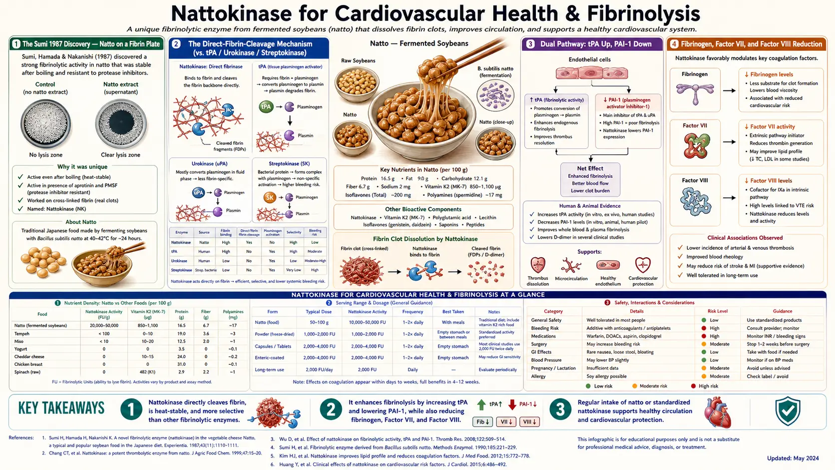
Nattokinase was first described in a 1987 paper by Dr. Hiroyuki Sumi and colleagues at Miyazaki Medical College in Japan, who reported a strong fibrinolytic (clot-dissolving) activity in natto. Popular accounts of the discovery describe Sumi screening traditional foods for fibrinolytic activity while working in Chicago in the early 1980s: in those accounts he dropped natto onto a fibrin plate (the standard agar substrate for testing clot-dissolving enzymes), expected to see the cleared zone characteristic of a strong fibrinolytic agent over a span of hours — and instead got the largest clear zone of any food he had ever tested, within just minutes. The enzyme he isolated and named "nattokinase" is unusual: unlike tPA, urokinase, and streptokinase — the three principal pharmaceutical thrombolytics, all of which work indirectly by activating plasminogen to plasmin — nattokinase acts directly on fibrin. It is also unusual in that it appears to retain measurable activity after oral absorption, one of the very few proteolytic enzymes for which this has been documented in human pharmacokinetic studies. This deep-dive walks through the discovery, the unique mechanism, the deep-vein-thrombosis prevention pilot data including the Cesarone long-flight trial, the fibrinogen and clotting-factor reductions reported in an open-label trial, the 3-year NAPS randomized trial that found no effect on atherosclerosis progression, comparison with aspirin and warfarin, and what the evidence does and does not show about its cardiovascular use.
Table of Contents
- The Sumi 1987 Discovery — Natto on a Fibrin Plate
- The Direct-Fibrin-Cleavage Mechanism (vs. tPA / Urokinase / Streptokinase)
- Dual Pathway: tPA and PAI-1 in Laboratory Studies
- Fibrinogen, Factor VII, and Factor VIII Reduction
- Deep Vein Thrombosis Prevention — The Cesarone Long-Flight Trial
- The Oral-Bioavailability Question
- Comparison with Aspirin and Warfarin
- Whole-Blood Viscosity and Red-Cell Aggregation
- Endothelial Function and Flow-Mediated Dilation
- Practical Cardiovascular Dosing Protocols
- Cautions — Anticoagulants, Antiplatelets, Surgery, Bleeding Disorders
- Key Research Papers
- Connections
- Featured Videos
The Sumi 1987 Discovery — Natto on a Fibrin Plate
The discovery of nattokinase is one of the more memorable stories in natural-product chemistry. Hiroyuki Sumi, a Japanese postdoctoral researcher at the University of Chicago Medical School in the early 1980s, was working in a lab studying thrombolysis — the dissolution of blood clots, which was becoming a clinically central problem as the developed world's leading cause of death shifted firmly to cardiovascular disease. tPA, urokinase, and streptokinase were the available pharmaceutical agents, all intravenous, all expensive, all with significant bleeding risk and short half-lives.
Sumi had a sideline interest: he was systematically screening traditional foods for fibrinolytic activity, on the theory that long-traditional foods that humans had selected for over centuries might contain useful bioactive compounds. The screening method was straightforward: a fibrin plate (purified fibrin polymerized on agar in a Petri dish) is the standard substrate for testing clot-dissolving activity. A drop of test material is placed on the surface; a clear zone develops over time as the fibrin polymer is dissolved. The diameter and rate of the clear zone, compared to a standard, gives the activity in fibrinolytic units (FU).
In the popular accounts, Sumi had screened over 200 traditional foods with mostly negative or weakly-positive results. Then he dropped natto — the sticky, stringy fermented soybean breakfast staple from his native Japan, an acquired-taste food he ate regularly — onto a fibrin plate. The clear zone developed within minutes, and was larger than any food he had previously tested. The 1987 Experientia paper, from Sumi’s group at Miyazaki Medical College, reported a fibrinolytic activity in natto of about 40 plasmin units per gram and launched the modern nattokinase literature.
Sumi isolated the active enzyme, named it "nattokinase" (the Greek suffix –kinase, indicating an enzyme), and characterized it as a 275-amino-acid serine protease produced during fermentation by Bacillus subtilis var. natto, the specific bacterial strain that converts cooked soybeans into the traditional natto food. The enzyme was assigned EC number 3.4.21.62 in the Enzyme Commission nomenclature, classifying it as a subtilisin-family serine endopeptidase.
What made the discovery particularly interesting was that natto had been a staple food in Japan for at least 1,000 years — documented in records back to the Heian period (794–1185 CE), with origin legends placing it considerably earlier — and Japan has long been notable for one of the world's lowest rates of cardiovascular mortality. The convergence of these facts — a uniquely concentrated dietary fibrinolytic enzyme, in a population with uniquely low cardiovascular disease — immediately raised the question of whether natto consumption was contributing causally to Japanese cardiovascular longevity. That question is still not fully resolved, but the epidemiologic data (Takayama study, JACC cohort) discussed in the stroke prevention deep-dive are consistent with a meaningful contribution.
The Direct-Fibrin-Cleavage Mechanism (vs. tPA / Urokinase / Streptokinase)
Most clinically used fibrinolytic agents do not actually dissolve fibrin themselves. They are plasminogen activators — they convert the inactive plasma zymogen plasminogen into the active enzyme plasmin, and it is plasmin that does the actual fibrin-cleaving work. The pharmaceutical thrombolytics fall into this category:
- Tissue plasminogen activator (tPA, alteplase) — recombinant human enzyme, the standard-of-care for acute ischemic stroke if administered within ~4.5 hours of symptom onset. Activates plasminogen specifically at fibrin surfaces.
- Urokinase — originally isolated from human urine, also activates plasminogen, but with less fibrin-specificity than tPA.
- Streptokinase — bacterial protein from Streptococcus, forms a complex with plasminogen that has plasminogen-activator activity. Cheap, used widely outside the US for myocardial infarction.
Nattokinase is different. It cleaves fibrin directly, in a manner similar to plasmin itself, without requiring plasminogen as an intermediate. It also activates plasminogen (more on that in the next section), but the direct fibrin-cleavage activity is the more distinctive feature. The cleavage sites on the fibrin alpha- and beta-chains are different from those preferred by plasmin, and the cleavage kinetics are different, but the net effect — depolymerization of the fibrin clot — is similar.
From a mechanistic standpoint this matters because direct fibrin cleavage proceeds even in conditions where plasminogen is depleted or where plasminogen activator inhibitor-1 (PAI-1) is elevated — both of which can limit the effectiveness of tPA and similar agents. From a clinical standpoint it matters because the direct cleavage mechanism is what allows oral nattokinase to have any plausible effect at all (an orally-absorbed plasminogen activator would face daunting bioavailability problems; an orally-absorbed enzyme that cleaves fibrin directly faces fewer).
Dual Pathway: tPA and PAI-1 in Laboratory Studies
The second mechanism — complementary to direct fibrin cleavage — is that nattokinase modulates the endogenous plasminogen-activator system in the direction of more fibrinolysis. Two specific changes are described, mainly in laboratory studies:
- tPA activity increases — in laboratory (in vitro) studies, nattokinase activates the production of tissue plasminogen activator (tPA). A 1990 report from Sumi’s group described a mild enhancement of plasma fibrinolytic activity and tPA production after oral nattokinase or natto. The Kurosawa 2015 single-dose crossover study in 12 healthy young men did not measure tPA; after 2,000 FU it found D-dimer raised at 6 and 8 hours and fibrin/fibrinogen degradation products raised at 4 hours, all within the normal range.
- PAI-1 activity decreases — plasminogen activator inhibitor-1 is the body's principal endogenous brake on fibrinolysis. PAI-1 binds and inactivates tPA. Elevated PAI-1 is independently associated with cardiovascular events, particularly myocardial infarction. In laboratory (in vitro) studies, nattokinase cleaves and inactivates PAI-1, removing this brake. In people, the Kurosawa 2015 study measured total PAI-1 and did not report a significant change, and the 3-year NAPS randomized trial (Hodis 2021) found no significant effect of nattokinase on any laboratory measure, which included coagulation and fibrinolysis factors.
The proposed net effect of these two changes is a shift of the fibrinolytic balance toward more endogenous clot dissolution. This is mechanistically distinct from the direct-fibrin-cleavage activity of nattokinase itself: the proposal is that even after the nattokinase molecules have been cleared from circulation, higher tPA and lower PAI-1 would continue to support endogenous fibrinolysis. Human trials have not confirmed this.
The clinical relevance: morning cardiovascular events (myocardial infarction, sudden cardiac death) are partly driven by the natural circadian peak of PAI-1 activity, which is highest in the early morning. Some practitioners suggest evening dosing of nattokinase specifically to support fibrinolytic activity through the high-risk pre-dawn hours; the evidence for this is suggestive rather than definitive.
Fibrinogen, Factor VII, and Factor VIII Reduction
The Hsia 2009 Nutrition Research paper is one of the more important nattokinase human trials. Hsia and colleagues ran an open-label, self-controlled trial (no placebo group) in 45 people from three groups (healthy volunteers, patients with cardiovascular risk factors, and dialysis patients), who each took two capsules of nattokinase (2,000 fibrinolytic units, FU, per capsule) daily for 2 months. The endpoints were not just fibrinolytic activity but the underlying coagulation cascade itself. The findings:
- Plasma fibrinogen decreased by approximately 7–10% across all three groups. Fibrinogen is the substrate from which clots are made; lower fibrinogen means less raw material for thrombus formation, lower blood viscosity, and lower cardiovascular risk (elevated fibrinogen is an independent cardiovascular risk factor).
- Factor VII activity decreased by 7–14% across the three groups (14% in the healthy group). Factor VII is part of the extrinsic coagulation pathway and is activated by tissue factor exposed at sites of vascular injury.
- Factor VIII activity decreased by 17–19% across the three groups. Factor VIII is part of the intrinsic coagulation pathway, the same factor that is deficient in hemophilia A.
The interpretation: nattokinase has effects on the coagulation cascade itself, not just on already-formed fibrin clots. The mechanism for these effects is not fully established — they may reflect direct proteolytic effects on the clotting factors themselves, indirect effects via the plasmin system, or hepatic-synthesis-rate effects via signaling. What matters clinically is the net direction: lower fibrinogen, lower factor VII, lower factor VIII, all of which trend in the direction of reduced thrombotic tendency. By contrast, the placebo-controlled NAPS trial (Hodis 2021), which gave 265 healthy adults 2,000 FU a day or placebo for a median of 3 years, found no significant effect of nattokinase on any laboratory measure, including coagulation and fibrinolysis factors.
This is also the source of the safety concern: a patient already on an anticoagulant or antiplatelet drug, which is suppressing coagulation through one mechanism, who adds nattokinase, which is suppressing coagulation through additional mechanisms, may end up at significantly elevated bleeding risk. See the cautions section below for the specific drug interactions and the pre-surgical stopping interval that is commonly described.
Deep Vein Thrombosis Prevention — The Cesarone Long-Flight Trial
The single best-known clinical use case for nattokinase is venous thromboembolism prevention, particularly in the long-haul-flight setting. The Cesarone 2003 trial published in Angiology (an Italian-led study, known as the LONFLIT-FLITE trial) is the pivotal study.
Design: 204 high-risk passengers on long-haul flights of 7–8 hours were randomized to placebo or to an oral branded nattokinase-containing preparation, which the abstract describes as “150 mg pinokinase” (the abstract does not list its ingredients or their doses separately). An exercise program was used in both groups. Ultrasound scans of the femoral, popliteal, tibial and superficial veins were performed before the flight and within 90 minutes after it.
Results:
- Placebo group: 5 deep vein thromboses (5.4%) and 2 superficial thromboses detected post-flight among the 92 who completed the study; the abstract reports that all events were asymptomatic.
- Treated group: 0 deep vein thromboses, 0 superficial thromboses.
- The difference was statistically significant (P < 0.025).
The Cesarone trial has been criticized on several grounds — the use of a combined product means the contribution of nattokinase alone cannot be cleanly separated; the sample size is modest; the endpoint (asymptomatic Doppler-detected calf thrombi) is a surrogate for clinically meaningful pulmonary embolism. But it remains the best randomized human evidence for nattokinase in venous thrombosis prevention, and the result has informed the off-label use of nattokinase as a long-haul-flight prophylactic in many integrative-medicine practices.
Practitioners who use it for flights describe 2,000–4,000 FU nattokinase taken 30–60 minutes before a long flight, alongside standard mechanical measures (compression stockings, hourly walking, hydration). For people with known hypercoagulable conditions (factor V Leiden, prothrombin G20210A, antiphospholipid syndrome), nattokinase has not been tested as a replacement for prescribed pharmaceutical anticoagulation; practitioners describe it at most as an adjunct under physician supervision.
For more on deep vein thrombosis itself, see our Deep Vein Thrombosis page.
The Oral-Bioavailability Question
The single most-asked question about nattokinase is whether an enzyme this large — a 275-amino-acid protein with a molecular weight around 27.7 kDa — can possibly survive gastric digestion and reach the systemic circulation in active form. Most ingested proteins are denatured by gastric acid and degraded by pepsin in the stomach, then further degraded by pancreatic proteases (trypsin, chymotrypsin) in the small intestine. The end products of normal dietary protein digestion are short peptides and free amino acids, which are absorbed via PEPT1 and amino-acid transporters in the small intestine.
For nattokinase to act systemically, intact (or at least catalytically active) enzyme must reach the circulation in measurable amounts. The evidence that this actually happens, while not as definitive as one would like, is reasonably strong:
- Human studies measure changes in downstream markers, and the enzyme itself, after oral nattokinase. In the Kurosawa 2015 crossover study, D-dimer and fibrin/fibrinogen degradation products rose 4–8 hours after a single 2,000 FU dose. The Ero 2013 pilot study measured nattokinase directly in serum (by an antibody-based ELISA test) in 11 healthy adults after a single 2,000 FU dose: levels rose significantly from 2 to 24 hours and peaked at about 13 hours, which the authors described as the first evidence that nattokinase can be measured directly in human blood.
- Rat absorption studies show measurable absorption of intact nattokinase through the intestinal wall, with detection in the lymphatic system and in serum.
- Enteric-coated formulations outperform uncoated formulations in head-to-head studies of downstream fibrinolytic markers, consistent with the interpretation that protecting the enzyme from gastric pH preserves activity.
- The enzyme is structurally robust — it is stable across a wide pH range (6–12), tolerates temperatures up to ~60°C, and has a structure (subtilisin family) known for resistance to proteolytic degradation.
The honest position is that the magnitude of absorption is small and probably highly variable from person to person and from formulation to formulation. But there is enough mechanistic and PK evidence to conclude that the downstream effects observed in clinical trials are not entirely placebo — some active enzyme does reach the systemic circulation, and that fraction is enhanced by enteric coating and proper formulation.
Comparison with Aspirin and Warfarin
Patients (and integrative-medicine practitioners) frequently ask whether nattokinase can substitute for low-dose aspirin or warfarin in their specific situation. The honest answer requires distinguishing several distinct clinical scenarios:
- Primary cardiovascular disease prevention in healthy adults with no atherosclerotic disease. Current guideline consensus is against routine low-dose aspirin for primary prevention in this population, because the bleeding risk roughly offsets the cardiovascular benefit. Some people consider nattokinase in this setting for pro-fibrinolytic support without the gastrointestinal (GI) bleeding risk profile of aspirin. The largest and longest randomized trial in exactly this group, the NAPS trial (Hodis 2021: 265 adults of median age 65 without cardiovascular disease, 2,000 FU a day or placebo for a median of 3 years), found no significant difference in the progression of carotid intima-media thickness or carotid arterial stiffness, and no significant effect on blood pressure or any laboratory measure; the authors concluded that nattokinase had a null effect on subclinical atherosclerosis progression in healthy people at low cardiovascular risk.
- Secondary prevention after established cardiovascular disease (post-MI, post-PCI, established coronary artery disease, CAD). Low-dose aspirin (and often clopidogrel for a period after stent placement) are evidence-based and life-saving in this population. There are no outcome data on nattokinase as a substitute. Adding nattokinase to prescribed antiplatelet therapy raises a bleeding concern: a published case report describes a cerebellar hemorrhage in a patient on aspirin who added nattokinase.
- Atrial fibrillation with elevated stroke risk (CHADS–VASc ≥ 2). Warfarin or DOAC (apixaban, rivaroxaban, edoxaban, dabigatran) is the standard of care and reduces stroke risk by approximately 60–70%. There are no outcome data on nattokinase as a substitute. Stopping warfarin in favor of "natural alternatives" gives up that reduction, leaving a stroke risk approximately 3x higher than on continued therapy.
- Deep vein thrombosis prophylaxis in long-distance travel. This is the one situation where nattokinase has supportive evidence from a randomized, placebo-controlled trial (the Cesarone trial above). That trial had no aspirin arm, so it does not compare nattokinase with aspirin.
- Active treatment of established deep vein thrombosis (DVT) or pulmonary embolism. Therapeutic anticoagulation with heparin or DOAC is the standard. Nattokinase is not an established treatment for VTE.
In summary: for healthy adults without cardiovascular disease, short studies report changes in clotting factors (Hsia 2009) and, in people with raised blood pressure, in blood pressure (Jensen 2016), while the 3-year NAPS randomized trial found no effect on atherosclerosis progression, blood pressure or laboratory measures; for long-distance travelers, one placebo-controlled trial reported fewer ultrasound-detected thromboses. There are no outcome data supporting nattokinase as a substitute for pharmaceutical anticoagulation in atrial fibrillation, mechanical heart valves, established VTE, or recent acute coronary syndrome, and a published case report describes a clot on a mechanical heart valve in a patient who replaced warfarin with nattokinase.
For more on aspirin and its evolving evidence base in primary vs. secondary prevention, see our Aspirin page.
Whole-Blood Viscosity and Red-Cell Aggregation
The Pais 2006 study in Clinical Hemorheology and Microcirculation measured the effect of nattokinase on whole-blood viscosity and on red-blood-cell aggregation in vitro. Findings:
- Low-shear whole-blood viscosity decreased significantly and in a dose-dependent way, at enzyme concentrations similar to those reached in earlier animal studies
- Erythrocyte aggregation (a measure of how readily red cells clump together at low shear rates, which contributes to microcirculatory sludging) decreased significantly and in a dose-dependent way
- The blood samples were incubated with nattokinase for 30 minutes in the laboratory; the authors called these preliminary data and wrote that additional studies and clinical trials are needed
These were test-tube findings; the study did not measure blood flow or capillary perfusion in people. The proposed applications are in peripheral vascular insufficiency — intermittent claudication, diabetic peripheral neuropathy with microcirculatory contribution, slow wound healing in the setting of compromised peripheral circulation — on the rationale that less red-cell clumping and lower low-shear viscosity would improve delivery of oxygen and nutrients to tissues at the end of the vascular tree.
Endothelial Function and Flow-Mediated Dilation
Flow-mediated dilation (FMD) is a standard non-invasive measure of endothelial function. The technique uses high-resolution ultrasound to measure brachial artery diameter at rest, then after a 5-minute occlusion of forearm blood flow (with a blood-pressure cuff inflated above systolic pressure), then again after release of the occlusion. The reactive hyperemia following cuff release causes endothelium-dependent dilation of the brachial artery; the percent change in diameter is a measure of endothelial-cell nitric oxide production capacity.
No human trial of nattokinase measuring FMD appears in PubMed; the published studies of nattokinase and the endothelium are animal and cell studies. The Jensen 2016 trial, an 8-week blood-pressure study in 79 adults with raised blood pressure, measured von Willebrand factor, a cardiovascular risk marker, and reported a decrease in the women taking nattokinase at P<0.1, which does not reach the conventional 0.05 threshold for statistical significance.
The clinical relevance is that endothelial dysfunction precedes plaque formation in atherosclerosis, and reversing endothelial dysfunction is one of the few interventions that has been shown to slow or reverse the atherosclerotic process. Whether nattokinase contributes in this direction has not been shown: the 3-year NAPS randomized trial found no difference from placebo in the progression of carotid intima-media thickness or carotid arterial stiffness. More established interventions include statins for severe dyslipidemia, angiotensin-converting enzyme inhibitors (ACE inhibitors) for hypertension, smoking cessation, and sustained aerobic exercise.
Practical Cardiovascular Dosing Protocols
- Primary cardiovascular maintenance: 2,000 FU once or twice daily is the amount commonly described. 2,000 FU a day is the dose used in many published trials, including the 3-year NAPS trial, which found no effect on atherosclerosis progression, blood pressure or laboratory measures.
- Active fibrinolytic support (post-event, hypercoagulable conditions under physician supervision): practitioners describe 4,000–6,000 FU twice daily.
- Long-haul flight prophylaxis: practitioners describe 2,000–4,000 FU 30–60 minutes before the flight, sometimes repeated for flights longer than 12 hours.
- Time of day: evening dosing is theoretically rational because endogenous fibrinolytic activity is lowest in the early-morning hours when most cardiovascular events occur. The evidence supporting this practice is suggestive rather than definitive.
- Form: enteric-coated capsules retain more activity through gastric pH than uncoated forms. A single standardized Japanese nattokinase extract is the most-studied preparation; it was used in the Kurosawa, Ero and Jensen studies cited on this page.
- Combination products: nattokinase is often paired with serrapeptase, lumbrokinase, bromelain, or rutin in proteolytic enzyme blends. Whether the combinations are additive or merely additive on price is not well-established.
- Onset: fibrinolytic markers changed within hours of a single dose in the Kurosawa study; the Jensen trial reported blood-pressure changes after 8 weeks of daily use, while the 3-year NAPS trial found no effect on blood pressure or on carotid atherosclerosis measures.
- Duration: the longest randomized trial, NAPS, gave 2,000 FU a day or placebo to 265 people for a median of 3 years; longer-term human safety data are not as comprehensive as for many pharmaceutical agents.
Cautions — Anticoagulants, Antiplatelets, Surgery, Bleeding Disorders
- Concurrent anticoagulant therapy is the principal contraindication. For people on warfarin, apixaban, rivaroxaban, edoxaban (Savaysa), dabigatran (Pradaxa), or therapeutic-dose heparin, adding nattokinase stacks clot-reducing effects, and the research raises a concern about bleeding, including major bleeding events. Published case reports describe a cerebellar hemorrhage in a patient taking aspirin who added nattokinase 400 mg daily for 7 days (Chang 2008), and a clot on a mechanical heart valve in a patient who replaced warfarin with nattokinase for nearly a year (Elahi 2015).
- Concurrent antiplatelet therapy — aspirin, clopidogrel, ticagrelor (Brilinta), prasugrel (Effient). The same bleeding concern applies (the cerebellar hemorrhage case above involved aspirin). The combination is occasionally used under physician supervision in specific scenarios; decisions about combining them belong with a clinician.
- Active bleeding disorders — hemophilia A or B, von Willebrand disease, thrombocytopenia (platelet count below 100), inherited platelet function disorders. Nattokinase is contraindicated.
- Recent or planned surgery — a stop at least 7 days before a planned procedure is the interval commonly described. Listing nattokinase among supplements on the pre-operative anesthesia clearance form lets the surgical team account for it. The same 7-day interval is described for dental procedures involving extraction or significant tissue trauma.
- Recent stroke, especially hemorrhagic stroke. Nattokinase is contraindicated in the acute post-stroke period regardless of stroke subtype, and is permanently contraindicated after intracerebral hemorrhage.
- Pregnancy and lactation. There is no good safety data in pregnancy or breastfeeding.
- Severe hypertension (greater than 180/110). Blood pressure in this range is a medical priority in its own right; nattokinase is not an established sole treatment at this level.
- Soy allergy. The enzyme is purified from fermentation broth and contains negligible soy protein in well-manufactured products, and preparations explicitly labeled soy-free are made for soy-allergic people; for immunoglobulin E-mediated soy allergy, any natto-derived product is a question for an allergist.
- Side effects in healthy adults are typically mild and dose-related: nausea, mild gastrointestinal discomfort, occasional bruising, infrequent menstrual flow increase in pre-menopausal women. Severe adverse events are rare in the published literature.
For more on the broader cardiovascular context, see our pages on Atherosclerosis, Coagulation Panel, and Deep Vein Thrombosis.
Key Research Papers
- Sumi H et al. (1987). A novel fibrinolytic enzyme (nattokinase) in the vegetable cheese natto; a typical and popular soybean food in the Japanese diet. Experientia 43(10):1110-1111. — PubMed PMID: 3478223
- Fujita M et al. (1993). Purification and characterization of a strong fibrinolytic enzyme (nattokinase) in the vegetable cheese natto, a popular soybean fermented food in Japan. Biochem Biophys Res Commun 197(3):1340-1347. — PubMed PMID: 8280151
- Cesarone MR et al. (2003). Prevention of venous thrombosis in long-haul flights with Flite Tabs. Angiology. — PubMed PMID: 14565628
- Hsia CH et al. (2009). Nattokinase decreases plasma levels of fibrinogen, factor VII, and factor VIII in human subjects. Nutrition Research. — PubMed PMID: 19358933
- Kurosawa Y et al. (2015). A single-dose of oral nattokinase potentiates thrombolysis and anti-coagulation profiles. Scientific Reports. — PubMed PMID: 26109079
- Pais E et al. (2006). Effects of nattokinase on red blood cell aggregation and whole blood viscosity. Clin Hemorheol Microcirc. — PubMed PMID: 16899918
- Tai MW, Sweet BV (2006). Nattokinase for prevention of thrombosis. Am J Health Syst Pharm. — PubMed
- Sumi H et al. (1990). Enhancement of the fibrinolytic activity in plasma by oral administration of nattokinase. Acta Haematol. — PubMed PMID: 2123064
- Ero MP et al. (2013). A pilot study on the serum pharmacokinetics of nattokinase in humans. Altern Ther Health Med. — PubMed PMID: 23709455
- Yatagai C et al. (2008). Nattokinase-promoted tissue plasminogen activator release from human umbilical vein endothelial cells. — PubMed PMID: 19996631
- Chen H et al. (2018). Nattokinase: a promising alternative in prevention and treatment of cardiovascular diseases. Biomark Insights. — PubMed PMID: 30013308
- Weng Y et al. (2017). Nattokinase: an oral antithrombotic agent for the prevention of cardiovascular disease. Int J Mol Sci. — PubMed PMID: 28264497
- Hodis HN et al. (2021). Nattokinase atherothrombotic prevention study: A randomized controlled trial. Clin Hemorheol Microcirc 78(4):339-353. — PubMed PMID: 33843667
- Jensen GS et al. (2016). Consumption of nattokinase is associated with reduced blood pressure and von Willebrand factor, a cardiovascular risk marker: results from a randomized, double-blind, placebo-controlled, multicenter North American clinical trial. Integr Blood Press Control 9:95-104. — PubMed PMID: 27785095
- Chang YY et al. (2008). Cerebellar hemorrhage provoked by combined use of nattokinase and aspirin in a patient with cerebral microbleeds. Intern Med 47(5):467-469. — PubMed PMID: 18310985
- Elahi MM et al. (2015). Consequence of patient substitution of nattokinase for warfarin after aortic valve replacement with a mechanical prosthesis. Proc (Bayl Univ Med Cent) 28(1):81-82. — PubMed PMID: 25552810
PubMed Topic Searches
Connections
- All Food
- Nattokinase Overview
- Nattokinase Benefits Hub
- Nattokinase for Blood Pressure
- Nattokinase for Stroke Prevention
- Vitamin K2 (Menaquinone-7) Connection
- Natto (Food)
- Aspirin
- Deep Vein Thrombosis
- Atherosclerosis
- Hypertension
- Coagulation Panel
- Coronary Calcium Score
- ApoB
- Lipid Panel
- Omega-3 Fatty Acids
- Vitamin K
- Bacillus Subtilis
- History