Eggs for Lutein, Zeaxanthin & Eye Health
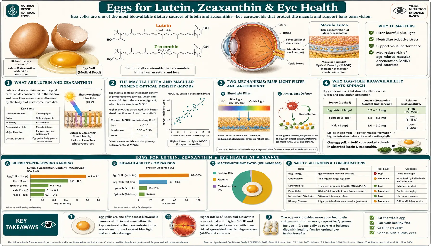
Lutein and zeaxanthin are two yellow xanthophyll carotenoids out of roughly 600 carotenoids found in nature. They are the only two carotenoids that selectively cross the blood-retinal barrier and accumulate in the macula lutea (Latin for "yellow spot") at the center of the retina, where they form the macular pigment that filters blue light and quenches singlet oxygen at the photoreceptor outer segments. Egg yolk is not the densest source of these carotenoids — spinach and kale contain 10-20× more per gram — but the egg-yolk lipid matrix makes them dramatically more bioavailable. The Chung 2004 trial showed egg-derived lutein produced higher serum responses than equivalent doses from spinach or supplements. The 2013 AREDS2 trial concluded that lutein and zeaxanthin could be an appropriate substitute for beta-carotene in the original AREDS formulation for age-related macular degeneration, because beta-carotene was linked to more lung cancers in former smokers. In the egg trials, six eggs a week for 12 weeks raised serum zeaxanthin and macular pigment optical density in women (Wenzel 2006), and one egg a day for 5 weeks raised serum lutein and zeaxanthin in older adults (Goodrow 2006).
Table of Contents
- Lutein and Zeaxanthin Explained
- The Macula Lutea and Macular Pigment Optical Density
- Two Mechanisms: Blue-Light Filter and Antioxidant
- Why Egg-Yolk Bioavailability Beats Spinach
- The AREDS2 Trial and Macular Degeneration
- Egg-Intervention Trials (Wenzel, Kelly, Goodrow)
- Cataract Prevention Evidence
- Blue Light, Screen Use, and MPOD
- Practical Intake: How Many Eggs the Studies Used
- Cautions and Special Populations
- Key Research Papers
- Connections
- Featured Videos
Lutein and Zeaxanthin Explained
Carotenoids are the family of fat-soluble plant pigments responsible for the yellow, orange, and red colors of carrots, peppers, tomatoes, marigolds, and autumn leaves. Plants synthesize them as accessory photosynthetic pigments and as photoprotectants — the same pigments that absorb excess light energy in the chloroplast to prevent oxidative damage to chlorophyll also work in human tissue to absorb light energy and quench reactive oxygen species. Of the ~600 known carotenoids, only about 20 appear in measurable concentration in human serum, and only two — lutein and zeaxanthin — selectively accumulate in the retina at the back of the eye.
Lutein and zeaxanthin are structural isomers (same molecular formula, different arrangement). Both are xanthophylls, meaning they contain hydroxyl groups that distinguish them from the hydrocarbon carotenes like beta-carotene and lycopene. The hydroxyl groups make them slightly polar, which gives them a different absorption pattern and a different cellular distribution than the pure-hydrocarbon carotenes.
A third closely related carotenoid, meso-zeaxanthin, is found in the human macula but not in most dietary sources. It is produced from lutein by an enzyme in the retinal pigment epithelium (RPE65 family) and concentrates at the very center of the fovea. Meso-zeaxanthin is essentially an internal conversion product, not a dietary nutrient in most populations, though some specialty supplements (including a marigold-derived lutein and zeaxanthin supplement) provide it directly.
In the macula, the three carotenoids segregate spatially: meso-zeaxanthin at the foveal center, zeaxanthin in the surrounding parafovea, and lutein in the peripheral macula. This spatial arrangement is exquisitely conserved across vertebrate retinas and strongly suggests the macular pigment evolved for a specific photoprotective purpose.
The Macula Lutea and Macular Pigment Optical Density
The macula lutea is the 5 mm-diameter central region of the retina that handles high-acuity central vision. It contains the highest density of cone photoreceptors and is the area you use to read, recognize faces, and focus on detail. The macula is anatomically distinguishable from the surrounding retina by its yellow color — the result of accumulated lutein and zeaxanthin in the inner retinal layers anterior to the photoreceptors.
The concentration of macular pigment is measured clinically as macular pigment optical density (MPOD), expressed as the log-base-10 of the ratio of incident to transmitted blue light at the peak absorbance wavelength (460 nm). Higher MPOD means more pigment present, which means more blue-light filtering and more antioxidant capacity. Typical MPOD in healthy adults ranges from 0.2 to 0.6, and the variance is largely driven by dietary intake of lutein and zeaxanthin, smoking history, and obesity status (smoking and obesity both reduce MPOD).
The retina presents MPOD a counterintuitive but elegant arrangement. The yellow macular pigment sits in the inner retinal layers, anterior to the photoreceptors. Light entering the eye must pass through the pigment before reaching the rods and cones that actually detect it. This means the pigment selectively absorbs short-wavelength blue light (the most energetic visible wavelengths, most likely to cause photochemical damage) before the light reaches the vulnerable photoreceptor outer segments. The macula is therefore protected by a built-in optical filter that is preferentially placed exactly where central vision is most concentrated and most metabolically active.
MPOD is measurable with a heterochromatic flicker photometer (HFP), a small clinical device that compares the brightness of a flickering blue/green light at the center of vision (where pigment is dense) versus the parafoveal region (where pigment is minimal). The technique is non-invasive, takes about 10 minutes, and produces a reproducible single number that quantifies an individual's macular pigment status.
Two Mechanisms: Blue-Light Filter and Antioxidant
The macular pigment protects the retina through two distinct biophysical mechanisms.
- Blue-light filtration. Lutein and zeaxanthin have peak absorbance around 460 nm, which is in the short-wavelength visible range that overlaps with the "blue light hazard" wavelengths (415-455 nm). Short-wavelength visible light has the highest photon energy in the visible spectrum and is most likely to produce free radicals when absorbed by retinal pigments. By absorbing this light before it reaches the photoreceptors, the macular pigment reduces the photochemical stress on the most metabolically vulnerable retinal cells. The effect is analogous to wearing yellow-tinted sunglasses internally and permanently.
- Singlet oxygen quenching. When visible light is absorbed by retinal pigments (lipofuscin in the RPE, melanin in the choroid), the absorbed energy occasionally transfers to molecular oxygen to produce singlet oxygen — an extremely reactive oxygen species that initiates lipid peroxidation chains in cell membranes. The macular carotenoids physically quench singlet oxygen, dissipating the energy as heat before it can damage neighboring tissue. This mechanism is biochemically similar to the photoprotective role carotenoids play in plant chloroplasts.
Both mechanisms protect the same vulnerable target: the polyunsaturated fatty acid-rich photoreceptor outer segments, which contain disproportionately high concentrations of docosahexaenoic acid (DHA, 22:6 n-3) and other long-chain omega-3 fatty acids. These fatty acids are essential for photoreceptor function but also highly susceptible to oxidative damage. The combination of high-energy light, high oxygen tension, and high omega-3 content makes the retina one of the most oxidatively stressed tissues in the body, and the macular pigment is the local antioxidant system that protects it.
Why Egg-Yolk Bioavailability Beats Spinach
On a per-gram basis, leafy greens absolutely dominate eggs for lutein and zeaxanthin content. One cup of cooked spinach contains roughly 20 mg of lutein + zeaxanthin. One large egg yolk contains only about 0.25 mg of lutein + zeaxanthin, so a cup of cooked spinach carries about 80 times as much as one yolk. Gram for gram the gap is smaller: USDA FoodData Central lists about 11,300 µg of lutein + zeaxanthin per 100 g of boiled spinach and 1,090 µg per 100 g of raw egg yolk, about 10 times as much.
And yet, the Chung et al. 2004 study published in the Journal of Nutrition delivered a startling result. Ten healthy men, after two weeks on a low-carotenoid diet, took each of four lutein sources in turn (a crossover design) for 9 days at a time: a lutein supplement, a lutein ester supplement, spinach, and lutein-enriched eggs, each giving 6 mg of lutein a day (5.5 mg lutein equivalents for the ester). Serum lutein responses on day 10 were:
- Spinach: no different from the two supplements
- Lutein supplement and lutein ester supplement: no different from spinach or from each other
- Lutein-enriched egg: a significantly higher serum response, after adjusting for baseline and dose, than any of the other three sources
The explanation: lutein and zeaxanthin are fat-soluble. To be absorbed by the small intestinal enterocytes, they must first be solubilized in mixed micelles formed by bile acids and dietary lipids. From a leafy-green source, the carotenoids are bound within the plant cell matrix (cellulose cell walls, chloroplast thylakoid membranes) and must be released by chewing and mechanical/chemical digestion before they can partition into micelles. From an egg yolk, the carotenoids are already dissolved in a natural lipid emulsion stabilized by phospholipids (lecithin) — essentially a pre-formed micelle. Absorption is much more efficient.
The practical implication: although spinach delivers far more total lutein per serving, a smaller dose from eggs is absorbed and reaches the macula more reliably. The two strategies are complementary, not competitive: leafy greens supply the high total dose, and eggs eaten alongside them add a lipid matrix that improves absorption of the green-vegetable carotenoids as well. Salad eaten with a low-fat dressing yields much less carotenoid absorption from the greens; adding eggs (or olive oil, or another fat source) to the meal can double or triple bioavailability.
The AREDS2 Trial and Macular Degeneration
The Age-Related Eye Disease Study (AREDS) was a landmark U.S. National Institutes of Health-funded clinical trial published in 2001 that established a specific combination of antioxidant vitamins and minerals (the "AREDS formula": Vitamin C 500 mg, Vitamin E 400 international units (IU), beta-carotene 15 mg, zinc 80 mg, copper 2 mg) as effective in slowing progression of age-related macular degeneration (AMD) in patients with intermediate or advanced disease. The trial randomized 3,640 participants and found a 25% reduction in progression to advanced age-related macular degeneration over five years in the active arm.
Two problems with the original AREDS formula emerged:
- High-dose beta-carotene was found in the contemporaneous Alpha-Tocopherol, Beta-Carotene Cancer Prevention (ATBC) and Beta-Carotene and Retinol Efficacy Trial (CARET) studies to increase lung cancer risk in current and former smokers, which raised a safety concern for AREDS patients who smoked or had recently smoked.
- Subsequent research suggested that lutein and zeaxanthin, not beta-carotene, were the carotenoids most directly relevant to macular pigment density and age-related macular degeneration pathophysiology — reflecting the fact that beta-carotene does not accumulate in the retina, while lutein and zeaxanthin selectively do.
AREDS2, published in JAMA in 2013, was designed to address both problems. It enrolled 4,203 people aged 50 to 85 at risk of progressing to advanced age-related macular degeneration and used a 2 × 2 factorial design: participants were randomly assigned to lutein 10 mg + zeaxanthin 2 mg, DHA 350 mg + eicosapentaenoic acid (EPA) 650 mg, both, or placebo, added on top of the AREDS formula. Participants could also accept a second random assignment to versions of the AREDS formula with the beta-carotene removed, the zinc dose lowered, or both. Results:
- Adding lutein + zeaxanthin did not significantly lower progression to advanced age-related macular degeneration in the main analysis (hazard ratio 0.90, P = .12). Removing beta-carotene had no apparent effect on progression, while more lung cancers occurred with beta-carotene than without it (23 [2.0%] vs 11 [0.9%]), mostly in former smokers. The authors concluded that lutein + zeaxanthin could be an appropriate carotenoid substitute in the AREDS formulation.
- Adding omega-3 fatty acids did not reduce progression (hazard ratio 0.97).
- Patients in the lowest tertile of dietary lutein/zeaxanthin intake at baseline benefited most from the substitution — supporting the hypothesis that the benefit operates through carotenoid repletion of macular pigment.
The AREDS2 formula (Vitamin C 500 mg, Vitamin E 400 international units, lutein 10 mg, zeaxanthin 2 mg, zinc 80 mg, copper 2 mg) became the new standard of care for intermediate and advanced age-related macular degeneration. It is sold as AREDS2-formula supplements, including generic versions in most pharmacies.
A 10-year follow-up (AREDS2 Report 28, 2022) of 3,882 participants, all of whom took the lutein/zeaxanthin version of the supplement after the trial ended, found that those originally assigned beta-carotene had nearly double the odds of lung cancer (odds ratio 1.82), while lutein/zeaxanthin showed no significant increase (odds ratio 1.15). Compared directly with beta-carotene, lutein/zeaxanthin was linked to less progression to late age-related macular degeneration (hazard ratio 0.85).
For more on age-related macular degeneration pathophysiology, see our Macular Degeneration page.
Egg-Intervention Trials (Wenzel, Kelly, Goodrow)
A series of controlled intervention trials have tested whether adding eggs to the diet raises serum lutein/zeaxanthin and macular pigment optical density.
- Wenzel et al. (2006) Journal of Nutrition — 24 women aged 24-59 were assigned to a placebo pill or to one of two egg groups eating 6 eggs per week for 12 weeks, with yolks carrying 331 µg or 964 µg of lutein + zeaxanthin each. Serum zeaxanthin, but not serum lutein, rose in both egg groups, and MPOD rose in both. Serum cholesterol did not change in the egg groups (total cholesterol and triglycerides rose in the placebo-pill group). This was the cleanest demonstration that egg intake could raise macular pigment in adult women.
- Goodrow et al. (2006) Journal of Nutrition — 33 men and women over 60, in a crossover design, ate one egg per day for five weeks and an egg substitute for another five weeks. Serum lutein increased by 26%, zeaxanthin by 38%. Critically, serum low-density lipoprotein (LDL) cholesterol and high-density lipoprotein (HDL) cholesterol did not change — this study was one of the first to demonstrate the lutein/zeaxanthin benefit was decoupled from any low-density lipoprotein response in an older population.
- Kelly et al. (2014) PLoS One — 100 healthy volunteers were randomized into 5 groups for 90 days: one normal egg a day, a lutein-enriched egg-yolk buttermilk drink, one lutein-enriched egg, one zeaxanthin-enriched egg, or no change to diet. Serum lutein rose 76% and 77% in the lutein-egg and drink groups, and serum zeaxanthin rose 430% in the zeaxanthin-egg group, but macular pigment density did not change in any group.
- Handelman et al. (1999) American Journal of Clinical Nutrition — the original demonstration that egg-yolk consumption raises plasma lutein and zeaxanthin in healthy adults. This trial laid the groundwork for the larger interventions that followed.
The dosing range across these trials was about 6 eggs per week to one egg per day, for 5 weeks to 90 days. Serum lutein and/or zeaxanthin rose in every trial; macular pigment rose in Wenzel 2006 but did not change in Kelly 2014, and Goodrow 2006 measured blood levels only. The consistent finding is that modest egg intake over weeks-to-months raises blood carotenoid levels — from ordinary dietary intake, not from pharmacological supplementation.
Cataract Prevention Evidence
Cataract — the age-related opacification of the lens of the eye — is the leading cause of treatable blindness globally. Modern cataract surgery (phacoemulsification with intraocular lens implantation) is among the safest and most successful surgical procedures, but prevention of cataract formation remains a worthwhile goal.
The lens, like the macula, is a highly oxidatively stressed tissue. It is continuously exposed to visible and ultraviolet light, contains a high concentration of long-lived proteins (crystallins) that cannot be replaced, and depends on a metabolically expensive antioxidant system to prevent oxidative damage to those proteins. Lutein and zeaxanthin are the only carotenoids that accumulate measurably in the lens (at concentrations far lower than in the macula, but still functionally relevant), where they protect the lens crystallins from photooxidation.
Observational data from the Nurses' Health Study and the Health Professionals Follow-Up Study (both large US cohort studies running for decades) have consistently shown that participants in the highest quintile of lutein/zeaxanthin intake have approximately 20-30% lower risk of cataract extraction surgery compared to those in the lowest quintile. The effect is modest in absolute terms but consistent across populations and dose-responsive within the observational range.
Interventional evidence for cataract prevention is weaker than for age-related macular degeneration — the trials have not been done at the scale or duration needed to establish causation. But the mechanism is plausible, the observational signal is consistent, and the dietary intervention (eating eggs and leafy greens) has no downside. The other oxidative-stress factors discussed for cataract are ultraviolet (UV) light exposure (reduced by sunglasses), smoking, and Vitamin C and Vitamin E intake.
Blue Light, Screen Use, and MPOD
The proliferation of LED screens (smartphones, laptops, tablets, modern televisions) has generated public concern about "blue light" exposure and its effects on the retina. The relevant facts:
- LED screens emit relatively high proportions of short-wavelength blue light compared to older CRT and fluorescent displays. Peak emission is typically around 450-460 nm.
- The absolute intensity of blue light from a screen is far lower than from sunlight. A laptop at typical viewing distance emits roughly 1% of the blue light dose received by simply being outdoors on a sunny day.
- Direct screen-induced retinal photodamage at typical brightness and viewing distance is not biologically plausible based on dose-response data from photochemistry of the retinal photoreceptors.
- The legitimate concern with evening screen use is suppression of melatonin secretion (which is exquisitely sensitive to blue light through the intrinsically photosensitive retinal ganglion cells), leading to disrupted sleep timing and quality — not direct retinal damage.
So while blue-light-blocking glasses and screen filters have become a substantial consumer product category, the rigorous evidence for their value in preventing retinal damage is weak. The clearer benefit is for sleep hygiene: warmer color temperature in the evening (whether via blue-blocking glasses or device night-mode settings) reduces melatonin suppression and improves sleep onset.
For long-term retinal health, the more meaningful intervention is maintaining adequate macular pigment density throughout life. Higher MPOD provides a built-in optical filter that operates 24 hours a day, in every light condition, without requiring any consumer product purchase. The dietary approach (eggs, leafy greens, fat-containing meals) is essentially free and durable.
Practical Intake: How Many Eggs the Studies Used
There is no formally established Recommended Dietary Allowance (RDA) for lutein and zeaxanthin. Most observational and intervention research suggests target intake in the range of 6-12 mg/day combined lutein + zeaxanthin for prevention of age-related macular degeneration and other oxidative retinal conditions. AREDS2 used 10 + 2 mg/day. The average American consumes approximately 1-2 mg/day, leaving most adults far below the protective range from diet alone.
Approximate dietary lutein + zeaxanthin content of common foods (per serving):
- Kale, 1 cup cooked: ~24 mg
- Spinach, 1 cup cooked: ~20 mg
- Collard greens, 1 cup cooked: ~12 mg
- Swiss chard, 1 cup cooked: ~12 mg
- Broccoli, 1 cup cooked: ~2 mg
- Brussels sprouts, 1 cup cooked: ~2 mg
- Green peas, 1 cup cooked: ~4 mg
- Pasture-raised egg yolk: ~0.4 mg per yolk (about 60% more than conventional eggs)
- Conventional egg yolk: ~0.25 mg per yolk
- Lutein-enriched eggs (marigold-fed hens): ~1.0 mg per yolk
- Avocado, 1/2 medium: ~0.4 mg
- Pistachios, 1 oz: ~0.4 mg
Approaches described for raising macular carotenoid intake combine (a) daily leafy greens for total dose, (b) eggs as a highly absorbable complement (the trials above used about 6 eggs a week to one egg a day), (c) fat with meals (olive oil, butter, avocado, or the eggs themselves) to improve micellar absorption of the green-vegetable carotenoids, and in some approaches (d) AREDS2 supplements for people with established age-related macular degeneration or a strong family history.
Cautions and Special Populations
- Smokers and the AREDS formula history. High-dose isolated beta-carotene raised lung cancer risk in smokers in the ATBC and CARET trials, and in AREDS2 more lung cancers occurred with the 15 mg beta-carotene of the original formula than without it (23 [2.0%] vs 11 [0.9%]), mostly in former smokers. The AREDS2 reformulation replaces beta-carotene with lutein/zeaxanthin; in the 10-year follow-up, lutein/zeaxanthin was not linked to a significant rise in lung cancer.
- Lutein/zeaxanthin from food is uniformly safe. No upper limit has been established because dietary intake at any realistic level has not been associated with adverse effects.
- Carotenoid yellowing. Very high intake of carotenoids (typically from carrot juice or pumpkin) can cause carotenodermia, a benign yellow-orange discoloration of the skin (especially the palms and soles). Lutein/zeaxanthin can also produce this effect at very high doses but it is rare from typical food intake. The discoloration resolves over weeks when intake is reduced.
- Drug interactions. No clinically significant drug interactions are established for lutein or zeaxanthin at typical intake levels.
- Obesity and reduced MPOD. Obese adults have measurably lower MPOD than lean adults at equivalent carotenoid intake, likely because adipose tissue sequesters fat-soluble carotenoids and reduces the proportion available for retinal accumulation. Weight loss can partially restore MPOD.
- Smoking and reduced MPOD. Active smoking dramatically reduces MPOD independent of dietary intake. The mechanism is thought to involve increased systemic oxidative stress that consumes circulating carotenoids before they can accumulate in the retina. Smoking cessation is the single largest modifiable intervention for retinal health.
- Macular degeneration progression beyond intermediate stage. The AREDS2 formula slows progression but does not reverse established age-related macular degeneration. Patients with neovascular ("wet") age-related macular degeneration typically require anti-VEGF intravitreal injections (ranibizumab, aflibercept, brolucizumab) as the primary intervention, with AREDS2 as adjunct.
Key Research Papers
- Chung HY, Rasmussen HM, Johnson EJ (2004). Lutein bioavailability is higher from lutein-enriched eggs than from supplements and spinach in men. J Nutr. 134(8):1887-93. — PubMed PMID: 15284371
- AREDS2 Research Group (2013). Lutein + zeaxanthin and omega-3 fatty acids for age-related macular degeneration: the Age-Related Eye Disease Study 2 (AREDS2) randomized clinical trial. JAMA. 309(19):2005-15. — PubMed PMID: 23644932
- Wenzel AJ, Gerweck C, Barbato D, Nicolosi RJ, Handelman GJ, Curran-Celentano J (2006). A 12-wk egg intervention increases serum zeaxanthin and macular pigment optical density in women. J Nutr. 136(10):2568-73. — PubMed PMID: 16988128
- Goodrow EF, Wilson TA, Houde SC, Vishwanathan R, Scollin PA, Handelman G, Nicolosi RJ (2006). Consumption of one egg per day increases serum lutein and zeaxanthin concentrations in older adults without altering serum lipid and lipoprotein cholesterol concentrations. J Nutr. 136(10):2519-24. — PubMed PMID: 16988120
- Kelly ER, Plat J, Haenen GR, Kijlstra A, Berendschot TT (2014). The effect of modified eggs and an egg-yolk based beverage on serum lutein and zeaxanthin concentrations and macular pigment optical density: results from a randomized trial. PLoS One. 9(3):e92659. — PubMed PMID: 24675775
- Chew EY, Clemons TE, Agrón E, Domalpally A, Keenan TDL, Vitale S, Weber C, Smith DC, Christen W; AREDS2 Research Group (2022). Long-term Outcomes of Adding Lutein/Zeaxanthin and ω-3 Fatty Acids to the AREDS Supplements on Age-Related Macular Degeneration Progression: AREDS2 Report 28. JAMA Ophthalmol. 140(7):692-698. — PubMed PMID: 35653117
- Handelman GJ, Nightingale ZD, Lichtenstein AH, Schaefer EJ, Blumberg JB (1999). Lutein and zeaxanthin concentrations in plasma after dietary supplementation with egg yolk. Am J Clin Nutr. 70(2):247-51. — PubMed PMID: 10426702
- Bone RA, Landrum JT, Cao Y, Howard AN, Alvarez-Calderon F (2007). Macular pigment response to a xanthophyll supplement of lutein, zeaxanthin, and meso-zeaxanthin. Vision Res. 47(20):2580-90. — Search PubMed
- SanGiovanni JP, Chew EY, Clemons TE, Ferris FL 3rd, Gensler G et al. (2007). The relationship of dietary carotenoid and vitamin A, E, and C intake with age-related macular degeneration in a case-control study: AREDS Report No. 22. Arch Ophthalmol. 125(9):1225-32. — PubMed PMID: 17846363
- Krinsky NI, Landrum JT, Bone RA (2003). Biologic mechanisms of the protective role of lutein and zeaxanthin in the eye. Annu Rev Nutr. 23:171-201. — Search PubMed
- Stringham JM, Hammond BR (2008). Macular pigment and visual performance under glare conditions. Optom Vis Sci. 85(2):82-8. — Search PubMed
- Christen WG, Liu S, Glynn RJ, Gaziano JM, Buring JE (2008). Dietary carotenoids, vitamins C and E, and risk of cataract in women: a prospective study. Arch Ophthalmol. 126(1):102-9. — PubMed PMID: 18195226
- Ma L, Dou HL, Wu YQ, Huang YM, Huang YB et al. (2012). Lutein and zeaxanthin intake and the risk of age-related macular degeneration: a systematic review and meta-analysis. Br J Nutr. 107(3):350-9. — PubMed PMID: 21899805
- Hammond BR Jr, Fletcher LM, Roos F, Wittwer J, Schalch W (2014). A double-blind, placebo-controlled study on the effects of lutein and zeaxanthin on photostress recovery, glare disability, and chromatic contrast. Invest Ophthalmol Vis Sci. 55(12):8583-9. — Search PubMed
- Beatty S, Koh HH, Phil M, Henson D, Boulton M (2000). The role of oxidative stress in the pathogenesis of age-related macular degeneration. Surv Ophthalmol. 45(2):115-34. — PubMed PMID: 11033038