Cor Pulmonale
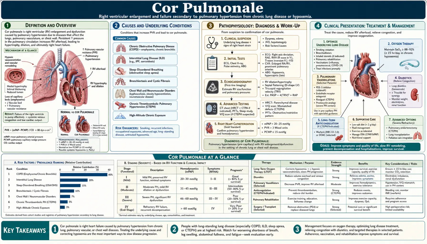
Cor pulmonale — literally "lung heart" in Latin — is right ventricular dysfunction or failure caused by pulmonary hypertension arising from lung disease or hypoxemia. Chronic obstructive pulmonary disease (COPD) is the leading cause in Western nations; supplemental oxygen is the only therapy proven to reduce mortality in the hypoxemic form; once established, it marks advanced underlying disease with poor prognosis.
Table of Contents
- Definition and Overview
- Causes and Underlying Conditions
- Pathophysiology
- Clinical Presentation
- Diagnosis: Imaging and Hemodynamics
- Acute Cor Pulmonale (Massive PE)
- Treatment: Targeting Underlying Disease
- Treatment: Volume Management and Vasodilators
- Prognosis
- Patient Self-Management and Monitoring
- Key Research Papers
- Featured Videos
Definition and Overview
Cor pulmonale is right ventricular (RV) dysfunction or failure caused by pulmonary hypertension (PH) due to lung disease or hypoxemia. The definition strictly excludes RV failure that arises from left ventricular disease: when left ventricular (LV) failure produces PH that then overloads the RV, the mechanism is "left heart disease" (WHO Group 2 PH), a categorically distinct entity. Only when the primary driver originates in the lungs or pulmonary vasculature — not the left heart — does the diagnosis of cor pulmonale apply.
The term translates literally as "lung heart" from Latin, reflecting this causal chain. Classification divides into two fundamentally different syndromes:
- Acute cor pulmonale: Massive pulmonary embolism is the most common cause. Sudden obstruction of more than 50% of the pulmonary vascular bed imposes acute RV pressure overload, causing rapid RV dilation and hemodynamic collapse. Treated as a PE emergency, not as chronic disease management.
- Chronic cor pulmonale: chronic obstructive pulmonary disease accounts for approximately 50% of cases in Western nations. Progressive hypoxic vasoconstriction, vascular remodeling, and destruction of the pulmonary capillary bed over months to years cause sustained elevation in pulmonary vascular resistance, with gradual RV hypertrophy, dilation, and eventual failure.
Epidemiologically, cor pulmonale affects roughly 6–7% of all US adults with chronic heart disease. Its prevalence is rising in parallel with the growing burden of chronic obstructive pulmonary disease and interstitial lung disease. Importantly, the development of cor pulmonale is a marker of advanced underlying lung disease — it signals that cardiopulmonary reserve is nearly exhausted and carries a poor prognosis independent of the specific underlying condition.
Causes and Underlying Conditions
Any condition that chronically elevates pulmonary vascular resistance through hypoxemia, vascular destruction, or mechanical obstruction can lead to cor pulmonale. The major cause categories are:
Obstructive Lung Diseases
This is the most common cause group in the United States and Europe. Chronic obstructive pulmonary disease is the single leading cause, driving cor pulmonale through a combination of chronic hypoxic pulmonary vasoconstriction, fixed vascular remodeling from tobacco-related endothelial injury, and destruction of the alveolar capillary bed in emphysema. Severe asthma and cystic fibrosis (late stage) also fall in this category.
Parenchymal Lung Diseases
Interstitial lung diseases obliterate the pulmonary vasculature through two mechanisms: fibrotic tissue physically encases and collapses small vessels, and hypoxemia from poor gas exchange triggers vasoconstriction. Idiopathic pulmonary fibrosis (IPF) carries particularly high PH prevalence (30–50% of patients). Other relevant conditions include sarcoidosis (WHO Group 5 PH), hypersensitivity pneumonitis, connective tissue disease-related interstitial lung disease, silicosis, and coal workers' pneumoconiosis.
Pulmonary Vascular Diseases
Chronic thromboembolic pulmonary hypertension (CTEPH) arises when organized, fibrotic thrombus fails to resolve after pulmonary embolism, mechanically obstructing the pulmonary arterial tree. It affects approximately 3–5% of PE survivors and is uniquely important because surgical pulmonary endarterectomy can be curative. Pulmonary arterial hypertension (Group 1 PH) produces cor pulmonale when untreated or inadequately treated, through primary obliterative vasculopathy of the small pulmonary arteries.
Hypoventilation Disorders
Obesity hypoventilation syndrome (OHS), historically called "Pickwickian syndrome," causes chronic hypercapnia and hypoxemia through blunted respiratory drive and chest wall restriction from excess weight. Recurrent nocturnal hypoxemia triggers sustained pulmonary vasoconstriction. Obstructive sleep apnea (OSA) produces repetitive nocturnal hypoxemia and hypercapnia; chronic exposure elevates baseline pulmonary artery pressure. Neuromuscular disease (respiratory muscle weakness causing hypoventilation) and kyphoscoliosis (restrictive chest wall physiology) operate through the same hypoxemia-vasoconstriction pathway. High altitude and chronic mountain sickness (Monge's disease) represent the environmental extreme of the same mechanism.
Pathophysiology
The sequence linking lung disease to RV failure passes through several identifiable steps: underlying lung disease → pulmonary hypertension → RV pressure overload → RV hypertrophy → RV dilation → RV failure.
Mechanisms of Pulmonary Hypertension in Lung Disease
Hypoxic pulmonary vasoconstriction (HPV) is the most important acute mechanism. When alveolar oxygen tension falls, smooth muscle cells in pulmonary arterioles contract, diverting blood away from poorly ventilated alveoli. In localized disease, this is actually beneficial — it reduces the mismatch between airflow and blood flow in the lung (ventilation-perfusion mismatch). In generalized lung disease with global hypoxemia, HPV affects the entire pulmonary vasculature, raising resistance throughout. Critically, HPV is largely reversible with supplemental oxygen in early disease — this is the basis for long-term oxygen therapy (LTOT) as the cornerstone of cor pulmonale management.
Vascular remodeling is the chronic, fixed mechanism. Sustained HPV drives smooth muscle cell proliferation, intimal thickening, and adventitial fibrosis in pulmonary arterioles. Once structural remodeling is established, the raised pulmonary vascular resistance is no longer fully reversible by supplemental oxygen — even optimal oxygenation cannot undo fixed anatomical narrowing.
Polycythemia compounds the hemodynamic burden. Chronic hypoxemia stimulates erythropoietin secretion, raising red blood cell mass and blood viscosity. Higher viscosity increases the work required to propel blood through the pulmonary vasculature, raising effective pulmonary vascular resistance further. Phlebotomy can reduce viscosity in symptomatic secondary erythrocytosis.
Mechanical destruction operates in emphysema (capillary bed destruction by alveolar wall loss) and fibrosis (vascular obliteration by scar tissue), reducing the total cross-sectional area of the pulmonary vasculature. With fewer parallel pathways for blood to traverse, resistance rises according to Poiseuille's law even at normal blood viscosity and flow.
Right Ventricular Adaptation and Failure
The RV responds to chronically elevated afterload through concentric hypertrophy — adding sarcomeres in parallel to increase wall thickness and normalize wall stress. Initially this is adaptive: RV output is maintained at the cost of increased oxygen demand. As pulmonary vascular resistance continues rising, the RV reaches a tipping point. Dilation begins (eccentric remodeling), the tricuspid annulus stretches producing regurgitation, and the interventricular septum shifts leftward (D-shaped LV on echocardiogram). Once RV wall tension consistently exceeds coronary perfusion pressure during systole, subendocardial ischemia develops — the RV cannot meet its own oxygen demand — and contractile failure follows. Ventricular interdependence means RV dilation impairs LV filling by leftward septal shift, further reducing cardiac output and systemic perfusion even when LV function itself is preserved.
Clinical Presentation
Symptoms of cor pulmonale are often masked by or intertwined with those of the underlying lung disease. The key clinical clue is deterioration beyond what the known lung disease would explain — worsening dyspnea that outpaces the pace of lung disease progression, or new fluid retention.
Symptoms
Dyspnea is universal but its change in character is the diagnostic clue: previously exertional dyspnea becoming dyspnea at rest, or dyspnea worsening faster than the lung disease alone would predict, signals that RV failure is developing. Peripheral edema — bilateral pitting edema of the ankles, calves, and in severe cases the thighs and sacrum — is the most visible manifestation of elevated venous pressure from RV failure. Ascites reflects third-spacing from hepatic venous congestion; right upper quadrant discomfort from hepatomegaly and capsular stretch may accompany it. Fatigue and severely reduced exercise tolerance result from a fixed, low cardiac output that cannot augment with exertion. Syncope with exertion indicates that cardiac output cannot meet the metabolic demand of exercise, causing cerebral hypoperfusion — a serious sign that hospitals treat as urgent.
Physical Examination Signs
Jugular venous distension (JVD) from elevated central venous pressure is a cardinal sign; cannon A-waves may appear in the setting of tricuspid regurgitation. Hepatojugular reflux (HJR) is positive — sustained abdominal pressure raises the jugular venous pressure as the failing RV cannot accommodate the increased venous return. Parasternal heave (a palpable sustained systolic impulse along the left lower sternal border) indicates RV enlargement. Loud pulmonic second heart sound (P2) (the pulmonic part of the second heart sound is accentuated and audible across the precordium) is the auscultatory hallmark of pulmonary hypertension. A tricuspid regurgitation murmur — holosystolic at the left sternal border, increasing with inspiration (Carvallo's sign) — reflects annular dilation from RV enlargement. Hepatomegaly with potential for pulsatile liver (from tricuspid regurgitation) and peripheral or central cyanosis from desaturation complete the picture in advanced disease.
Diagnosis: Imaging and Hemodynamics
No single test diagnoses cor pulmonale; the workup confirms RV dysfunction, quantifies pulmonary pressures, and characterizes the underlying lung disease. Echocardiography is the gateway non-invasive test; right heart catheterization provides definitive hemodynamic confirmation.
Electrocardiogram
The electrocardiogram (ECG) shows several signs of right heart strain, though no single finding is highly sensitive. P pulmonale (peaked P waves >2.5 mm in leads II, III, aVF) indicates right atrial enlargement. Right axis deviation (axis beyond +90°) reflects RV dominance. RV strain pattern (ST depression and T-wave inversion in V1–V4) indicates RV pressure overload. Right bundle branch block (RBBB), complete or incomplete, and S1Q3T3 pattern (particularly in acute cor pulmonale from massive PE) round out the electrocardiogram profile.
Echocardiography
Transthoracic echocardiography is the first-line non-invasive assessment. Key measurements include: right ventricular systolic pressure estimated from the tricuspid regurgitation jet velocity; RV size and function (tricuspid annular plane systolic excursion — how far the base of the right ventricle moves with each beat — <16 mm or tissue Doppler S′ <10 cm/s indicate RV dysfunction); interventricular septal flattening (D-shaped LV in systole indicates pressure overload; flattening in both systole and diastole indicates combined pressure and volume overload); and pericardial effusion, which carries a poor prognostic significance. Echocardiography also evaluates LV function to exclude Group 2 PH from left heart disease.
Chest Radiograph and Pulmonary Function Tests
The chest X-ray reveals enlarged central pulmonary arteries (PA diameter >29 mm), peripheral vascular pruning (absent markings in the outer third of the lung fields), and cardiomegaly with RV prominence. Signs of the underlying disease — hyperinflation and flat diaphragms in chronic obstructive pulmonary disease, honeycombing and reticulation in IPF — provide etiologic context. Pulmonary function tests characterize the underlying disease pattern: obstructive (chronic obstructive pulmonary disease, asthma) or restrictive (IPF, kyphoscoliosis).
Right Heart Catheterization
Right heart catheterization is the definitive test. Per the 2022 ESC/ERS guidelines, pulmonary hypertension is now defined as mean pulmonary artery pressure (mPAP) >20 mmHg (the prior threshold was >25 mmHg). Pre-capillary PH — the hemodynamic pattern of cor pulmonale from lung disease — requires mPAP >20 mmHg, pulmonary vascular resistance >2 Wood units, and pulmonary capillary wedge pressure (PCWP) ≤15 mmHg. The PCWP threshold distinguishes cor pulmonale (pre-capillary; PCWP ≤15 mmHg; driven by lung disease) from PH due to left heart disease (post-capillary; PCWP >15 mmHg) — a critical distinction with major therapeutic implications.
Acute Cor Pulmonale (Massive PE)
Acute cor pulmonale from massive pulmonary embolism is a distinct clinical emergency with different pathophysiology, time course, and treatment from the chronic form.
Mechanism and Recognition
When a massive clot burden suddenly obstructs more than 50% of the pulmonary vascular bed, the RV faces acute pressure overload it has no time to adapt to (unlike the chronic form, where slow remodeling allows partial compensation). Acute RV dilation develops within hours, producing hemodynamic collapse. The key echocardiographic findings that distinguish acute cor pulmonale from PE are the RV:LV ratio >0.9 and McConnell's sign — regional RV dysfunction with free wall akinesis but preserved apical motility — which is highly specific for massive PE rather than chronic RV failure.
Treatment of Massive PE
Systemic thrombolysis (a clot-dissolving drug, tissue plasminogen activator (alteplase), 100 mg over 2 hours) is indicated for hemodynamically unstable massive PE. A 2004 meta-analysis of 11 randomized trials (748 patients) found that, in trials that included hemodynamically unstable PE, thrombolysis lowered recurrent PE or death from 19.0% with heparin to 9.4% (about one event prevented for every 10 patients treated); in trials that excluded these patients there was no benefit (5.3% vs 4.8%), and the authors found no evidence of benefit in unselected PE patients. Catheter-directed therapy (CDT) — including ultrasound-assisted catheter-directed thrombolysis — is used for submassive PE with RV dysfunction without overt hemodynamic collapse. The SEATTLE II study was single-arm (150 patients, no comparison group): with a low dose of 24 mg of tissue plasminogen activator through the catheter, the right-to-left ventricle size ratio on chest CT fell from 1.55 to 1.13 within 48 hours, no patient had bleeding in the brain, and 15 patients (10%) had moderate or severe bleeding. Surgical embolectomy on cardiopulmonary bypass is reserved for cases where thrombolysis is contraindicated or has failed. Anticoagulation alone (unfractionated heparin immediately) is appropriate for submassive PE without hemodynamic compromise, where the RV is stressed but not yet failing.
Treatment: Targeting Underlying Disease
The primary therapeutic strategy in chronic cor pulmonale is aggressive management of the underlying pulmonary disease. Treating the cause of the elevated pulmonary vascular resistance — not just the RV failure — is what offers the best opportunity to slow progression and improve prognosis.
Chronic Obstructive Pulmonary Disease
Treatment of the lung disease includes bronchodilator inhalers (long-acting muscarinic antagonists, long-acting beta agonists, and inhaled corticosteroids as set out in the GOLD guidelines) and pulmonary rehabilitation; most important is stopping smoking — the only intervention that slows the decline in forced expiratory volume in one second (the amount of air a person can blow out in the first second) in chronic obstructive pulmonary disease. Lung volume reduction surgery (LVRS) improves exercise capacity and survival in selected emphysema patients with upper-lobe predominant disease and low exercise capacity (NETT trial).
IPF and Other Interstitial Lung Diseases
Antifibrotic drugs (nintedanib, pirfenidone) slow IPF progression by approximately 50% in trials (INPULSIS, ASCEND) but do not reverse established pulmonary hypertension. Treating the fibrotic process limits further vascular obliteration.
Sleep Apnea and Hypoventilation Syndromes
Continuous positive airway pressure (CPAP) for obstructive sleep apnea is among the most actionable interventions in this category: eliminating nocturnal hypoxemic episodes reduces repetitive pulmonary vasoconstriction, and longitudinal data show measurable reduction in pulmonary artery pressures over months. Bilevel positive airway pressure (including volume-assured pressure support ventilation) for obesity hypoventilation syndrome addresses both nocturnal hypercapnia and hypoxemia; combined with substantial weight loss, dramatic improvement in PH and cor pulmonale is possible — unlike most other causes, this is potentially reversible.
CTEPH
Pulmonary endarterectomy (PEA) is potentially curative for operable CTEPH, removing organized thrombus from the proximal pulmonary arteries and normalizing pressures in many patients. For inoperable CTEPH, riociguat (a soluble guanylate cyclase stimulator) is U.S. Food and Drug Administration-approved and reduces pulmonary vascular resistance and improves functional capacity.
Supplemental Oxygen
Long-term oxygen therapy (LTOT) is the most important evidence-based intervention for hypoxic cor pulmonale. The landmark MRC Trial (1981) and Nocturnal Oxygen Therapy Trial (NOTT, 1980) established that continuous supplemental oxygen (>15–18 hours/day, target blood oxygen saturation (SpO2) 88–92%) reduces mortality and slows the rise in pulmonary vascular resistance in hypoxemic chronic obstructive pulmonary disease. Oxygen reverses the reversible component of HPV — it cannot undo established vascular remodeling, but it removes the ongoing vasoconstriction stimulus. In chronic obstructive pulmonary disease patients who retain carbon dioxide (hypercapnia), oxygen is targeted at a saturation of 88–92% (not higher), because higher levels can suppress the hypoxic respiratory drive and worsen carbon dioxide retention.
Treatment: Volume Management and Vasodilators
Beyond targeting the underlying disease, symptomatic management of RV failure focuses on fluid balance optimization and careful consideration of vasoactive agents.
Diuretics
Loop diuretics (furosemide, torsemide) are the primary treatment for fluid overload, peripheral edema, and ascites. The therapeutic goal is euvolemia — resolution of peripheral edema without inducing intravascular volume depletion, which can reduce RV preload and precipitate hemodynamic deterioration. The RV in cor pulmonale is preload-dependent; excessive diuresis is a common cause of acute decompensation. Careful titration with monitoring of electrolytes (particularly potassium and magnesium) is essential. Aldosterone antagonists (spironolactone) reduce aldosterone-driven sodium retention but require caution in renal insufficiency due to hyperkalemia risk.
Digoxin
Digoxin has limited and inconsistent evidence in cor pulmonale. It is a weak positive inotrope and can be considered in refractory RV failure, but hypoxemia and hypokalemia both sensitize the myocardium to digoxin toxicity — common conditions in cor pulmonale patients — requiring careful dosing and electrolyte monitoring.
Pulmonary Vasodilators: A Cautionary Note
PAH-specific vasodilators (phosphodiesterase-5 inhibitors, endothelin receptor antagonists, prostacyclin analogues, riociguat) are not routinely recommended for cor pulmonale arising from chronic obstructive pulmonary disease or interstitial lung disease (WHO Group 3 PH). Unlike Group 1 PAH, where the vascular obliteration is the primary problem, Group 3 cor pulmonale involves ventilated but poorly perfused lung areas. Pulmonary vasodilators increase flow to these poorly ventilated areas, worsening ventilation-perfusion mismatch and arterial hypoxemia. No survival benefit has been proven in randomized trials for this indication, and potential harm from worsened oxygenation is real. Pulmonary hypertension due to interstitial lung disease is now an exception for one drug: in the 2021 INCREASE trial (326 patients), whose report noted that no therapy was approved for this condition at the time, inhaled treprostinil improved the 6-minute walk distance by 31.12 m compared with placebo at 16 weeks, and clinical worsening occurred in 22.7% versus 33.1% of patients; inhaled treprostinil was approved for this use in 2021. The trial did not show a survival benefit, and this caution still applies to cor pulmonale from chronic obstructive pulmonary disease. The exception is CTEPH and Group 1 PAH, where these drugs are specifically indicated and the mechanism is different.
Phlebotomy
For symptomatic secondary polycythemia with hematocrit consistently above 55%, phlebotomy to a target hematocrit of 45–50% reduces blood viscosity and effective pulmonary vascular resistance. This does not improve prognosis but can relieve symptoms of hyperviscosity (headache, plethora, impaired cognition) in selected patients.
Prognosis
Cor pulmonale signals that underlying lung disease has consumed most of the available cardiopulmonary reserve. Prognosis is primarily determined by the underlying condition and its responsiveness to treatment, but the development of RV failure universally worsens the trajectory.
Chronic Obstructive Pulmonary Disease with Cor Pulmonale
Five-year survival is approximately 30% once RV failure is established. In the MRC Trial (1981), 87 patients with chronic bronchitis or emphysema, severe low blood oxygen and a history of heart failure were randomized to oxygen for at least 15 hours a day or no oxygen: 19 of the 42 oxygen-treated patients died within five years, compared with 30 of the 45 controls; in men the survival advantage did not appear until 500 days had passed. The NOTT Trial (1980) randomized 203 patients to continuous oxygen or 12-hour nocturnal oxygen: overall mortality with nocturnal oxygen was 1.94 times that with continuous oxygen. These trials are the basis of the current standard of LTOT >15–18 hours daily.
IPF with Cor Pulmonale
Pulmonary hypertension in IPF carries a particularly grim prognosis. In a 2006 study of 79 patients with advanced IPF assessed for lung transplantation, pulmonary hypertension (mean pulmonary artery pressure above 25 mmHg) was present in 31.6%. One-year mortality was 28.0% with pulmonary hypertension versus 5.5% without, and the 6-minute walk distance was 143.5 m versus 365.9 m. Higher pressures were linked to a greater risk of death, while lung function (forced vital capacity and diffusing capacity) did not predict outcomes in that study.
Prognostic Markers
Key prognostic indicators include echocardiographic tricuspid annular plane systolic excursion <16 mm, RV:LV ratio >1, raised heart-strain blood tests (B-type natriuretic peptide or N-terminal pro-B-type natriuretic peptide), reduced six-minute walk distance, reduced diffusing capacity of the lung for carbon monoxide (how well the lungs pass gas into the blood), and a low resting arterial oxygen level (partial pressure of oxygen in arterial blood). Pericardial effusion on echocardiography is a particularly ominous finding.
Lung Transplantation
Bilateral sequential lung transplantation is the definitive treatment for refractory end-stage lung disease with cor pulmonale. Patients with CTEPH who have undergone successful pulmonary endarterectomy can experience dramatic, near-complete recovery of RV function — one of the most gratifying outcomes in pulmonary medicine. For IPF and chronic obstructive pulmonary disease with cor pulmonale, timing matters: candidacy for transplant depends on being referred while still in a functional window that permits it, and median post-transplant survival of 5–7 years compares favorably to the trajectory of advanced cor pulmonale on medical therapy.
Patient Self-Management and Monitoring
Daily monitoring and steady use of the therapies with proven mortality benefit are the main ways people living with cor pulmonale stay stable and catch decompensation early. The practices below are the ones care plans describe.
Oxygen Therapy Adherence
Long-term oxygen therapy is the only drug treatment proven to extend life in hypoxemic cor pulmonale. In the MRC trial oxygen was given for at least 15 hours a day, and the current standard is 15–18 hours every day. Nighttime use alone falls short: in the NOTT trial, mortality with 12-hour nocturnal oxygen was 1.94 times that with continuous oxygen. Oxygen equipment (flow settings, tubing, concentrator) only delivers the prescribed dose when it is working properly. Air travel takes planning: commercial aircraft cabin pressure is equivalent to approximately 6,000–8,000 feet altitude, and high-risk patients need in-flight supplemental oxygen arranged through the airline in advance. High-altitude destinations worsen hypoxic vasoconstriction acutely and can precipitate decompensation even in stable patients.
Daily Weight and Edema Monitoring
Daily weight is taken at the same time each morning after urinating. A gain of more than 2 pounds in a single day or 5 pounds in a week indicates fluid retention; in care plans this is the point for prompt contact with the care team or for starting a pre-agreed diuretic action plan. Weekly photographs of ankle swelling give an objective record of change. Swelling or weight beyond a person's usual baseline is a sign of fluid build-up that care teams treat as needing attention before the next scheduled appointment.
Activity Pacing and Pulmonary Rehabilitation
Energy conservation strategies — planning activities for times of day when energy is highest, spacing tasks with rest periods, using assistive devices — help maintain function within the limits of a fixed cardiac output. Supervised pulmonary rehabilitation programs improve functional status, dyspnea, and six-minute walk distance in chronic obstructive pulmonary disease-related cor pulmonale through supervised exercise training, education, and psychosocial support.
Vaccination and Infection Prevention
Annual influenza vaccination is standard in chronic lung disease. For pneumococcal vaccination, the U.S. Centers for Disease Control and Prevention (CDC) recommendations (updated February 25, 2026) call for one of the pneumococcal conjugate vaccines PCV15, PCV20 or PCV21 for all adults 50 years or older; when PCV15 is used, a dose of the pneumococcal polysaccharide vaccine PPSV23 follows one year later, and after PCV20 or PCV21 a PPSV23 dose is not indicated. The CDC notes that adults younger than 50 may also be recommended pneumococcal vaccines because of risk conditions. Respiratory infections are among the most common precipitants of acute cor pulmonale decompensation in patients with chronic lung disease.
Smoking Cessation and Sodium Restriction
Smoking cessation is the single most impactful modifiable intervention in chronic obstructive pulmonary disease-related cor pulmonale — it is the only strategy proven to slow the underlying decline in forced expiratory volume in one second that drives disease progression. Nicotine replacement, varenicline, and behavioral support all improve quit rates. For patients with recurrent fluid overload, sodium restriction to less than 2 grams per day reduces the kidney's tendency to retain fluid and may allow lower diuretic doses.
Key Research Papers
- Medical Research Council Working Party. Long term domiciliary oxygen therapy in chronic hypoxic cor pulmonale complicating chronic bronchitis and emphysema (MRC Trial). Lancet. 1981;1(8222):681–686. — PubMed PMID: 6110912
- Nocturnal Oxygen Therapy Trial Group. Continuous or nocturnal oxygen therapy in hypoxemic chronic obstructive lung disease (NOTT). Ann Intern Med. 1980;93(3):391–398. — PubMed PMID: 6776858
- Weitzenblum E, Sautegeau A, Ehrhart M, et al. Long-term oxygen therapy can reverse the progression of pulmonary hypertension in patients with chronic obstructive pulmonary disease. Am Rev Respir Dis. 1985;131(4):493–498. — PubMed PMID: 3922267
- Seeger W, Adir Y, Barbera JA, et al. Pulmonary hypertension in chronic lung diseases. J Am Coll Cardiol. 2013;62(25 Suppl):D109–116. — PubMed PMID: 24355635
- Vonk-Noordegraaf A, Haddad F, Chin KM, et al. Right heart adaptation to pulmonary arterial hypertension. J Am Coll Cardiol. 2013;62(25 Suppl):D22–33. — PubMed PMID: 24355638
- Benza RL, Park MH, Keogh A, Girgis RE. Management of pulmonary arterial hypertension with a focus on combination therapies. J Heart Lung Transplant. 2007;26(5):437–446. — PubMed PMID: 17449411
- Strange G, Playford D, Stewart S, et al. Pulmonary hypertension: prevalence and mortality in the Armadale echocardiography cohort. Heart. 2012;98(24):1805–1811. — PubMed PMID: 22760869
- Simonneau G, Montani D, Celermajer DS, et al. Haemodynamic definitions and updated clinical classification of pulmonary hypertension. Eur Respir J. 2019;53(1):1801913. — PubMed PMID: 30545968
- Humbert M, Kovacs G, Hoeper MM, et al. 2022 ESC/ERS Guidelines for the diagnosis and treatment of pulmonary hypertension. Eur Heart J. 2022;43(38):3618–3731. — PubMed PMID: 36017548
- McLaughlin VV, Archer SL, Badesch DB, et al. ACCF/AHA 2009 expert consensus document on pulmonary hypertension. J Am Coll Cardiol. 2009;53(17):1573–1619. — PubMed PMID: 19389575
- Vestbo J, Hurd SS, Agusti AG, et al. Global strategy for the diagnosis, management, and prevention of chronic obstructive pulmonary disease (GOLD 2013). Am J Respir Crit Care Med. 2013;187(4):347–365. — PubMed PMID: 22878278
- Barst RJ, Gibbs JS, Ghofrani HA, et al. Updated evidence-based treatment algorithm in pulmonary arterial hypertension. J Am Coll Cardiol. 2009;54(1 Suppl):S78–84. — PubMed PMID: 19555861
- Wan S, Quinlan DJ, Agnelli G, Eikelboom JW. Thrombolysis compared with heparin for the initial treatment of pulmonary embolism: a meta-analysis of the randomized controlled trials. Circulation. 2004;110(6):744–749. — PubMed PMID: 15262836
- Piazza G, Hohlfelder B, Jaff MR, et al. A Prospective, Single-Arm, Multicenter Trial of Ultrasound-Facilitated, Catheter-Directed, Low-Dose Fibrinolysis for Acute Massive and Submassive Pulmonary Embolism: The SEATTLE II Study. JACC Cardiovasc Interv. 2015;8(10):1382–1392. — PubMed PMID: 26315743
- Waxman A, Restrepo-Jaramillo R, Thenappan T, et al. Inhaled Treprostinil in Pulmonary Hypertension Due to Interstitial Lung Disease. N Engl J Med. 2021;384(4):325–334. — PubMed PMID: 33440084
- Lettieri CJ, Nathan SD, Barnett SD, Ahmad S, Shorr AF. Prevalence and outcomes of pulmonary arterial hypertension in advanced idiopathic pulmonary fibrosis. Chest. 2006;129(3):746–752. — PubMed PMID: 16537877
- U.S. Centers for Disease Control and Prevention. Pneumococcal Vaccine Recommendations (updated February 25, 2026). — cdc.gov
PubMed Topic Searches
The following PubMed topic searches retrieve current peer-reviewed literature on Cor Pulmonale. Each link opens a live PubMed query.
- PubMed: Cor pulmonale COPD pulmonary hypertension treatment
- PubMed: Right ventricular failure chronic lung disease
- PubMed: Hypoxic pulmonary vasoconstriction oxygen therapy
- PubMed: Cor pulmonale echocardiography right heart function
- PubMed: Cor pulmonale COPD prognosis survival
- PubMed: Long term oxygen therapy hypoxemic COPD
- PubMed: Obesity hypoventilation cor pulmonale CPAP
- PubMed: Acute cor pulmonale massive pulmonary embolism
- PubMed: Pulmonary hypertension lung disease diagnosis
- PubMed: Right ventricular dysfunction pulmonary fibrosis