⚠ Evidence warning: Clinical trials or other evidence do not support the claim that a leucomethylthioninium trial has enrolled people with corticobasal syndrome. This page keeps it as a report of what people, websites or other media say.
Corticobasal Syndrome (CBS)
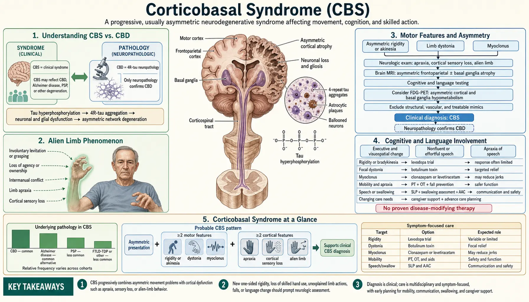
Corticobasal syndrome (CBS) is a rare, progressive neurodegenerative disorder characterized by profoundly asymmetric motor and cognitive impairment. Its signature features — alien limb phenomenon, apraxia, cortical sensory loss, and asymmetric rigidity — arise from degeneration of the cerebral cortex and basal ganglia on one side more than the other. Crucially, CBS is a clinical syndrome: the identical presentation can be caused by several distinct underlying pathologies, most commonly corticobasal degeneration tau, but also progressive supranuclear palsy (PSP) tau, frontotemporal lobar degeneration with TDP-43 inclusions (FTLD-TDP), Alzheimer's disease pathology, or rarely prion disease. This clinicopathological dissociation makes CBS both diagnostically challenging and a window into how different protein aggregates converge on the same clinical picture.
- Understanding CBS vs. Corticobasal degeneration
- Alien Limb Phenomenon
- Motor Features and Asymmetry
- Cognitive and Language Involvement
- Eye Movement and Other Signs
- Underlying Pathology and Tau Biology
- Imaging and Diagnosis
- Epidemiology and Prognosis
- Treatment and Supportive Care
- Key Research Papers
- Featured Videos
- Connections
Understanding CBS vs. Corticobasal degeneration
For many years clinicians used the term corticobasal degeneration to describe both the clinical syndrome and the autopsy-confirmed 4R-tau pathology. Advances in neuropathology forced a crucial renaming. Post-mortem studies show that fewer than half of patients with the classic CBS clinical picture actually have corticobasal degeneration (CBD)-tau at autopsy. The rest harbor progressive supranuclear palsy-tau, FTLD-TDP-43 type A, Alzheimer's disease, or other rare proteinopathies.
Current terminology therefore separates:
- CBS (corticobasal syndrome) — the clinical diagnosis made during life, based on a defined set of motor, sensory, and cognitive features.
- Corticobasal degeneration — the specific 4R-tau pathology confirmed at autopsy or, increasingly, inferred by research-grade tau positron emission tomography (PET) imaging.
This distinction matters enormously for clinical trials and emerging tau-directed therapies: a trial enrolling "CBS" patients is actually enrolling a mixed-pathology group, diluting any signal from a therapy targeting one specific protein. The Armstrong 2013 diagnostic criteria define two tiers, possible and probable corticobasal degeneration, that aim to enrich for the underlying pathology; the disease itself is confirmed only at autopsy. The Movement Disorder Society's 2017 criteria take a similar approach for progressive supranuclear palsy (probable, possible, and suggestive).
From the patient perspective, reports of typical CBS onset run from the late 50s to about 70, usually on one side of the body, and progresses relentlessly over 6–8 years. There is no proven disease-modifying treatment, but symptom management can meaningfully preserve function and quality of life.
Alien Limb Phenomenon
The alien limb phenomenon is the most evocative and diagnostically striking feature of CBS, reported in approximately 50–60% of patients at some point in the disease course. Asking directly finds more: in a clinic series using structured examinations, 25 of 30 patients (83%) reported one or more alien limb symptoms and 28 of 30 (93%) had significant apraxia, with the left hand preferentially affected in 23 of 30; apraxia severity did not track with alien limb. The affected limb — nearly always an arm, sometimes a leg — behaves as if it has a will of its own. Patients describe the experience in vivid, often distressing terms: the arm rises involuntarily from the lap, reaches across to grasp objects the patient did not intend to pick up, or interferes with the voluntary movements of the opposite hand (intermanual conflict).
Three distinct subtypes of alien limb have been described:
- Frontal type: The limb engages in groping, grasping, and utilization behavior — picking up and using objects in the environment without conscious intention. This reflects disinhibition of the supplementary motor area and loss of voluntary motor suppression.
- Callosal type: The affected hand acts in opposition to the patient's intention — for instance, undoing a button as fast as the other hand fastens it (intermanual conflict). This results from corpus callosum degeneration disrupting interhemispheric coordination.
- Posterior (sensory) type: The limb floats upward spontaneously (levitation), especially with eyes closed or during distraction, due to severe cortical proprioceptive loss. The patient does not know where the limb is in space and cannot actively suppress its drift.
Neuroimaging correlates of alien limb include atrophy of the supplementary motor area, anterior cingulate, and callosal body. The phenomenon is highly specific for cortical rather than subcortical disease — it does not occur in pure Parkinson's disease, multiple system atrophy (MSA), or progressive supranuclear palsy. Its presence in a patient with asymmetric parkinsonism strongly tips the diagnosis toward CBS.
Motor Features and Asymmetry
The defining motor hallmark of CBS is profound asymmetry. Whereas progressive supranuclear palsy and multiple system atrophy predominantly cause axial (midline, symmetric) degeneration, CBS destroys the cortex and basal ganglia far more on one side. Patients typically present with one-sided clumsiness — they notice that one hand has become slow, stiff, and inaccurate — often years before any bilateral involvement emerges. This unilateral onset creates what some describe as "hemi-Parkinson plus cortical signs."
Key motor features include:
- Asymmetric rigidity: Limb rigidity, usually cogwheel quality, markedly worse on the initially affected side. Unlike Parkinson's disease, where rigidity responds to levodopa, rigidity in CBS is largely levodopa-resistant.
- Akinesia and bradykinesia: Slowness and poverty of movement, again asymmetric. Finger tapping and hand movements are reduced in amplitude and speed on the affected side.
- Dystonia: Fixed or action-triggered posturing, commonly an inverted, clenched hand or plantar flexion of the foot. The "clenched fist" in CBS is a direct result of cortical degeneration causing loss of inhibitory control over distal motor units. Dystonia can be painful.
- Myoclonus: Cortical myoclonus — brief, high-amplitude, action- or reflex-triggered jerks of the affected limb — occurs in up to 30–50% of patients. It is elicited by tapping the fingers or touching the hand and can be confirmed on electroencephalogram (EEG) (preceding cortical discharge). This distinguishes it from the subcortical myoclonus of other disorders.
- Postural instability and falls: Less prominent early on than in progressive supranuclear palsy (which causes backward falls in the first year), but emerges as the disease progresses bilaterally.
- Limb apraxia: See below — a cardinal cortical feature.
The combination of limb rigidity, dystonia, and myoclonus on one side alongside cortical signs on the same side creates a distinctive clinical picture that points toward CBS in a patient with asymmetric parkinsonism.
Cognitive and Language Involvement
CBS is not a "pure motor" disorder. Cortical degeneration — particularly of the parietal and frontal lobes contralateral to the more affected limb — produces distinctive cognitive and language impairments that are often the first features patients notice before the motor syndrome is fully developed.
Limb apraxia is a cardinal sign. Apraxia is the loss of ability to perform learned skilled movements despite intact strength, sensation, and coordination. In CBS, both limb-kinetic apraxia (clumsy, inaccurate distal movements) and ideomotor apraxia (inability to pantomime tool use — "show me how you would use a hammer") are present. The patient who cannot wave goodbye, use an imaginary scissors, or mimic brushing teeth despite understanding the request and having no arm weakness has ideomotor apraxia. This is a cortical sign that separates CBS from pure basal ganglia disorders.
Cortical sensory loss is highly characteristic: astereognosis (inability to identify objects by touch — a key in the palm cannot be recognized with eyes closed) and agraphesthesia (inability to identify numbers or letters written on the palm). Light touch and pinprick are often preserved — this is not a peripheral sensory loss but a failure of cortical processing. Cortical sensory loss on one hand in a patient with that same hand's motor impairment is a strong CBS pointer.
Language syndromes depend on which pathology underlies CBS. CBS caused by corticobasal degeneration tau more commonly causes frontal executive dysfunction and non-fluent/agrammatic primary progressive aphasia (PNFA). CBS caused by Alzheimer's disease pathology more frequently causes posterior cortical atrophy syndrome — visuospatial dysfunction, alexia, dyscalculia — or logopenic progressive aphasia. The presence of language impairment does NOT exclude CBS; it points toward the underlying cause.
Behavioral change — apathy, disinhibition, perseveration — occurs as degeneration spreads to the prefrontal cortex. This can resemble the behavioral variant of frontotemporal dementia, particularly when FTLD-TDP underlies the CBS presentation. Memory, by contrast, is usually relatively spared early compared to Alzheimer's disease.
Eye Movement and Other Signs
Eye movement abnormalities in CBS are qualitatively different from those in progressive supranuclear palsy. While progressive supranuclear palsy produces a characteristic vertical supranuclear gaze palsy — inability to look up or down — CBS produces cortical oculomotor deficits:
- Saccadic pursuit: Smooth pursuit eye movements (tracking a moving target) become broken and jerky (cogwheel or saccadic pursuit) due to parietal cortex degeneration. Normal smooth pursuit requires intact parieto-occipital cortex.
- Apraxia of eyelid opening: Difficulty initiating voluntary eyelid opening, despite intact orbicularis function (unlike blepharospasm). Patients must concentrate hard or use a finger to raise the lid — this reflects failure of levator premotor cortex activation.
- Asymmetric reflexive saccades: Saccades to targets may be hypometric or slowed to one side. A full vertical gaze palsy argues more for underlying progressive supranuclear palsy pathology than corticobasal degeneration.
Other notable signs include:
- Dysarthria: Hypophonic, effortful speech, sometimes with apraxia of speech (distorted articulation without consistent phonemic errors, a feature of cortical motor speech area degeneration).
- Dysphagia: Appears later than in progressive supranuclear palsy, but can cause aspiration risk as the disease progresses.
- Hyperreflexia and Babinski signs: Corticospinal tract involvement produces upper motor neuron signs on the affected side, distinguishing CBS from purely extrapyramidal disorders.
- Stimulus-sensitive myoclonus: A tap on the affected hand triggers a jerk visible to the examiner, a direct bedside marker of cortical hyperexcitability.
Underlying Pathology and Tau Biology
When the underlying cause is corticobasal degeneration, the molecular signature is abnormal accumulation of 4-repeat (4R) tau — the same tau isoform that accumulates in progressive supranuclear palsy. In healthy neurons, tau exists in six isoforms defined by whether exon 10 is included (4R) or excluded (3R) during mRNA splicing; a 1:1 ratio is normally maintained. In corticobasal degeneration, 4R-tau predominates due to altered splicing, leading to hyperphosphorylated tau filaments that aggregate into distinctive structures:
- Astrocytic plaques: The pathological hallmark of corticobasal degeneration — fan-shaped tau deposits in the distal processes of astrocytes, forming stellate clusters. These are distinct from the tufted astrocytes seen in progressive supranuclear palsy.
- Neuronal threads: Tau-positive threads in the white matter neuropil, disrupting axonal conduction.
- Ballooned (achromatic) neurons: Swollen, pale neurons that have lost their Nissl substance, indicative of severe cytoskeletal disruption. Found most prominently in frontoparietal cortex.
- Coiled bodies: Tau inclusions in oligodendrocytes (compare with multiple system atrophy, where inclusions are α-synuclein in oligodendrocytes — multiple system atrophy's glial cytoplasmic inclusions, GCIs).
Electron microscope studies of brain tissue from three people with corticobasal degeneration found tau filaments that were identical between the cases and built on a previously unseen four-layered fold, distinct from those in Alzheimer's disease, Pick's disease and chronic traumatic encephalopathy. A genome study of 152 autopsy-proven cases and 3,311 controls linked the disease to the tau gene MAPT plus lnc-KIF13B-1 and SOS1, and found genetic risk shared with progressive supranuclear palsy at MOBP.
The anatomical distribution of corticobasal degeneration pathology mirrors the asymmetric clinical syndrome: maximal degeneration in the frontoparietal cortex, substantia nigra, and subthalamic nucleus, all substantially worse on the side contralateral to the more-affected limb. This spread likely begins unilaterally and propagates via corticobasal and thalamocortical circuits before eventually affecting both sides.
Tau positron emission tomography imaging using the radiotracer flortaucipir (18F-AV-1451) now allows visualization of tau burden in living patients. Corticobasal degeneration shows a characteristic asymmetric frontoparietal cortical and striatal tau positron emission tomography signal that correlates with clinical severity. While not yet diagnostic-grade, tau positron emission tomography is transforming both clinical trials and the understanding of the relationship between CBS symptom onset and tau spread.
Imaging and Diagnosis
No single imaging finding definitively diagnoses CBS or its underlying pathology in life, but a characteristic pattern on structural magnetic resonance imaging (MRI) is highly supportive:
- Asymmetric perirolandic atrophy: Loss of cortical volume in the frontoparietal (sensorimotor and supplementary motor) cortex, markedly worse on the side contralateral to the more-affected limb. This is the most recognizable structural magnetic resonance imaging finding in CBS and is best seen on coronal T1 sequences; a 2011 series found perirolandic atrophy in CBS whatever the underlying pathology.
- Asymmetric parietal and frontal atrophy: Superior parietal lobule atrophy contralateral to the affected side — the region subserving complex sensorimotor integration and spatial awareness — correlates with apraxia and cortical sensory loss severity.
- Asymmetric putaminal signal: In some cases, T2/FLAIR signal change or volume loss in the posterior putamen is seen on the side contralateral to the more-affected limb, reflecting striatonigral degeneration.
Fluorodeoxyglucose positron emission tomography (FDG-PET), a metabolic scan, demonstrates asymmetric hypometabolism in the frontoparietal cortex and contralateral basal ganglia, providing additional diagnostic support and sometimes detecting metabolic changes before atrophy is visible on magnetic resonance imaging. Asymmetry on this scan correlating with limb-symptom asymmetry is one of the best supportive imaging biomarkers for CBS.
The Armstrong criteria (2013) remain the most widely used clinical diagnostic framework. They describe four clinical forms of corticobasal degeneration — corticobasal syndrome, frontal behavioral-spatial syndrome, the nonfluent/agrammatic variant of primary progressive aphasia, and progressive supranuclear palsy syndrome — and set out two sets of criteria for the pathology:
- Probable corticobasal degeneration: insidious onset and gradual progression for at least 1 year, age at onset ≥50, no similar family history or known tau mutation, and either probable CBS (asymmetric presentation, ≥2 of limb rigidity/akinesia, limb dystonia, limb myoclonus, plus ≥2 of orobuccal or limb apraxia, cortical sensory deficit, alien limb) or frontal behavioral-spatial syndrome or the aphasia variant with at least 1 CBS feature.
- Possible corticobasal degeneration: similar criteria with no limits on age or family history; tau mutations are allowed, CBS may be symmetric, and broader features are allowed, including a progressive supranuclear palsy syndrome with vertical gaze palsy, or a frontal behavioral syndrome.
- Definite: not a tier in these criteria; pathological confirmation at autopsy is what definitively diagnoses corticobasal degeneration.
Distinguishing CBS from progressive supranuclear palsy clinically can be challenging; an asymmetric patient with prominent alien limb and apraxia favors CBS, while symmetric axial disease with early backward falls and vertical gaze palsy favors progressive supranuclear palsy. Both are 4R-tauopathies and sometimes overlap pathologically.
Epidemiology and Prognosis
CBS is the rarest of the three major atypical parkinsonian syndromes. Prevalence estimates range from 1.0 to 2.5 per 100,000 population: a Scottish study found 2.05 per 100,000 at regional level, with national rates peaking at ages 70–79, and a 2022 systematic review pooled 2.3 per 100,000, roughly 3 times below progressive supranuclear palsy at 7.1. Age of onset is typically between 60 and 70 years, with most series reporting a mean onset around 63–67 years. Unlike Parkinson's disease (which shows a slight male predominance) and progressive supranuclear palsy (approximately equal sex ratio), some studies have suggested a slightly higher prevalence in women for CBS, though the data are inconsistent across centers and likely affected by ascertainment bias; the Scottish study and the systematic review both found no clear sex differences.
Survival from symptom onset to death is typically 6–8 years, making CBS intermediate between progressive supranuclear palsy (6–7 years) and multiple system atrophy (6–9 years) in terms of pace. However, underlying pathology influences prognosis: CBS with underlying Alzheimer's disease pathology tends to run a slightly longer course than corticobasal degeneration-tau. The cause of death is most commonly aspiration pneumonia (from dysphagia and reduced cough reflex), pulmonary embolism (immobility), or sepsis from urinary tract infections.
Functional decline follows a characteristic arc: the first 1–2 years are dominated by unilateral motor clumsiness and the alien limb phenomenon, often misdiagnosed as focal dystonia, stroke, or early Parkinson's disease. Years 2–4 see progressive bilateral motor and cognitive involvement. By years 4–6, most patients are wheelchair-dependent, severely dysarthric, and require full-time care. Dysphagia and aspiration risk become the dominant management concern in the final 1–2 years.
There are currently no validated prognostic biomarkers that predict rate of progression in individual patients. Cerebrospinal fluid (CSF) neurofilament light chain (NfL) is elevated in CBS (as in all atypical parkinsonisms) and correlates with disease severity, but is not specific for CBS or the underlying pathology.
Treatment and Supportive Care
There is no approved disease-modifying therapy for CBS or its underlying pathologies. Multiple clinical trials have enrolled CBS/corticobasal degeneration populations in recent years, driven by advances in tau biology and tau-targeting strategies, but none has demonstrated clinical benefit to date.
Symptomatic pharmacological treatment:
- Levodopa trial: A levodopa/carbidopa trial (up to 1,000 mg levodopa/day for at least 3 months) is standard practice. Response is typically poor — fewer than 25% of CBS patients have a meaningful motor response, compared to the nearly universal response in Parkinson's disease. A partial response does not exclude CBS but favors a Parkinson's disease-spectrum pathology. The trial is described as adequate only once that dose has been reached.
- Botulinum toxin injections: The most consistently useful pharmacological intervention in CBS. Injected into the forearm flexors, hand intrinsics, or lower leg, botulinum toxin reduces dystonia severity, relieves pain from the clenched fist, and can improve hand function and hygiene. Also used for blepharospasm and drooling (submandibular gland injection). Typically repeated every 3–4 months.
- Clonazepam: Effective for cortical myoclonus, reducing jerk frequency and amplitude. Starting dose 0.25–0.5 mg at bedtime, titrated as tolerated. Sedation and fall risk are the main concerns.
- Levetiracetam or valproate: Second-line options for myoclonus when clonazepam is not tolerated or insufficient.
- Selective serotonin reuptake inhibitors (SSRIs): For behavioral symptoms — apathy, irritability, depression, pseudobulbar affect.
Rehabilitation and allied health:
- Physiotherapy: Fall prevention exercises, gait training with an assistive device, management of postural instability. Particularly important given the fall risk from the asymmetric motor syndrome.
- Occupational therapy: Adaptive equipment for the unaffected hand; strategies to compensate for apraxia; home safety assessments.
- Speech-language pathology: Dysphagia assessment and modified-texture diet, augmentative and alternative communication (AAC) devices as dysarthria progresses, apraxia of speech therapy.
- Neuropsychology: Cognitive assessment, strategies for apraxia compensation, support for caregiver education.
Research pipeline: Anti-tau strategies under investigation include antisense oligonucleotides (ASOs) targeting tau mRNA, small-molecule tau aggregation inhibitors, and active and passive immunotherapy. Leucomethylthioninium and multiple tau antibody trials are reported to have enrolled CBS/progressive supranuclear palsy patients. CBD Solutions and the CurePSP Foundation maintain registries and trial pipelines that interested clinicians and patients can access.
Key Research Papers
- Armstrong MJ, Litvan I, Lang AE, et al. Criteria for the diagnosis of corticobasal degeneration. Neurology. 2013;80(5):496–503. — Landmark criteria paper setting out 4 clinical forms of corticobasal degeneration and 2 sets of criteria, probable and possible corticobasal degeneration; the standard reference for clinical trial enrollment. — PubMed PMID: 23359374
- Boeve BF, Lang AE, Litvan I. Corticobasal degeneration and its relationship to progressive supranuclear palsy and frontotemporal dementia. Ann Neurol. 2003;54(Suppl 5):S15–S19 — Comprehensive review defining the clinical overlap between CBS, PSP, and FTLD, establishing the 4R-tau spectrum concept. — PubMed PMID: 12833363
- Dickson DW, Bergeron C, Chin SS, et al. Office of rare diseases neuropathologic criteria for corticobasal degeneration. J Neuropathol Exp Neurol. 2002;61(11):935–946. — Defines the neuropathological criteria for CBD, including astrocytic plaques, ballooned neurons, and tau thread distribution. — PubMed PMID: 12430710
- Murray R, Neumann M, Forman MS, et al. Cognitive and motor assessment in autopsy-proven corticobasal degeneration. Neurology. 2007;68(16):1274–1283 — Autopsy-confirmed series of 15 patients with corticobasal degeneration: a specific pattern of cognitive impairment at presentation, with a movement abnormality in less than half. — PubMed PMID: 17438218
- Kouri N, Murray ME, Hassan A, et al. Neuropathological features of corticobasal degeneration presenting as corticobasal syndrome or Richardson syndrome. Brain. 2011;134(11):3264–3275 — Compares corticobasal degeneration presenting as CBS (11 cases) or as Richardson syndrome, the progressive supranuclear palsy picture (15 cases), and shows differing tau density by region. — PubMed PMID: 21933807
- Lee SE, Rabinovici GD, Mayo MC, et al. Clinicopathological correlations in corticobasal degeneration. Ann Neurol. 2011;70(2):327–340 — Series with 40 CBS patients of known histopathology: corticobasal degeneration in 35%, Alzheimer's disease in 23%, progressive supranuclear palsy in 13%, frontotemporal lobar degeneration with TDP inclusions in 13%; perirolandic atrophy whatever the pathology. — PubMed PMID: 21823158
- Südmeyer M, Pieperhoff P, Ferrea S, et al. Longitudinal deformation-based morphometry reveals spatio-temporal dynamics of brain volume changes in patients with corticobasal syndrome. PLoS One. 2012;7(7):e41873 — Repeated scans of 6 patients and 11 controls: increasing volume loss in premotor, primary motor and superior parietal areas, with asymmetric atrophy in the basal ganglia. — PubMed PMID: 22848644
- Lewis-Smith DJ, Wolpe N, Ghosh BCP, Rowe JB. Alien limb in the corticobasal syndrome: phenomenological characteristics and relationship to apraxia. J Neurol. 2020;267(4):1147–1157 — Structured examinations of 30 patients with CBS: alien limb symptoms in 83%, apraxia in 93%, with no significant link between the two in severity. — PubMed PMID: 31919566
- Whitwell JL, Jack CR Jr, Boeve BF, et al. Imaging correlates of pathology in corticobasal syndrome. Neurology. 2010;75(21):1879–1887 — 24 autopsied CBS patients: widespread atrophy points toward frontotemporal lobar degeneration with TDP-43 or Alzheimer's disease, while focal premotor and supplemental motor atrophy suggests corticobasal degeneration or progressive supranuclear palsy, whose patterns were similar. — PubMed PMID: 21098403
- Josephs KA, Whitwell JL, Dickson DW, et al. Voxel-based morphometry in autopsy proven PSP and CBD. Neurobiol Aging. 2008;29(2):280–289 — Voxel-based morphometry of autopsy-confirmed CBD vs. PSP reveals distinct spatial distribution of atrophy, useful for distinguishing the two 4R-tauopathies on imaging. — PubMed PMID: 17097770
- Höglinger GU, Respondek G, Stamelou M, et al. Clinical diagnosis of progressive supranuclear palsy: the Movement Disorder Society criteria. Mov Disord. 2017;32(6):853–864. — Movement Disorder Society criteria for PSP also address the CBS-PSP overlap variants, providing the framework for separating CBS-PSP from CBS-CBD clinically. — PubMed PMID: 28467028
- Tetzloff KA, Graff-Radford J, Martin PR, et al. Regional distribution, asymmetry, and clinical correlates of tau uptake on [18F]AV-1451 PET in atypical Alzheimer's disease. J Alzheimers Dis. 2018;62(4):1713–1724 — Tau PET in 18 people with posterior cortical atrophy and 19 with logopenic aphasia; tau deposition closely related to clinical presentation. The study does not include CBS. — PubMed PMID: 29614676
- Zhang W, Tarutani A, Newell KL, et al. Novel tau filament fold in corticobasal degeneration. Nature. 2020;580(7802):283–287. — PubMed PMID: 32050258
- Kouri N, Ross OA, Dombroski B, et al. Genome-wide association study of corticobasal degeneration identifies risk variants shared with progressive supranuclear palsy. Nat Commun. 2015;6:7247. — PubMed PMID: 26077951
- Swallow DMA, Counsell CE. Prevalence of progressive supranuclear palsy and corticobasal syndrome in Scotland. Neuroepidemiology. 2022;56(4):291–297. — PubMed PMID: 35654007
- Swallow DMA, Zheng CS, Counsell CE. Systematic review of prevalence studies of progressive supranuclear palsy and corticobasal syndrome. Mov Disord Clin Pract. 2022;9(5):604–613. — PubMed PMID: 35844273
PubMed topic searches:
Corticobasal syndrome diagnosis corticobasal degeneration tau pathology Alien limb phenomenon CBS apraxia treatment