⚠ Evidence warning: Clinical trials or other evidence do not support the claim that the micro-dystrophin gene therapy delandistrogene moxeparvovec includes Becker muscular dystrophy patients in ongoing extension trials. This page keeps it as a report of what people, websites or other media say.
Becker Muscular Dystrophy (BMD)
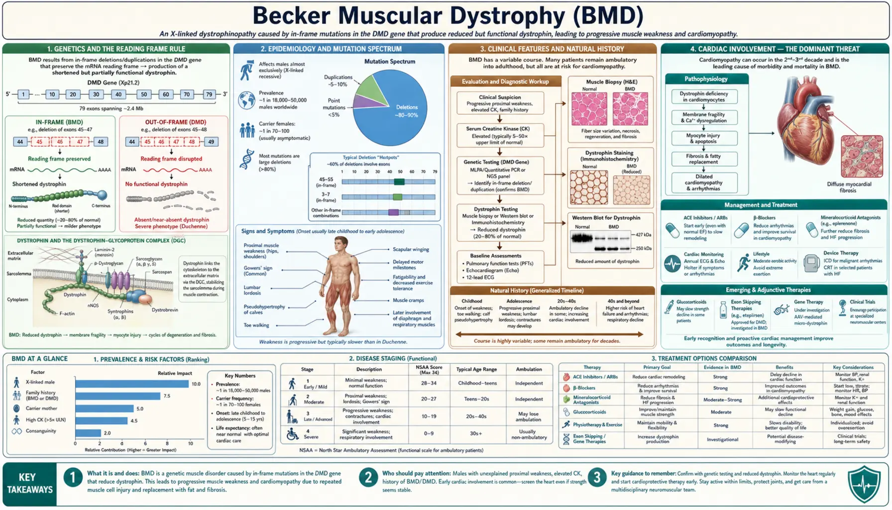
Becker muscular dystrophy (BMD) is an X-linked recessive neuromuscular disease caused by in-frame mutations in the DMD gene (Xp21.2) that allow production of a truncated but partially functional dystrophin protein. Because some dystrophin reaches the sarcolemma and stabilizes the dystrophin-glycoprotein complex, the phenotype is significantly milder than Duchenne muscular dystrophy — with onset typically in adolescence or early adulthood and ambulation often preserved into the fifth or sixth decade. However, dilated cardiomyopathy in BMD is frequently more severe relative to skeletal muscle involvement and can dominate the clinical picture, making cardiac surveillance a cornerstone of management.
Interactive Visualization Muscle Contraction — release the calcium Expose the actin binding sites and watch myosin heads ratchet through the cross-bridge cycle to shorten the sarcomere. Launch →Table of Contents
Genetics and the Reading Frame Rule
Becker muscular dystrophy and Duchenne muscular dystrophy both arise from mutations in the same gene — the DMD gene located at Xp21.2. At 2.4 megabases and 79 exons, the DMD gene is the largest known human gene. The disorder follows an X-linked recessive inheritance pattern, meaning that males with a single mutant allele are affected and females carrying one mutant allele are generally carriers, though some develop symptoms.
The fundamental distinction between BMD and DMD was explained by the Monaco Reading Frame Rule, published in 1988. In-frame deletions or duplications — those that preserve the translational reading frame — allow the ribosome to continue reading the mRNA and produce a dystrophin protein that, while internally deleted or shortened, retains partial function. These truncated proteins can still partially anchor the dystrophin-glycoprotein complex at the muscle cell membrane, producing the milder BMD phenotype. Out-of-frame mutations, by contrast, introduce a premature stop codon downstream, resulting in no stable dystrophin protein and the severe DMD phenotype.
The most common BMD mutations are large in-frame exon deletions, particularly those spanning exons 45–47 or 45–51, a region often called the "BMD hotspot." Some duplications also produce in-frame transcripts. Rare point mutations (missense or splice-site) can also yield partially functional protein. On Western blot, BMD muscle shows dystrophin at reduced quantity and abnormal molecular weight compared with normal, in contrast to the virtual absence seen in DMD. Patients with dystrophin levels of 10–40% of normal tend toward very mild phenotypes, while those with 3–10% overlap the severe BMD range.
Genotype-phenotype correlation is imperfect — the same exon 45–51 deletion can produce ambulatory patients in their sixties in one family and early wheelchair dependence in another. Modifier genes including SPP1 (osteopontin) and LTBP4 are under active investigation as contributors to this variability. Brain involvement is less common in BMD than in DMD, in part because the shorter Dp140 and Dp71 dystrophin isoforms, which are expressed in the brain and are disrupted in most DMD deletions, are usually at least partially preserved by in-frame BMD mutations. It is not rare, however: a 2026 systematic review found decreased cognitive function or cognitive dysfunction reported in 41% of people with BMD across all ages.
Epidemiology and Mutation Spectrum
BMD is considerably less common than DMD. Prevalence estimates range from approximately 1 in 18,000 to 1 in 31,000 live male births, translating to roughly 5,000–6,000 patients in the United States at any given time. The disease occurs worldwide without clear ethnic predisposition.
Approximately 80% of BMD cases result from in-frame exon deletions, 10% from duplications, and 10% from point mutations (missense, nonsense, or splice-site changes that nonetheless allow some functional dystrophin). The single most common mutation — an in-frame deletion of exons 45–51 — accounts for approximately 30% of all BMD cases and is associated with a particularly mild phenotype: many affected males remain ambulatory well into their sixties and seventies.
Approximately 30% of BMD cases arise from de novo mutations with no prior family history, a figure similar to DMD. Genetic counseling is therefore important even when no family history is apparent. Female carriers of DMD gene mutations have an approximately 8% lifetime risk of developing dilated cardiomyopathy themselves, even when skeletal muscle function is entirely normal. Heart muscle changes in carriers are common: a 1996 Italian study that followed 197 female carriers of Duchenne or Becker mutations (aged 5 to 60) with yearly heart tests for 3 to 10 years found early (preclinical) or obvious heart muscle involvement in 84.3%, rising from 54.5% of carriers aged 5 to 16 to 90.2% of those older than 16, with no difference between Duchenne and Becker carriers. Findings like these are the basis for the cardiac screening of female carriers regardless of symptoms.
A clinically important subset of patients with DMD gene mutations present as X-linked dilated cardiomyopathy (XLDCM) — a condition characterized by isolated or predominant cardiomyopathy with minimal or absent skeletal muscle weakness. XLDCM is typically caused by mutations that selectively affect the muscle-specific promoter region or exon 1 of the DMD gene, impairing cardiac dystrophin expression while leaving enough skeletal muscle isoform function intact to prevent obvious weakness. For this reason, a DMD gene test can uncover the cause of unexplained dilated cardiomyopathy in a young male.
Clinical Features and Natural History
BMD typically presents between ages 5 and 25, in contrast to DMD where weakness is usually evident by ages 2–5. The range is wide — some mildly affected males are not diagnosed until they present with exertional myoglobinuria or cardiomyopathy in their thirties or forties. Others have a more severe course that resembles late-onset DMD.
The distribution of weakness mirrors DMD — proximal muscles of the hip girdle and proximal lower extremities are affected first. Patients commonly notice difficulty rising from the floor (some use the Gowers maneuver, pushing up on their thighs with their hands), climbing stairs, or running. The pace of progression is substantially slower than DMD, and ambulation is typically preserved into the forties or sixties in most BMD patients. Approximately 30% require wheelchair assistance by age 40–50; the remainder may remain ambulatory much longer.
Selective quadriceps wasting with relative preservation of hip abductors is a characteristic pattern in BMD that differs subtly from DMD and can be a useful clinical clue. Calf hypertrophy, caused by fibrofatty replacement of degenerating muscle fibers, is present in many BMD patients as in DMD. Gait abnormalities — waddling, toe-walking, and a Trendelenburg pattern — are milder than in DMD. Contractures develop later and are less severe; clinically significant scoliosis is uncommon.
Cognitive involvement is less frequent in BMD than in DMD, where disruption of the short Dp71 isoform expressed widely in the brain contributes to a higher rate of intellectual disability and attention problems. Many BMD patients have normal intelligence, but the 2026 systematic review found decreased cognitive function or cognitive dysfunction reported in 41% across all ages. Creatine kinase (CK) is markedly elevated, typically 5,000–20,000 international units (IU)/L in younger patients; the level may paradoxically decline in adulthood as progressive muscle wasting reduces the total mass of muscle available to leak the enzyme. Exertional rhabdomyolysis — episodes of acute muscle breakdown triggered by unaccustomed intense exercise or febrile illness — is a common and sometimes dramatic presenting feature in mildly affected adult males, producing dark cola-colored urine (myoglobinuria) and sharply elevated creatine kinase.
A 2026 systematic review of Becker natural history (121 publications) pooled 36 studies of the general BMD population. By age 41 and over, 93.6% had muscle weakness, 69.4% heart involvement, 55.6% scoliosis, 47.4% had lost the ability to walk and 33.3% needed breathing support (ventilation). Among people who reached each milestone, the mean age was 12.5 years at first symptoms, 31.9 years at heart involvement, 33.3 years at loss of walking, 35.7 years at the start of ventilation and 55.6 years at death. The review stresses how widely the disease varies from person to person.
Cardiac Involvement — The Dominant Threat
Dilated cardiomyopathy develops in 70–80% of BMD patients by age 40, making it one of the most clinically significant features of the disease and a major driver of morbidity and mortality. Importantly, cardiac involvement in BMD does not track closely with skeletal muscle disease severity. A patient who is ambulatory and functionally independent may simultaneously harbor severe left ventricular dysfunction — a phenomenon called the cardiomyopathy-skeletal mismatch — that makes BMD cardiac disease potentially more dangerous per unit of skeletal disability than DMD, where respiratory and orthopedic complications often dominate before cardiac disease becomes the limiting factor.
The cardiac pathology in BMD is the same as in DMD: left ventricular dilatation, global systolic dysfunction with reduced ejection fraction, and progressive diastolic dysfunction. Cardiac magnetic resonance imaging (MRI) characteristically demonstrates posterolateral left ventricular (LV) wall fibrosis on late gadolinium enhancement sequences, reflecting replacement of dystrophin-deficient cardiomyocytes by scar tissue. This fibrosis pattern may appear earlier in BMD relative to the degree of skeletal involvement. Electrocardiographic findings mirror DMD — a tall R-wave in lead V1 and deep Q-waves in inferolateral leads are characteristic.
X-linked dilated cardiomyopathy represents the extreme of cardiac-dominant BMD: affected males may have entirely normal or minimally elevated creatine kinase and no skeletal weakness, presenting to cardiology with unexplained dilated cardiomyopathy (DCM) in their twenties or thirties.
Heart involvement is frequent even when the muscles seem barely affected. A 1996 study from Padua examined 28 men with subclinical or benign (mild) BMD using ECG and echocardiography and found heart muscle involvement in 72% of those with subclinical disease and 60% of those with benign disease. The damage appeared to start early in the right ventricle and later involve the left ventricle. The authors suggested that men with mild BMD, unaware of their heart risk and still able to do strenuous exercise, may put mechanical stress on dystrophin-deficient heart cells. Without awareness of this entity, the underlying genetic cause and appropriate family counseling may be missed for years.
Cardiac screening guidelines recommend electrocardiogram (ECG) and echocardiography beginning at age 10 (or at diagnosis if older), repeated annually. Cardiac magnetic resonance imaging every 2–3 years provides superior characterization of fibrosis burden and early LV dysfunction not yet apparent on echo. Guidelines also include cardiac evaluation for all symptomatic female carriers, and many now recommend baseline cardiac assessment for all carriers regardless of symptoms.
Treatment follows heart failure guidelines with disease-specific modifications. Angiotensin-converting enzyme inhibitors (enalapril, lisinopril) and beta-blockers (carvedilol, metoprolol succinate) are started early — often even before the ejection fraction falls below the normal range — because early intervention has been shown to slow progression in DMD and is assumed to apply to BMD. Mineralocorticoid receptor antagonists such as eplerenone add further cardioprotection. Patients with sustained ventricular tachycardia or ejection fraction persistently below 35% are candidates for implantable cardioverter-defibrillator (ICD) placement. For ambulatory BMD patients with end-stage cardiomyopathy who retain adequate skeletal muscle function, cardiac transplantation is a legitimate and increasingly utilized option, with outcomes comparable to other transplant indications.
Quadriceps Myopathy and Exercise Intolerance
Selective quadriceps wasting is one of the clinical hallmarks that distinguishes BMD from other muscular dystrophies. Affected patients develop progressive weakness and visible atrophy of the anterior thigh muscles — particularly the rectus femoris and vastus lateralis — while other proximal muscle groups, including the hip abductors, may be relatively spared early in the disease. Patients notice this as difficulty climbing stairs, rising from a low chair or squat, and instability on uneven terrain well before global gait disturbance develops. The selective pattern can mimic an isolated quadriceps myopathy, and BMD belongs in the differential diagnosis for any male presenting with this pattern.
Exertional rhabdomyolysis and myoglobinuria are particularly common in mildly affected BMD adults and in some patients represent the initial presentation before a diagnosis of BMD is established. Episodes are typically triggered by unusually strenuous or unaccustomed exercise — a single intense workout, a long hike, or a competitive sport — or by febrile illness. During an episode, creatine kinase may spike above 100,000 international units/L and the urine turns dark brown (cola or tea colored) from myoglobin. The primary complication is acute kidney injury from myoglobin-induced tubular toxicity, requiring aggressive intravenous (IV) hydration and monitoring of renal function. Some patients carry a medical alert identification noting their diagnosis and the anesthetic contraindications.
Succinylcholine is absolutely contraindicated in BMD, as in all muscular dystrophies. The depolarizing neuromuscular blocking agent can trigger life-threatening hyperkalemia and rhabdomyolysis in these patients. Volatile inhalational anesthetic agents carry a similar risk. Anesthesia teams use the diagnosis to choose drugs before any surgical procedure, and a preoperative creatine kinase measurement provides a useful baseline. Non-depolarizing agents and total intravenous anesthesia are preferred.
Physical therapy guidance in BMD aims to maintain function while avoiding the muscle damage of high-intensity eccentric exercise. Moderate-intensity aerobic exercise (swimming, cycling, walking) is safe and beneficial. Resistance training at approximately 60% of maximum voluntary contraction is supported by evidence in related conditions and is commonly part of exercise programs. Aquatic therapy reduces joint stress and is well tolerated. High-intensity plyometric (jumping) or heavy eccentric loading is the kind of exercise linked to the muscle damage described above. Occupational therapy assessment helps preserve independence in daily activities as strength declines.
Diagnosis
The diagnostic workup for suspected BMD begins with serum creatine kinase measurement. Creatine kinase is elevated in virtually all affected males — typically 5,000–20,000 international units/L in younger patients — and an unexpectedly high creatine kinase in an otherwise healthy-appearing male commonly leads to genetic evaluation. In adults with advanced muscle wasting, creatine kinase may be lower due to reduced total muscle mass; a normal creatine kinase in a middle-aged male with known family history does not exclude the diagnosis.
Genetic testing has become the definitive first-line diagnostic modality. Multiplex ligation-dependent probe amplification (MLPA) detects exon deletions and duplications across the entire DMD gene and identifies approximately 80–90% of pathogenic variants. For patients with a negative MLPA, full sequencing of the DMD gene identifies the remaining point mutations and small indels. Once a deletion or duplication is found, reading frame analysis — determining whether the mutation is in-frame or out-of-frame — is essential to predict and understand the phenotype. The same exon deletion can be BMD in one patient and DMD in another depending on the precise boundaries and reading frame consequences.
Muscle biopsy is reserved for patients in whom genetic testing is inconclusive or when the diagnosis remains in question. Histology in BMD shows the hallmarks of a dystrophic myopathy — fiber size variation, internal nuclei, necrosis and regeneration — but these changes are less severe than in DMD. The critical finding is dystrophin immunostaining: in BMD, dystrophin is present but reduced in intensity and sometimes patchy in distribution, in contrast to DMD where staining is absent or nearly absent. Western blot demonstrates reduced quantity and abnormal (lower) molecular weight of the dystrophin protein band, directly reflecting the internally deleted protein.
Cardiac evaluation — electrocardiogram and echocardiography — is part of the workup at diagnosis in all patients regardless of age, because cardiac disease may already be present or may progress independently of skeletal muscle disease. Electromyography reveals a myopathic pattern (short-duration, low-amplitude, polyphasic motor unit potentials) and normal nerve conduction studies, helping exclude neuropathic causes of weakness such as Charcot-Marie-Tooth disease or spinal muscular atrophy. The differential diagnosis includes other limb-girdle muscular dystrophies (particularly the sarcoglycanopathies, now named LGMD R3–R6 and formerly LGMD2C–2F, where dystrophin is normal), XLDCM without skeletal weakness, sporadic dilated cardiomyopathy, inflammatory myopathies such as polymyositis (which typically show elevated inflammatory markers and respond to steroids), and mitochondrial myopathy.
Management
BMD management is multidisciplinary, encompassing neurology, cardiology, physical medicine and rehabilitation, genetic counseling, and psychosocial support. The goal is to preserve function, prevent complications, and maintain quality of life across what may be a decades-long disease course.
No exon-skipping therapies are specifically approved for BMD by the U.S. Food and Drug Administration (FDA). Exon-skipping drugs make the cell skip one exon so that an out-of-frame Duchenne mutation becomes in-frame. By the reading frame rule above, an in-frame change is expected to give a shorter, partly working, Becker-like dystrophin — the aim is to turn Duchenne into a milder disease. People with BMD already have an in-frame mutation. The exon-skipping drugs approved for specific DMD genotypes (eteplirsen for exon 51 skipping, golodirsen and viltolarsen for exon 53 skipping, casimersen for exon 45 skipping) are approved for Duchenne muscular dystrophy only. A micro-dystrophin gene therapy (delandistrogene moxeparvovec) is also approved in the United States (first in 2023); its current label (August 2026) covers patients 4 years of age and older with Duchenne muscular dystrophy who are ambulatory (can walk) and have a confirmed DMD gene mutation. The label does not cover Becker muscular dystrophy, and it carries a boxed warning that acute serious liver injury, including life-threatening and fatal acute liver failure, has occurred with the therapy. Exon skipping in BMD remains an area of active clinical investigation. The recently approved micro-dystrophin gene therapy (delandistrogene moxeparvovec) includes BMD patients in ongoing extension trials.
Sevasemten (EDG-5506) is an experimental pill that slows the contraction of fast muscle fibres, which in animal models of Duchenne and Becker muscular dystrophy reduced the contraction injury that leads to inflammation, scarring and muscle loss. In a 2026 open-label phase 1b study, 12 walking adults with BMD (aged 18 to 55) took it for up to 24 months. It was well tolerated, with only mild or moderate side effects and none serious; blood markers of muscle injury fell within 4 weeks and stayed lower for up to 24 months, and physical function on the North Star Ambulatory Assessment stayed stable. The study had no placebo group and was run by the drug's developer; further trials are ongoing.
Corticosteroids (prednisone or deflazacort) have a well-established role in slowing progression in DMD but evidence in BMD is more limited. For patients with a severe or intermediate phenotype — particularly those who are losing ambulation — a trial of corticosteroids at individualized dosing is a reasonable consideration after shared decision-making about side effects. Typical dosing extrapolated from DMD protocols (prednisone 0.75 mg/kg/day or deflazacort 0.9 mg/kg/day) is used; weekend-only regimens reduce but do not eliminate side effects.
Cardiac management is the highest-priority therapeutic domain given the frequency and severity of BMD cardiomyopathy. Early initiation of angiotensin-converting enzyme inhibitors (ACE inhibitors) and beta-blockers — even when ejection fraction remains preserved — is supported by evidence from DMD studies and expert consensus. Diuretics manage fluid overload. Aldosterone antagonists (eplerenone or spironolactone) provide additive cardioprotection. Patients with ejection fraction below 35% or life-threatening arrhythmia are referred for implantable cardioverter-defibrillator (ICD) implantation. Ambulatory BMD patients with end-stage cardiomyopathy who retain reasonable skeletal muscle function are appropriate candidates for cardiac transplantation; this is increasingly recognized as a viable and appropriate intervention.
Care programs typically schedule physical and occupational therapy reviews at regular intervals — every 6–12 months — to monitor functional status, adjust orthotic devices, and update exercise prescriptions. Ankle-foot orthoses help manage foot drop when it develops. Surgical correction of contractures is rarely necessary given the slower progression compared with DMD. Scoliosis surveillance is appropriate but surgical intervention is uncommon. Psychosocial screening for depression and anxiety is important; the late-onset, slowly progressive nature of BMD means many patients have established careers, relationships, and independent lives before significant disability develops, and the progressive course requires ongoing psychological adjustment.
Genetic counseling is essential for the patient and family. The X-linked pattern means that affected males pass the mutation to all daughters (who become carriers) but to none of their sons. Carrier females have a 50% chance of having an affected son and a 50% chance of having a carrier daughter with each pregnancy. Preimplantation genetic testing and prenatal diagnosis are available options. Carrier testing is offered to female relatives in the maternal line, followed by cardiac evaluation when carrier status is confirmed.
Key Research Papers
- Monaco AP et al. "An explanation for the phenotypic differences between patients bearing partial deletions of the DMD locus." Genomics. 1988;2(1):90–5. — PubMed PMID: 3384440
- Hoffman EP et al. "Characterization of dystrophin in muscle-biopsy specimens from patients with Duchenne's or Becker's muscular dystrophy." N Engl J Med. 1988;318(21):1363–8. — PubMed PMID: 3285207
- Melacini P et al. "Myocardial involvement is very frequent among patients affected with subclinical Becker's muscular dystrophy." Circulation. 1996;94(12):3168–75. — PubMed PMID: 8989125
- Finsterer J et al. "Cardiac involvement in Becker muscular dystrophy." Can J Cardiol. 2008;24(10):786–92. — PubMed PMID: 18841259
- Bushby KM, Gardner-Medwin D. "The clinical, genetic and dystrophin characteristics of Becker muscular dystrophy. I. Natural history." J Neurol. 1993;240(2):98–104. — PubMed PMID: 8437027
- Matthews E, Brassington R, Kuntzer T, Jichi F, Manzur AY. "Corticosteroids for the treatment of Duchenne muscular dystrophy." Cochrane Database Syst Rev. 2016;2016(5):CD003725. — PubMed PMID: 27149418
- Politano L et al. "Development of cardiomyopathy in female carriers of Duchenne and Becker muscular dystrophies." JAMA. 1996;275(17):1335–8. — PubMed PMID: 8614119
- Emery AE. "The muscular dystrophies." Lancet. 2002;359(9307):687–95. — PubMed PMID: 11879882
- McNally EM et al. "Contemporary cardiac issues in Duchenne muscular dystrophy." Circulation. 2015;131(18):1590–8. — PubMed PMID: 25940966
- Mickle AT, Johnston KM, Ricchetti-Masterson KL, Kennedy AR, Korpach SG, Gooch KL. "The natural history of Becker muscular dystrophy: A systematic literature review." J Neuromuscul Dis. 2026;13(5):1140–1158. — PubMed PMID: 41671094
- Phan H, Barthel B, Madden M, Silverman JA, Bronson A, Thaler E, Kilburn NR, Amato M, Massaro M, MacDougall J, Russell AJ, Donovan J. "Effects of sevasemten (EDG-5506) on safety, biomarkers, and functional measures in adults with Becker muscular dystrophy: results of a phase 1b, open-label study." EBioMedicine. 2026;129:106335. — PubMed PMID: 42308588