Glucose-6-Phosphate Dehydrogenase (G6PD) Deficiency Test
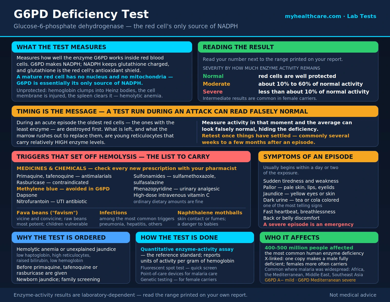
The glucose-6-phosphate dehydrogenase (G6PD) deficiency test is a simple blood test that measures how well one particular enzyme — glucose-6-phosphate dehydrogenase, or glucose-6-phosphate dehydrogenase — is working inside your red blood cells. That enzyme is a quiet bodyguard: it helps protect red cells from a kind of chemical wear-and-tear called oxidative stress. When someone is born with too little of it, most days feel completely normal. But certain foods, medicines, and infections can suddenly overwhelm those unprotected red cells and make them burst, a process called hemolysis. This page explains what the enzyme does, why the test is ordered, and — most importantly — the practical list of triggers to avoid if you or a family member turns out to be deficient. Glucose-6-phosphate dehydrogenase deficiency is the most common enzyme deficiency in the world, affecting hundreds of millions of people, and the vast majority live full, healthy lives simply by knowing what to steer clear of.
Table of Contents
- What Glucose-6-Phosphate Dehydrogenase Is
- What Glucose-6-Phosphate Dehydrogenase Deficiency Is
- Why the Test Is Ordered
- Triggers to Avoid
- Symptoms of an Episode
- How the Test Is Done
- Understanding Your Results
- Living With Glucose-6-Phosphate Dehydrogenase Deficiency
- When to Talk to a Doctor
- Key Research Papers
- Connections
- Featured Videos
What Glucose-6-Phosphate Dehydrogenase Is
Glucose-6-phosphate dehydrogenase (G6PD) is an enzyme — a tiny protein machine that speeds up a chemical reaction. It sits at the start of a metabolic route called the pentose phosphate pathway, and its main job is to produce a molecule called reduced nicotinamide adenine dinucleotide phosphate (NADPH). Think of reduced nicotinamide adenine dinucleotide phosphate as the fuel that keeps your cells' antioxidant defenses charged up.
Inside a red blood cell, that defense matters more than almost anywhere else. Red cells spend their entire life carrying oxygen, which makes them constantly exposed to reactive, damaging byproducts. To stay intact, they rely on an antioxidant called glutathione, and glutathione can only do its job if it is continuously "recharged" using reduced nicotinamide adenine dinucleotide phosphate. Here is the catch: mature red blood cells have no nucleus and no mitochondria, so glucose-6-phosphate dehydrogenase is essentially their only source of that protective reduced nicotinamide adenine dinucleotide phosphate. Other cells in the body have backup systems; the red cell does not.
When glucose-6-phosphate dehydrogenase is working normally, a red cell can shrug off oxidative stress the way a well-oiled machine handles a rough patch. When glucose-6-phosphate dehydrogenase is deficient, the antioxidant shield runs low. Hemoglobin inside the cell gets damaged and clumps into deposits called Heinz bodies, the cell membrane stiffens and gets injured, and the spleen removes these damaged cells from circulation. If enough cells are lost at once, the result is hemolytic anemia — the red-cell count drops faster than the body can replace it.
What Glucose-6-Phosphate Dehydrogenase Deficiency Is
Glucose-6-phosphate dehydrogenase deficiency is an inherited condition in which the body makes a version of the enzyme that either doesn't work well or breaks down too quickly. It is the most common human enzyme deficiency in the world, with an estimated 400 to 500 million people affected. Despite that huge number, many people never know they have it, because without an oxidative trigger their red cells hold up just fine.
The gene for glucose-6-phosphate dehydrogenase sits on the X chromosome, which shapes who is affected. Because males have only one X chromosome, a single altered gene copy leaves them fully deficient. Females have two X chromosomes, so they are more often carriers with a mix of normal and deficient red cells; some female carriers have essentially normal enzyme levels, while others — especially those who inherit an altered copy from both parents — can be as affected as males. In practice this means the condition shows up most visibly in men and boys, but women can absolutely be affected too.
Deficiency is far more common in people whose ancestry traces to regions where malaria was historically widespread: much of Africa, the Mediterranean (Italy, Greece, Sardinia), the Middle East, and Southeast Asia. That geographic pattern is not a coincidence. Carrying a glucose-6-phosphate dehydrogenase variant appears to offer partial protection against severe malaria, because the malaria parasite has a harder time surviving inside enzyme-deficient red cells. Over many generations, in places where malaria killed large numbers of people, that survival edge caused glucose-6-phosphate dehydrogenase variants to become common — the same evolutionary bargain seen with sickle cell trait and thalassemia. It is a striking example of a trait that can be a liability under some conditions and an advantage under others.
There are hundreds of different glucose-6-phosphate dehydrogenase variants, and they are not all equally severe. Doctors often group them by how much enzyme activity remains:
- Mild to moderate variants (for example, the "glucose-6-phosphate dehydrogenase A−" form common in people of African descent) leave enough enzyme that hemolysis is usually self-limited — the newest red cells still carry decent enzyme levels, so an episode tends to stop on its own once the trigger is removed.
- Severe variants (for example, "glucose-6-phosphate dehydrogenase Mediterranean") leave very little enzyme, so triggers can cause more dramatic hemolysis, and fava beans in particular can be dangerous.
- Rare chronic forms cause ongoing low-grade hemolysis even without an obvious trigger. These are uncommon and are managed by a hematologist.
Why the Test Is Ordered
A doctor may order a glucose-6-phosphate dehydrogenase test for several practical reasons:
- To find the cause of hemolytic anemia or unexplained jaundice. If blood work shows red cells are being destroyed — low hemoglobin, a high reticulocyte count, low haptoglobin, and elevated bilirubin — glucose-6-phosphate dehydrogenase deficiency is one of the causes worth checking.
- To screen before certain medications. Some drugs are known to trigger hemolysis in glucose-6-phosphate dehydrogenase-deficient people. Antimalarial medicines like primaquine and tafenoquine, and the cancer-related drug rasburicase, are usually preceded by a glucose-6-phosphate dehydrogenase test precisely to avoid a dangerous reaction.
- As part of a newborn jaundice workup. Glucose-6-phosphate dehydrogenase deficiency is a recognized cause of significant jaundice in the first days of life. In regions where the condition is common, newborns may be screened routinely; elsewhere, it is checked when a baby's jaundice is unusually early, severe, or slow to resolve.
- Family or ancestry-based screening. If a close relative has glucose-6-phosphate dehydrogenase deficiency, or if someone belongs to a group where it is common, testing before an episode ever happens lets a person learn their trigger list in advance.
Triggers to Avoid
This is the part that matters most day to day. For most people with glucose-6-phosphate dehydrogenase deficiency, staying well is mainly about avoiding the things that set off hemolysis. The big categories are fava beans, certain drugs, infections, and naphthalene mothballs. Keep a copy of this list, and share your diagnosis with every doctor, dentist, and pharmacist you see.
Fava beans ("favism")
Fava beans (also called broad beans) contain natural compounds — vicine and convicine — that generate exactly the kind of oxidative stress glucose-6-phosphate dehydrogenase-deficient red cells cannot handle. Eating them can trigger sudden, sometimes severe hemolysis, a reaction so classic it has its own name: favism. Fresh or raw beans tend to be the most potent, but dried beans and even fava-containing products can cause trouble in people with severe variants. Children are especially vulnerable. If you have a severe form of the deficiency, the safest approach is to avoid fava beans entirely.
Medications and chemicals
A number of drugs can trigger hemolysis. Dose matters, and the exact risk of some medicines is still debated, so treat this as a "flag it and check" list rather than an absolute rulebook — always confirm with your pharmacist against a current, dedicated glucose-6-phosphate dehydrogenase drug list before starting anything new. Commonly implicated agents include:
- Antimalarials — primaquine and tafenoquine are the classic triggers; these are usually given only after a glucose-6-phosphate dehydrogenase test.
- Rasburicase — used to prevent tumor lysis syndrome; considered contraindicated in glucose-6-phosphate dehydrogenase deficiency.
- Methylene blue — ironically both a trigger and, in other settings, a treatment, so it is avoided in glucose-6-phosphate dehydrogenase deficiency.
- Dapsone — used for certain infections and skin conditions.
- Nitrofurantoin — a common urinary-tract-infection antibiotic.
- Some sulfa drugs (sulfonamides) — such as sulfamethoxazole and sulfasalazine.
- Phenazopyridine — a urinary pain reliever that turns urine orange.
- High-dose intravenous vitamin C — large intravenous (IV) doses have triggered hemolysis; ordinary dietary amounts are not a concern.
Infections
This one surprises people: ordinary infections are among the most common triggers of hemolysis in glucose-6-phosphate dehydrogenase deficiency. The body's inflammatory response to a virus or bacteria produces oxidative stress that vulnerable red cells feel. Pneumonia, hepatitis, and other infections have all been linked to episodes. You can't always avoid getting sick, but knowing this helps you and your doctor recognize why anemia might appear during an illness.
Naphthalene (mothballs)
Naphthalene, the classic mothball chemical, is a potent trigger — through skin contact, or by breathing the fumes in a closed closet or drawer, or when clothing and bedding stored with mothballs are worn. This is a particular danger for babies whose clothes were stored this way. If someone in the household has glucose-6-phosphate dehydrogenase deficiency, skip naphthalene mothballs entirely and use cedar or other alternatives.
Symptoms of an Episode
When hemolysis is triggered, symptoms usually begin within a day or two of the exposure and reflect both the loss of red cells and the breakdown products flooding the bloodstream. Watch for:
- Sudden tiredness and weakness — a wave of fatigue that comes on faster than ordinary tiredness.
- Pallor — unusually pale skin, lips, or inner eyelids.
- Jaundice — yellowing of the whites of the eyes or the skin, from the bilirubin released when red cells break down.
- Dark urine — tea-colored, cola-colored, or reddish-brown, caused by hemoglobin spilling into the urine. This is one of the most telling signs.
- A fast heartbeat and shortness of breath — the body's response to a sudden drop in oxygen-carrying red cells.
- Back or belly discomfort in more severe episodes.
In newborns, the main sign is jaundice appearing in the first days of life, sometimes severe enough to need treatment. A severe hemolytic episode at any age is a medical emergency — dark urine plus pallor and fatigue after a known trigger warrants prompt medical attention.
How the Test Is Done
The glucose-6-phosphate dehydrogenase test is done on a small blood sample, usually drawn from a vein in the arm (or from a heel prick in newborns). In the lab, the sample is analyzed to see how much functional enzyme the red cells contain. A few methods are used:
- Quantitative enzyme-activity assay. This is the reference-standard test. It measures the rate at which the enzyme generates reduced nicotinamide adenine dinucleotide phosphate and reports a number, typically in units of activity per gram of hemoglobin. It tells you not just whether the enzyme is low, but how low.
- Fluorescent spot test. A quicker, qualitative screen that shows whether reduced nicotinamide adenine dinucleotide phosphate is being made. It is good for flagging clearly deficient samples but is less precise for in-between results.
- Point-of-care tests. Newer bedside devices, developed largely so that antimalarial treatment can be given safely in regions where laboratory testing is limited.
- Genetic testing. Sometimes used to identify the exact variant, and especially helpful for female carriers whose enzyme-activity results can look deceptively normal.
A critical timing caveat. There is a real trap in testing during an acute hemolytic episode. During a crisis, the oldest red cells — the ones with the least enzyme — are the first to be destroyed. What is left behind, and what the bone marrow rushes out to replace them, are young red cells (reticulocytes) that carry relatively high enzyme levels. Measure enzyme activity in that moment and the average can look falsely normal, hiding the deficiency. For this reason, testing is often repeated once things have settled — commonly several weeks to a few months after an episode — to get a true reading. If your first test was done during a flare and came back normal but the clinical picture still points to glucose-6-phosphate dehydrogenase deficiency, ask about retesting when you are stable.
Understanding Your Results
Results are reported against your laboratory's own reference range, so always read your number next to the range printed on your report. In broad terms:
- Normal enzyme activity means your red cells are well protected; glucose-6-phosphate dehydrogenase deficiency is unlikely to be your explanation for hemolysis. (Remember the timing caveat above — a "normal" result taken during an active episode may need to be repeated.)
- Deficient (low) activity confirms the diagnosis. Labs often describe severity by how much activity remains compared with normal — roughly, severe deficiency leaves less than about 10 percent of normal activity, while moderate deficiency leaves somewhere in the range of 10 to 60 percent.
- Intermediate or borderline results are common in female carriers, whose blood is a mosaic of normal and deficient cells. Here a genetic test may give a clearer answer than an activity number alone.
Your doctor interprets the number alongside the rest of the picture — your symptoms, your ancestry, a complete blood count, reticulocyte count, bilirubin, lactate dehydrogenase (LDH), and haptoglobin — and, where it matters, by identifying the specific variant, since a "glucose-6-phosphate dehydrogenase A−" result and a "glucose-6-phosphate dehydrogenase Mediterranean" result carry very different levels of caution.
Living With Glucose-6-Phosphate Dehydrogenase Deficiency
Here is the reassuring headline: most people with glucose-6-phosphate dehydrogenase deficiency live entirely normal lives. There is no medicine to "fix" the enzyme, and none is needed for most people. Management is built almost entirely on avoiding triggers — the fava beans, drugs, and chemicals listed above — and on treating the occasional episode if one happens.
Practical steps that make a real difference:
- Learn and keep your trigger list. Carry a copy on your phone or in your wallet, and know which variant you have if possible.
- Tell every clinician. Mention your glucose-6-phosphate dehydrogenase deficiency to any doctor, dentist, or pharmacist before you accept a new prescription, so they can check it against a glucose-6-phosphate dehydrogenase-safe list.
- Consider a medical alert card or bracelet, especially if you have a severe variant, so the diagnosis is known in an emergency.
- Know what an episode looks like. Recognizing dark urine, pallor, and jaundice early lets you get help before anemia becomes severe.
When a hemolytic episode does occur, the first and most important step is to stop the trigger — discontinue the offending drug, remove the exposure. Most episodes with milder variants are self-limited and resolve with rest and fluids as the body replaces the lost red cells. Severe episodes may require hospital care and, occasionally, a blood transfusion. Newborns with significant jaundice are treated with phototherapy and, rarely, exchange transfusion. People with the uncommon chronic forms are followed by a hematologist and may be advised to take folic acid to support ongoing red-cell production.
When to Talk to a Doctor
Reach out to a healthcare professional if:
- You notice unexplained jaundice, dark urine, sudden pallor, or unusual fatigue — particularly after eating fava beans, starting a new medication, or during an infection. A combination of these after a known trigger deserves prompt attention.
- You are about to start an antimalarial (such as primaquine or tafenoquine), dapsone, or rasburicase — ask whether you should be tested for glucose-6-phosphate dehydrogenase deficiency first.
- A newborn in the family develops jaundice, especially if it appears early or is severe, and there is a family history or relevant ancestry.
- You have a family history of glucose-6-phosphate dehydrogenase deficiency or belong to a group in which it is common and want to know your status before any episode occurs.
- You have already been diagnosed and want a current, trustworthy drug-safety list and a medical alert card.
This page is educational and does not replace personal medical advice. If you think you or your child is having a hemolytic episode, contact a clinician or seek urgent care.
Key Research Papers
- Cappellini MD, Fiorelli G. Glucose-6-phosphate dehydrogenase deficiency. The Lancet. 2008;371(9606):64–74. — the widely cited clinical review of the condition, its genetics, and its triggers. — doi:10.1016/S0140-6736(08)60073-2
- Luzzatto L, Ally M, Notaro R. Glucose-6-phosphate dehydrogenase deficiency. Blood. 2020;136(11):1225–1240. — a modern, comprehensive overview of diagnosis and management. — doi:10.1182/blood.2019000944
- Beutler E. Glucose-6-phosphate dehydrogenase deficiency: a historical perspective. Blood. 2008;111(1):16–24. — traces how the disorder was discovered through drug-induced hemolysis studies. — doi:10.1182/blood-2007-04-077412
- Nkhoma ET, Poole C, Vannappagari V, et al. The global prevalence of glucose-6-phosphate dehydrogenase deficiency: a systematic review and meta-analysis. Blood Cells Mol Dis. 2009;42(3):267–278. — pooled estimates confirming how common the deficiency is worldwide. — doi:10.1016/j.bcmd.2008.12.005
- Howes RE, Piel FB, Patil AP, et al. G6PD deficiency prevalence and estimates of affected populations in malaria endemic countries: a geostatistical model-based map. PLoS Med. 2012;9(11):e1001339. — maps the overlap between G6PD deficiency and historically malarial regions. — doi:10.1371/journal.pmed.1001339
- Ruwende C, Hill A. Glucose-6-phosphate dehydrogenase deficiency and malaria. J Mol Med. 1998;76(8):581–588. — evidence that the deficiency offers partial protection against severe malaria. — doi:10.1007/s001090050253
- Luzzatto L, Nannelli C, Notaro R. Glucose-6-phosphate dehydrogenase deficiency. Hematol Oncol Clin North Am. 2016;30(2):373–393. — clinical guidance on variants, testing pitfalls, and care. — doi:10.1016/j.hoc.2015.11.006
- Youngster I, Arcavi L, Schechmaster R, et al. Medications and glucose-6-phosphate dehydrogenase deficiency: an evidence-based review. Drug Saf. 2010;33(9):713–726. — sorts the drug-safety evidence, showing which agents are truly risky versus over-cautioned. — doi:10.2165/11536520-000000000-00000
- Chu CS, Bancone G, Nosten F, et al. Primaquine-induced haemolysis in females heterozygous for G6PD deficiency. Malar J. 2018;17:101. — why female carriers with borderline results still need caution with oxidative drugs. — doi:10.1186/s12936-018-2248-y
- Ley B, Luter N, Espino FE, et al. The challenges of introducing routine G6PD testing into radical cure: a workshop report. Malar J. 2015;14:377. — the case for point-of-care testing before antimalarial treatment. — doi:10.1186/s12936-015-0896-8
- Gómez-Manzo S, Marcial-Quino J, Vanoye-Carlo A, et al. Glucose-6-phosphate dehydrogenase: update and analysis of new mutations around the world. Int J Mol Sci. 2016;17(12):2069. — catalogs the many variants that explain the range of severity. — doi:10.3390/ijms17122069