Rabies
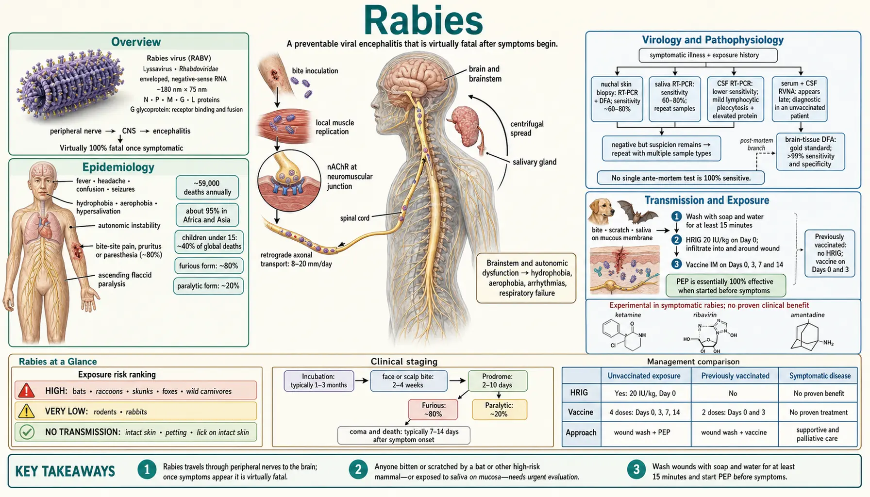
Table of Contents
Overview
Rabies is a viral encephalitis caused by the rabies virus (RABV), a member of the genus Lyssavirus within the family Rhabdoviridae. The virion is immediately recognizable under electron microscopy by its characteristic bullet-shaped morphology — approximately 180 nm long by 75 nm wide. Once clinical symptoms appear, rabies is virtually 100% fatal, making it one of the very few infectious diseases with a near-universal case fatality rate.
The World Health Organization (WHO) says rabies causes tens of thousands of deaths every year, mainly in Asia and Africa; its 2021 Global Health Estimates model puts the figure at 44,203 deaths a year. In these regions, domestic dog bites account for approximately 99% of all human rabies cases. In the United States and other high-income countries, canine rabies has been effectively eliminated through mass dog vaccination, but the virus persists in wildlife reservoirs — particularly bats, raccoons, foxes, and skunks.
Despite this grim prognosis, rabies is almost entirely preventable. Post-exposure prophylaxis (PEP) — a combination of thorough wound washing, human rabies immunoglobulin (HRIG), and a vaccine series — is essentially 100% effective when started before the onset of symptoms. The single most important immediate action after any potential exposure is washing the wound vigorously with soap and water for at least 15 minutes, which can dramatically reduce the viral load at the inoculation site.
- Causative agent: Rabies virus (RABV), genus Lyssavirus, family Rhabdoviridae
- Virion: enveloped, single-stranded negative-sense RNA, bullet-shaped (~180 nm × 75 nm)
- Case fatality: virtually 100% once symptomatic — a 2025 review counted only about 34 well-documented survivors worldwide (alive 6 months after symptoms began)
- Global deaths: tens of thousands a year, mainly in Asia and Africa (WHO 2021 Global Health Estimates: 44,203 a year)
- Primary transmission: dog bites (~99% of global human cases)
- USA status: eliminated from domestic dogs; persists in wildlife (bats, raccoons, foxes, skunks)
- Most critical immediate action (WHO first aid): washing the wound with soap and water for at least 15 minutes — reduces infection risk significantly
- Prevention: PEP is essentially 100% effective if started before symptom onset
Epidemiology
The World Health Organization describes rabies as causing tens of thousands of deaths annually, mainly in Asia and Africa, with 40% of them in children under 15; its 2021 Global Health Estimates model gives 44,203 deaths a year. A 2015 modelling study by Hampson and colleagues estimated about 59,000 deaths a year from dog-transmitted rabies, 59.6% of them in Asia and 36.4% in Africa. However, because rabies is severely underreported — particularly in rural areas of endemic countries where laboratory confirmation is rarely available — the true burden is estimated to be 2 to 10 times higher than official figures suggest.
Global Distribution
- India: Bears the single largest national burden, accounting for roughly 36% of global rabies deaths — an estimated 20,000 deaths per year
- Sub-Saharan Africa: Second largest burden, with widespread dog-mediated transmission and limited access to PEP
- Southeast Asia: Philippines, Indonesia, and Vietnam report significant dog-mediated cases; Thailand has dramatically reduced cases through dog vaccination programs
- The Americas: Dog-mediated rabies largely controlled; bat-mediated transmission is the dominant exposure route; vampire bat transmission remains a serious issue in parts of Latin America
- Europe: Most of Western Europe is rabies-free in domestic animals; wildlife (fox) rabies eliminated in much of Europe through oral bait vaccination programs; Eastern Europe and Russia still have fox/raccoon dog reservoirs
United States
The USA reports fewer than 10 human rabies deaths per year (CDC), and contact with infected bats is the leading cause. The last confirmed dog-to-human rabies transmission from a dog bitten in the USA occurred in 2007. This achievement — eliminating canine rabies — is one of the great public health successes of the 20th century, accomplished through widespread mandatory pet vaccination laws and stray dog control. Key US epidemiological points:
- Wildlife reservoirs dominate — CDC notes rabies is found in wildlife throughout the country except Hawaii: bats (nationwide), raccoons (Eastern seaboard), skunks (Central plains and California), foxes (Alaska and Texas)
- Around 100,000 Americans are vaccinated against rabies each year after a potential rabies exposure (CDC)
- Bat exposures frequently go unrecognized — the puncture wound from a bat bite is often too small to notice
- Cost of a full PEP course in the USA: approximately $3,000–$10,000
Vulnerable Populations
- Children under 15 account for approximately 40% of global rabies deaths — they are more likely to be bitten and less likely to report exposures to adults
- Rural populations with limited access to PEP and health infrastructure bear disproportionate burden
- People without health insurance or in countries without subsidized PEP face high out-of-pocket costs that lead to treatment avoidance
PEP Impact
According to the World Health Organization's current fact sheet, over 29 million people worldwide receive human rabies vaccine each year.
Virology and Pathophysiology
Understanding how rabies virus invades, travels through, and ultimately destroys the nervous system explains both why it is so lethal and why early intervention is so critical.
Viral Structure
The rabies virion is a single-stranded, negative-sense RNA virus enclosed in a lipid envelope derived from the host cell membrane. The genome encodes exactly five proteins:
- Nucleoprotein (N): encapsidates the RNA genome; primary target for diagnostic assays
- Phosphoprotein (P): cofactor for the RNA polymerase complex; modulates innate immune evasion
- Matrix protein (M): bridges the envelope and nucleocapsid; controls budding
- Glycoprotein (G): the only surface-exposed protein; mediates receptor binding and membrane fusion; the primary target of virus-neutralizing antibodies and the key virulence determinant
- RNA-dependent RNA polymerase (L): the large polymerase protein; performs transcription and replication
Cell Entry and Receptor Binding
The glycoprotein (G protein) binds to at least three identified receptors on neurons and other cells:
- Nicotinic acetylcholine receptor (nAChR): present at neuromuscular junctions — the initial site of viral uptake after a bite
- Neural cell adhesion molecule (NCAM / CD56): expressed on neurons and muscle cells
- p75 neurotrophin receptor (p75NTR): expressed on peripheral neurons; may facilitate retrograde transport
After binding, the virus enters cells by receptor-mediated endocytosis. Acidification of the endosome triggers membrane fusion, releasing the nucleocapsid into the cytoplasm where transcription begins.
Retrograde Axonal Transport
This step explains the incubation period and its variability. After initial replication at the bite site (in muscle cells), the virus is taken up by peripheral nerve endings and transported retrogradely up the axon toward the spinal cord and brain at a rate of approximately 8–20 mm per day. This transport uses the host cell's dynein motor protein complex along microtubules.
Incubation period depends on the distance from the bite to the central nervous system (CNS) and the viral load inoculated:
- Bites on the face or scalp: incubation as short as 2–4 weeks
- Bites on the trunk or upper extremities: typically 1–3 months
- Bites on the feet or distal extremities: can be months to years — the longest documented incubation is 19 years
- Typical range overall: 1–3 months
Central Nervous System Invasion and Spread
Once the virus reaches the spinal cord, it spreads rapidly throughout the central nervous system via transsynaptic transmission. One of the virus's most remarkable (and deadly) adaptations is that it minimizes cytopathic effect in neurons — rather than killing cells outright (which would trigger inflammation and potentially alert the immune system), it modulates apoptosis pathways to keep neurons alive long enough to amplify and spread virus throughout the brain.
After central nervous system amplification, the virus spreads centrifugally along efferent nerves to peripheral organs — most critically to the salivary glands (enabling transmission via biting), and also to the cornea, skin hair follicles, and the heart. This centrifugal spread to the skin and cornea is the basis for ante-mortem biopsy-based diagnosis.
Cause of Death
Death in rabies results from:
- Autonomic nervous system dysfunction — cardiac arrhythmias, blood pressure instability
- Respiratory center dysfunction leading to apnea and respiratory failure
- Cerebral edema
- Hypothalamic dysfunction — hyperthermia, diabetes insipidus
Notably, gross neuropathological findings are often surprisingly mild given the severity of clinical disease — relatively little neuronal loss compared to other viral encephalitides. The dysfunction is more physiological (ion channel disruption, synaptic failure) than structural.
Transmission and Exposure
Rabies is a zoonotic disease — it requires transmission from an infected animal to a human. Understanding the routes and risk levels of different exposures is essential for making correct PEP decisions.
Primary Transmission Routes
- Dog bites: Account for ~99% of human rabies cases worldwide; the overwhelmingly dominant route in Africa, Asia, and Latin America
- Bat bites in the USA: The primary domestic exposure route; bat bites are often undetected because they leave tiny puncture marks; people may be entirely unaware they were bitten
- Other animal bites: Cats, raccoons, foxes, skunks, wolves, jackals, and primates can transmit rabies; rodents and rabbits almost never transmit rabies to humans
- Mucous membrane exposure: Contact of saliva with eyes, nose, or mouth from a rabid animal is a recognized (though less common) exposure route
- Corneal and organ transplants: Rare documented clusters of transmission; donors with undiagnosed rabies encephalitis have transmitted virus via corneal grafts and solid organ transplants
Routes That Do NOT Transmit Rabies
- Contact with intact (unbroken) skin
- Blood, urine, or feces from a rabid animal
- Petting an animal or being licked on intact skin
- Casual human-to-human contact with a rabies patient (no documented human-to-human transmission outside organ transplantation)
The Bat Exposure Problem
Bats deserve special mention because they are responsible for nearly all recent human rabies deaths in the United States and Canada, and exposures are frequently unrecognized. U.S. Centers for Disease Control and Prevention (CDC) and public health guidelines specify that a person who was sleeping in a room where a bat was found — and cannot rule out that a bite occurred — should receive PEP. Bat teeth are small enough that a bite may leave no visible wound and cause no pain. This "sleep bat exposure" rule has been controversial but is based on documented cases where people died of rabies after an apparently incidental bat encounter.
PEP Decision Framework
When evaluating a potential exposure, clinicians consider:
- Was this a bite (breaks skin), scratch (breaks skin), or mucous membrane contact? — All warrant evaluation
- Is the animal available for observation (dogs/cats)? A healthy dog/cat observed for 10 days without signs can stop PEP
- What species? Bats, raccoons, skunks, foxes, and most wild carnivores are considered high-risk; rodents and rabbits are very low risk
- What is the geographic area? Local wildlife rabies prevalence matters
- Was the exposure provoked or unprovoked? An unprovoked attack raises suspicion of rabies
Clinical Presentation
Rabies progresses through distinct phases after the incubation period. Recognizing the early signs — particularly the pathognomonic paresthesia at the bite site — can prompt urgent PEP in the rare window that might still be effective, and allows appropriate palliative planning.
Prodromal Phase (2–10 days)
The prodrome is nonspecific and easily mistaken for influenza or other common illnesses:
- Fever, headache, malaise, fatigue, anorexia, nausea and vomiting
- Paresthesias, pruritis, or pain at the healed bite site — present in approximately 80% of cases; this is the most suggestive early sign, and clinicians treat it as a reason for immediate suspicion of rabies in anyone with a prior animal bite history
- Anxiety, irritability, insomnia
Furious (Encephalitic) Rabies (~80% of cases)
This is the classic presentation most people associate with rabies:
- Hydrophobia (pathognomonic): violent involuntary spasms of the pharynx, larynx, and diaphragm triggered by attempts to swallow liquids — or even by seeing, hearing, or thinking about water. The patient is conscious, aware, and terrified by these episodes. The physiological basis is virus-induced dysfunction of the nucleus ambiguus and respiratory centers.
- Aerophobia (highly specific): similar spasms triggered by a puff of air directed at the face; very specific for rabies among the differential diagnosis of viral encephalitides
- Hypersalivation — the patient cannot swallow saliva
- Agitation, combativeness, bizarre behavior, confusion, hallucinations
- Autonomic instability: hyperthermia, tachycardia, hypertension, pupillary dilation
- Priapism in males
- Seizures
- Lucid intervals alternating with agitation
- Progression to coma and death typically within 7–14 days of symptom onset if untreated
Paralytic (Dumb) Rabies (~20% of cases)
The paralytic form is frequently misdiagnosed as Guillain-Barré syndrome or other causes of acute flaccid paralysis:
- Ascending flaccid paralysis beginning at the bitten limb
- Sphincter dysfunction
- Less prominent agitation and hydrophobia (may still develop late)
- Sensory loss in some cases
- Longer clinical course than furious form before coma and death
- More common with bat-mediated transmission and with certain viral strains
- Important differential: Guillain-Barré, poliomyelitis, other viral encephalitides
Hydrophobia and Aerophobia: Why They Occur
These signs deserve emphasis because they are nearly pathognomonic. Hydrophobia is not a fear of water in the psychological sense — it is a physical laryngospasm triggered by the act of swallowing. The virus infects brainstem nuclei controlling the pharynx and larynx. The pain and terror of these spasms cause patients to refuse all liquids, leading to dehydration. Aerophobia — identical spasms triggered by a draft of air — is even more specific for rabies, and when present it is a reason for immediate laboratory testing.
Diagnosis
Because there is no reliable blood test that diagnoses rabies in the early incubation phase, and because the virus does not generate a typical antibody response until very late in the disease course, diagnosis is challenging. Multiple sample types and testing methods are used, and no single test is 100% sensitive.
Ante-Mortem Diagnosis (Living Patient)
- Nuchal skin biopsy (RT-PCR and direct fluorescent antibody / DFA): A full-thickness punch biopsy from the nape of the neck containing hair follicles is the most sensitive ante-mortem test. Virus travels centrifugally to cutaneous nerve fibers around hair follicles; sensitivity ~60–80% in symptomatic disease
- Saliva RT-PCR: Detects viral RNA; sensitivity 60–80%; multiple samples increase yield
- Cerebrospinal fluid (CSF) RT-PCR: Lower sensitivity than saliva or skin biopsy; cerebrospinal fluid typically shows mild lymphocytic pleocytosis and elevated protein
- Serum and cerebrospinal fluid rabies virus-neutralizing antibodies (RVNA): Diagnostic only in unvaccinated patients; antibodies usually appear late in the disease course; their presence in cerebrospinal fluid of an unvaccinated patient is highly specific for rabies
- Corneal impression smear DFA: Historically used; lower sensitivity; less commonly used now
Important: No single ante-mortem test reliably rules out rabies early in disease. Negative results are therefore repeated with multiple sample types when clinical suspicion remains high.
Post-Mortem Diagnosis
- Direct fluorescent antibody (DFA) test on brain tissue: The gold standard for rabies diagnosis worldwide; performed on fresh unfixed brain tissue (hippocampus, cerebellum, brainstem, cortex); sensitivity and specificity >99% when performed correctly
- Negri bodies on H&E staining: Eosinophilic intracytoplasmic inclusions in neurons (classically in hippocampal pyramidal neurons and Purkinje cells of the cerebellum); pathognomonic when present but found in only 50–80% of confirmed cases; now largely replaced by DFA
- RT-PCR on brain tissue: Highly sensitive; useful when fresh tissue is unavailable or when strain typing is needed
- Mouse inoculation test (MIT): Historical gold standard; still used in some reference laboratories for viral isolation
Differential Diagnosis
Rabies has to be told apart from other viral encephalitides (HSV, enterovirus, West Nile, Japanese encephalitis), Guillain-Barré syndrome (in paralytic form), tetanus, delirium tremens, and hysteria. The combination of animal bite history + bite-site paresthesias + hydrophobia + aerophobia is essentially diagnostic.
Treatment and PEP
The cornerstone of rabies management is prevention of disease through prompt post-exposure prophylaxis. Once symptoms appear, no treatment has been proven to prevent death, and care is palliative.
Immediate First Aid After Any Exposure
The World Health Organization's first-aid guidance after a bite or scratch from a possibly rabid animal is to "immediately and always wash the wound with water and soap for at least 15 minutes and seek medical attention." This single step reduces the probability of infection and is the most important immediate action. An antiseptic such as povidone-iodine, ethanol (70%), or tincture of iodine is applied afterwards if available. Wounds are not stitched closed straight away when this can be avoided — stitching may push virus deeper into the tissues.
Post-Exposure Prophylaxis (PEP) — Unvaccinated Person
PEP consists of two simultaneous components administered as soon as possible after exposure:
- Human Rabies Immunoglobulin (HRIG), 20 international units (IU)/kg:
- Is infiltrated directly into and around the wound(s) to neutralize virus locally before it enters nerve endings
- Any remaining volume that cannot be anatomically infiltrated into the wound is given IM at a distant site (away from the vaccine injection site)
- Given only on Day 0 — giving it later reduces its effectiveness and may interfere with vaccine-induced immunity
- Is never given in the same syringe or at the same body site as the vaccine
- If HRIG is not available on Day 0, it is given as soon as possible, up to 7 days after the first vaccine dose
- Rabies vaccine series — 4 doses IM: Days 0, 3, 7, and 14 (reduced from the former 5-dose series by a 2010 recommendation of the U.S. Advisory Committee on Immunization Practices (ACIP)). For people with weakened immune systems, ACIP kept PEP as a 5-dose vaccine series with 1 dose of rabies immunoglobulin.
When given before symptom onset, this regimen is essentially 100% effective. There are no documented treatment failures when PEP was correctly administered and started before symptoms appeared.
PEP — Previously Vaccinated Person
If the exposed person has previously completed a full pre-exposure or post-exposure vaccine series (confirmed by documented history or adequate RVNA titer):
- Wound washing remains critical
- No HRIG is needed
- Only 2 doses of vaccine are needed (Days 0 and 3)
Treatment of Symptomatic Rabies
Once symptoms of rabies encephalitis develop, no antiviral treatment has been proven to alter the course of disease. Management is supportive and, in most cases, palliative:
- Intensive care unit (ICU) admission with close monitoring of cardiorespiratory status
- Sedation and analgesia (benzodiazepines, opioids) — essential for comfort and to control agitation and spasms
- Mechanical ventilation as needed
- Treatment of autonomic instability (cardiac arrhythmias, blood pressure fluctuations)
- Anticonvulsants for seizures
- Hydration via intravenous (IV) (oral hydration is impossible due to hydrophobia)
Experimental agents that have been tested without proven clinical benefit include ribavirin, interferon-alpha, and ketamine. Their use remains investigational.
HRIG Supply and Access
A major barrier to PEP effectiveness globally is the cost and availability of HRIG. In the USA, a full PEP course including HRIG costs $3,000–$10,000. In many endemic countries, HRIG is simply unavailable or unaffordable. Equine rabies immunoglobulin (ERIG) — cheaper and more widely available — is used instead in many resource-limited settings; it carries a slightly higher risk of serum sickness but is still highly effective.
Milwaukee Protocol
The Milwaukee Protocol is an intensive-care approach first used in 2004 on an unvaccinated teenager who survived symptomatic rabies. Later reviews found no evidence that it works (see Subsequent Experience below), and a 2016 critical appraisal called it a "failed protocol" whose therapies could not be substantiated.
Background and Rationale
In November 2004, Dr. Rodney Willoughby Jr., a pediatric infectious disease physician at Children's Hospital of Wisconsin in Milwaukee, faced an apparently hopeless case: a 15-year-old girl named Jeanna Giese who had been bitten by a bat and developed full clinical rabies without having received any pre- or post-exposure vaccination.
Willoughby's reasoning was that rabies kills through neurological dysfunction — it disrupts ion channels, neurotransmitter systems, and autonomic regulation — rather than through massive irreversible neuronal destruction. If the brain could be pharmacologically protected while the immune system mounted a response, the patient might clear the virus.
The Protocol
- Medically induced coma using ketamine (N-methyl-D-aspartate (NMDA) receptor antagonist with potential direct antiviral activity) and midazolam — to reduce cerebral metabolic demand and limit excitotoxic neuronal death
- Antiviral therapy with ribavirin and amantadine — chosen for their theoretical antiviral activity against RNA viruses; neither has convincingly demonstrated in vivo efficacy against RABV
- Intensive supportive care — mechanical ventilation, hemodynamic monitoring, seizure control, nutritional support
- Continued until the patient showed evidence of mounting an immune response (rising RVNA titers) and viral markers declined
Jeanna Giese: The First Survivor
Giese survived and was discharged from the hospital after 76 days, with moderate neurological deficits including ataxia and cognitive impairment. Over subsequent years, she regained substantial function and graduated from college. Her survival was extraordinary: no unvaccinated human had previously survived symptomatic rabies in recorded medical history.
Subsequent Experience
A 2025 review counted at least 64 cases in which versions of the Milwaukee Protocol failed, and found that over the past two decades no detailed report had documented evidence of its efficacy. Analysis of the survivors reveals an important pattern: each survivor had evidence of partial or pre-existing immune response to rabies virus (either from unrecognized prior vaccination, or an unusually robust innate immune response). Patients with no immune response consistently died.
Current Status
The Milwaukee Protocol is not recommended as standard of care by any major infectious disease or neurology guideline due to its very low success rate, the enormous resource commitment required for intensive care unit management, and the lack of meaningful benefit in immunologically naive patients; a 2025 review concluded that the time had come to abandon it. It may be considered on an individualized basis for patients who:
- Show evidence of mounting a rabies-specific immune response (rising RVNA titers)
- Have access to a tertiary care intensive care unit capable of sustained aggressive management
- Have families who understand the extremely poor prognosis and wish to pursue all possible options
The primary legacy of the Milwaukee Protocol is scientific: it demonstrated for the first time that human survival is biologically possible, that immune response is the key determinant of outcome, and it prompted ongoing research into immunomodulatory and antiviral approaches.
Prevention and Pre-Exposure Prophylaxis
Rabies is one of the most preventable lethal infectious diseases, through a combination of pre-exposure vaccination for high-risk individuals, animal vaccination programs, wildlife reservoir management, and accessible PEP.
Pre-Exposure Prophylaxis (PrEP)
Since 2022, the U.S. Advisory Committee on Immunization Practices (ACIP) recommends PrEP as a 2-dose vaccine series given into the muscle on Days 0 and 7; this replaced the older 3-dose schedule (Days 0, 7, and 21 or 28). It is recommended for:
- Veterinarians and veterinary staff
- Animal handlers and wildlife biologists
- Travelers spending extended time in rabies-endemic areas, particularly in rural regions
- Bat researchers and spelunkers (cavers)
- Rabies laboratory workers
- International travelers on extended itineraries where PEP may not be accessible within 24 hours
Key advantage of PrEP: if a subsequent exposure occurs, no HRIG is needed and only 2 vaccine doses (Days 0 and 3) are required — simpler, cheaper, and effective even in settings where HRIG is unavailable.
Pet and Livestock Vaccination
Rabies vaccination of dogs and cats is a core vaccine in the USA and most developed countries, required by law in most states. This single intervention eliminated dog-mediated human rabies in the USA. Mass dog vaccination campaigns in endemic countries have similarly dramatic results — Thailand reduced its annual human rabies deaths from >200 in the 1980s to <5 today through sustained canine vaccination. The World Health Organization (WHO) estimates that vaccinating 70% of the dog population in endemic areas is sufficient to eliminate the dog-to-human transmission cycle.
Oral Wildlife Bait Vaccines
A recombinant vaccinia-vectored oral rabies vaccine is distributed in wildlife bait (fishmeal-based) dropped from aircraft across wide geographic areas. The USA has used this approach since 1990 to vaccinate raccoons, coyotes, and gray foxes. At the Canadian border the result has not been permanent: a 2016 Canadian report describes Canada eliminating raccoon rabies that had spread in from the south and staying free of it from about 2009 to 2014, before new incursions were detected in Ontario, Quebec and New Brunswick; the responses have included surveillance, trap-vaccinate-release and oral bait vaccination campaigns. Similar programs in Europe eliminated fox rabies from most of Western Europe.
Wildlife Avoidance and Exposure Prevention
- Handling wild animals is a direct exposure risk, especially animals that appear sick, disoriented, or unusually approachable (unusual daytime activity in nocturnal species like raccoons or bats is a warning sign)
- For a bat found in the home, CDC's advice is to "contact animal control or your health department to safely capture it for rabies testing," and not to release the bat until a public health expert has been consulted — testing matters most when anyone may have been asleep in the room
- Secured trash cans discourage raccoon contact
- Vaccinated pets create a buffer between wildlife reservoirs and human exposure
- In endemic areas abroad, PEP and HRIG may not be available at the destination — the situation the PrEP travel recommendation above covers
World Health Organization Zero by 2030
The World Health Organization, in partnership with the Food and Agriculture Organization (FAO), World Organisation for Animal Health (OIE/WOAH), and the Global Alliance for Rabies Control, has set a target of zero human deaths from dog-mediated rabies by 2030. Mathematical modeling demonstrates this is achievable through: sustained mass dog vaccination to cover >70% of dog populations in endemic countries, accessible and affordable PEP (including intradermal regimens that reduce costs by 60–80%), and elimination of PEP stockout gaps.
Prognosis
The prognosis for rabies is starkly binary: almost certain survival with timely PEP, or near-certain death once symptoms appear.
With Timely PEP
Post-exposure prophylaxis correctly administered before symptom onset is essentially 100% effective. There are no documented treatment failures when:
- The wound was thoroughly washed with soap and water
- HRIG was infiltrated into the wound on Day 0
- The full vaccine series was completed
- Treatment began before symptom onset
Time from exposure to PEP initiation matters enormously. While PEP has worked up to weeks after exposure (because the virus may still be in peripheral nerves during the long incubation), delay increases risk — particularly for bites close to the head and neck.
Once Symptomatic
A 2025 review counted only about 34 well-documented survivors of symptomatic rabies (alive 6 months after symptoms began), many with serious lasting nerve and brain damage. Most of these rare survivors:
- Had partial pre-existing immunity from prior vaccination or unrecognized exposure
- Were treated with some form of the Milwaukee Protocol
- Retained significant neurological deficits (ataxia, cognitive impairment, behavioral changes)
Without prior immunity and without aggressive intensive care unit management, survival is essentially unheard of. The virus's strategy of minimizing early cytopathic effect while spreading widely through the central nervous system means by the time symptoms appear, infection is already disseminated far beyond any point where local antiviral measures could be effective.
Key Prognostic Factors
- Time to PEP initiation — the most modifiable factor
- Location of bite — bites on the face/head/neck have shorter incubation and less time to initiate PEP; poorer prognosis without PEP
- Viral inoculum — deep bites with heavy salivary contamination deliver more virus
- Pre-existing immunity — the most important determinant of survival in symptomatic disease
- Viral strain — some strains (e.g., certain bat strains) may be less virulent in humans
The Central Message
Every hour between an animal exposure and wound washing plus PEP initiation increases risk. Rabies is one of those rare diseases where acting immediately — before waiting to see if symptoms develop — saves lives. Once symptoms appear, the window has closed. PEP is started on the basis of exposure, not on the basis of symptoms.
Recent Research
Despite being one of the oldest known diseases (described in ancient Mesopotamian law codes and Greek medical texts), rabies research is advancing rapidly across multiple fronts.
Monoclonal Antibody Alternatives to HRIG
Human rabies immunoglobulin (HRIG) is expensive, produced from pooled human plasma, and in limited supply. Two neutralizing monoclonal antibodies — RVC20 (binding the glycoprotein antigenic site I) and RVC58 (binding antigenic site III) — form a cocktail that, in a 2016 laboratory study, neutralized all 35 rabies virus strains and 25 related lyssaviruses tested, with higher potency than commercial human rabies immunoglobulin, and protected hamsters from a lethal rabies challenge. The World Health Organization's current fact sheet lists rabies monoclonal antibodies (RmAb), as an alternative to rabies immunoglobulin, among the components of post-exposure prophylaxis when indicated.
Intradermal PEP Regimens
World Health Organization now endorses intradermal (ID) PEP regimens (such as the 2-site ID regimen using one-fifth the standard intramuscular dose per injection point) as equivalent to the standard IM regimen in immunogenicity. Intradermal PEP reduces the total vaccine cost by 60–80%, a transformational change for resource-limited settings. Regulatory approval of ID regimens is expanding globally.
Next-Generation Vaccines for Dog Vaccination Programs
Thermostable, single-dose, oral rabies vaccines for mass canine vaccination are under development. Current oral bait vaccines for wildlife work well but are not suitable for the scale of mass dog vaccination needed in Africa and Asia. Improved vectors and adjuvant systems aim to produce durable immunity from a single dose without a cold chain.
Antiviral Compounds
High-throughput screening has identified several promising antivirals with in vitro activity against RABV:
- Favipiravir (T-705): Broad-spectrum RNA virus polymerase inhibitor with demonstrated in vivo activity in mouse models; clinical trials would need to precede human use
- Dasatinib: An approved kinase inhibitor found to reduce RABV replication in cell culture by targeting cellular kinase pathways required for viral spread
- Interferon lambda: May offer better central nervous system penetration and less systemic toxicity than interferon alpha
mRNA Vaccine Platforms
The same mRNA technology used in COVID-19 vaccines is being applied to rabies. Early-phase trials of mRNA rabies vaccines show strong immunogenicity with fewer doses and potentially more flexible production. The ability to rapidly manufacture mRNA vaccines could be critical for addressing outbreak strains.
Global Zero by 2030
Mathematical modeling published in PLOS Neglected Tropical Diseases confirms that the World Health Organization 2030 target is achievable with current tools — mass dog vaccination reaching 70% coverage combined with accessible PEP supply chains. The primary barrier is not scientific but logistical and financial: sustained political commitment, funding for vaccines, and last-mile delivery of PEP in endemic settings. Several countries including the Philippines and Tanzania are now in the advanced stages of national elimination programs.
Key Research Papers
- Hampson K, Coudeville L, Lembo T, et al. Estimating the global burden of endemic canine rabies. PLoS Negl Trop Dis. 2015;9(4):e0003709. — PubMed PMID: 25881058
- Rupprecht CE, Briggs D, Brown CM, et al. Use of a reduced (4-dose) vaccine schedule for postexposure prophylaxis to prevent human rabies. MMWR Recomm Rep. 2010;59(RR-2):1–9. — PubMed PMID: 20300058
- Willoughby RE Jr, Tieves KS, Hoffman GM, et al. Survival after treatment of rabies with induction of coma. N Engl J Med. 2005;352(24):2508–2514. — PubMed PMID: 15958806
- Jackson AC. Current and future approaches to the therapy of human rabies. Antiviral Res. 2013;99(1):61–67. — Search PubMed · Search PubMed
- Dietzschold B, Schnell M, Koprowski H. Pathogenesis of rabies. Curr Top Microbiol Immunol. 2005;292:45–56. — PubMed PMID: 15981467 · Search PubMed
- Hanlon CA, Niezgoda M, Rupprecht CE. Postexposure prophylaxis for prevention of rabies in dogs. Am J Vet Res. 2002;63(8):1096–1100. — PubMed PMID: 12171160
- Warrell MJ, Warrell DA. Rabies: the clinical features, management and prevention of the classic zoonosis. Clin Med (Lond). 2015;15(1):78–81. — PubMed PMID: 25650205 · Search PubMed
- Hemachudha T, Ugolini G, Wacharapluesadee S, et al. Human rabies: neuropathogenesis, diagnosis, and management. Lancet Neurol. 2013;12(5):498–513. — PubMed PMID: 23602163
- Fooks AR, Cliquet F, Finke S, et al. Rabies. Nat Rev Dis Primers. 2017;3:17091. — PubMed PMID: 29188797 · Search PubMed
- WHO Expert Consultation on Rabies, third report. World Health Organ Tech Rep Ser. 2018;(1012):1–183. PubMed Search
- WHO position paper: rabies vaccines and immunoglobulins. Intradermal PEP regimens. 2018. PubMed Search
- Jackson AC. Demise of the Milwaukee Protocol for Rabies. Clin Infect Dis. 2025;81(4):e229–e232. — PubMed PMID: 40139921
- Zeiler FA, Jackson AC. Critical Appraisal of the Milwaukee Protocol for Rabies: This Failed Approach Should Be Abandoned. Can J Neurol Sci. 2016;43(1):44–51. — PubMed PMID: 26639059
- De Benedictis P, Minola A, Rota Nodari E, et al. Development of broad-spectrum human monoclonal antibodies for rabies post-exposure prophylaxis. EMBO Mol Med. 2016;8(4):407–421. — PubMed PMID: 26992832
- Rao AK, Briggs D, Moore SM, et al. Use of a Modified Preexposure Prophylaxis Vaccination Schedule to Prevent Human Rabies: Recommendations of the Advisory Committee on Immunization Practices - United States, 2022. MMWR Morb Mortal Wkly Rep. 2022;71(18):619–627. — PubMed PMID: 35511716
- Stevenson B, Goltz J, Massé A. Preparing for and responding to recent incursions of raccoon rabies variant into Canada. Can Commun Dis Rep. 2016;42(6):125–129. — PubMed PMID: 29770016
- World Health Organization. Rabies (fact sheet), 17 September 2026. — who.int fact sheet: Rabies
- U.S. Centers for Disease Control and Prevention. About Rabies; Preventing Rabies from Bats. — cdc.gov: About Rabies · cdc.gov: Preventing Rabies from Bats
PubMed Topic Searches
The following PubMed topic searches retrieve current peer-reviewed literature on Rabies.
- PubMed: Rabies virus pathogenesis
- PubMed: Rabies postexposure prophylaxis
- PubMed: Rabies global burden mortality
- PubMed: Rabies bat transmission human
- PubMed: Rabies vaccine pre-exposure prophylaxis
- PubMed: Milwaukee protocol rabies survival
- PubMed: Rabies encephalitis furious paralytic
- PubMed: Rabies Negri bodies neuropathology
- PubMed: Rabies wound washing first aid
- PubMed: Rabies HRIG immunoglobulin PEP
- PubMed: Rabies zero by 2030 dog vaccination
- PubMed: Rabies diagnosis ante-mortem RT-PCR