Erythema Migrans — The Lyme Rash Guide
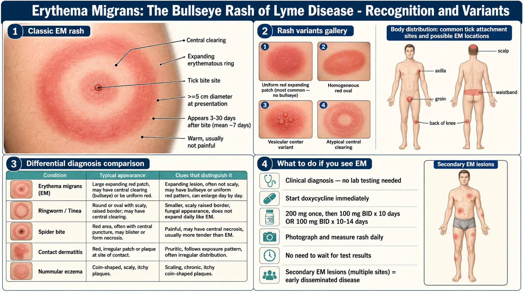
Table of Contents
- What EM Is — and Why It Matters Most
- Classic Bullseye vs. Other Forms
- Timing — When It Appears and How Fast It Grows
- What It Feels Like
- Where It Appears — the "Missed Rash" Problem
- Photographing and Measuring the Rash
- Common Look-Alikes
- Multiple EM Lesions — Early Disseminated Disease
- Why the Guideline Treats the Rash as the Diagnosis
- When the Guideline Calls for Clinical Diagnosis Without Testing
- What Happens If EM Is Missed
- Common Questions at a Clinic Visit — What the Evidence Says
- After Antibiotics Begin
- Key Research Papers
- Connections
- Featured Videos
What EM Is — and Why It Matters Most
Erythema migrans (EM) is the expanding skin rash that appears at the site of a tick bite that transmitted Borrelia burgdorferi. It is the most common manifestation of early Lyme disease. The CDC states that EM occurs in approximately 70–80% of infected people, beginning 3 to 30 days after the bite. The 2020 IDSA/AAN/ACR guideline recommends that a person with potential tick exposure in a Lyme-endemic area who has one or more skin lesions compatible with EM be diagnosed clinically rather than by laboratory testing.
Because the rash is on the skin, it is often the first sign of infection that people notice themselves. The guideline’s listed regimens for EM are short oral courses — 10 days of doxycycline, or 14 days of amoxicillin or cefuroxime axetil — and in a randomized trial of 180 patients with EM, 10 days of doxycycline worked as well as 20 days. Untreated, the infection can spread and later cause Lyme arthritis, carditis, facial palsy, or neuroborreliosis.
The rash is caused by the bacteria migrating outward through the skin from the bite site. As spirochetes spread radially through the dermis, the immune system follows, creating an expanding ring of inflammation. That is literally what erythema migrans means: "migrating redness."
Classic Bullseye vs. Other Forms
Everybody has seen the stock photo — a round red ring with a pale center and a dark dot in the middle. That classic bullseye is the familiar image, but in the United States it is not the most common form, and EM is not pathognomonic: other rashes can look similar.
A 2007 JAMA analysis of 53 studies (8,493 patients) found central clearing in only 19% of EM lesions in Lyme-endemic areas of the United States (95% CI 11–32%), against 79% in Europe and 80% in non-endemic U.S. areas. In a U.S. vaccine-trial cohort of 118 patients whose EM was confirmed by culture or PCR, early lesions commonly showed homogeneous or central redness rather than a red ring with partial central clearing. Forms described in these studies and in the CDC’s image gallery include:
- Solid / homogeneous red oval. The most common form in U.S. studies. A uniform pink-to-red patch, often oval rather than round, with no central clearing. Looks like a flat, painless bruise that happens to be red instead of purple.
- Uniform red plaque. Slightly raised, smooth, warm but not hot. Easy to mistake for a mild allergic reaction.
- Bluish or purplish hue. The CDC’s gallery includes bluish-hued and red-blue lesions, and some lesions have a purplish discoloration in the center. How EM is recognized on darker skin is covered below.
- Central vesicle or crust. A small blister or scab at the bite point inside an expanding red area, which can be mistaken for a spider bite.
- Multiple concentric rings. A target-like pattern of two or three rings; less common in U.S. lesions.
- Multiple EM lesions. Several separate red patches scattered on trunk, limbs, and face, each one representing a site where bacteria seeded through the bloodstream. See the disseminated section below.
EM on darker skin. A 2026 analysis of the dermatology resources used at 50 top U.S. medical schools notes that cutaneous Lyme disease presents differently on light and dark skin, and found that only 3 of 47 EM images (6.4%) in 16 resources showed dark skin. Black patients are less often diagnosed at the rash stage: in a multicenter pediatric study, Black children with Lyme disease were less likely to have an EM diagnosed (adjusted odds ratio 0.34) and more likely to have a swollen joint (3.68); in a Maryland specialty clinic, the median time to appropriate antibiotic treatment for Black patients with EM was 26 days, against 4 days for White patients, and two of the Black patients whose rashes the study illustrates had first been diagnosed with an allergic reaction and with cellulitis. A 2000 Maryland surveillance study suggested that failure to recognize early EM in African Americans may explain their higher rates of late manifestations.
Size and growth carry more weight than shape. The CDC’s surveillance case definition describes EM as a lesion that begins as a red macule or papule and expands over days to weeks to form a large round lesion, often with partial central clearing, and requires a single primary lesion of at least 5 cm (2 inches) across; the CDC notes that this definition is for surveillance and is not intended for making a clinical diagnosis. The 2007 JAMA analysis concluded that no single element of the history or examination is highly sensitive on its own.
Timing — When It Appears and How Fast It Grows
The CDC states that EM begins at the site of the bite 3 to 30 days after the tick bite, with an average of about 7 days. A small bump or redness that appears immediately at a tick bite and resembles a mosquito bite is common; the CDC notes that this irritation generally goes away in 1–2 days and is not a sign of Lyme disease. EM takes days to develop.
The CDC describes EM as expanding gradually over several days and reaching up to 12 inches (30 cm) or more across. In a Slovenian study of patients with a single EM, lesion diameter grew by a median of about 1.2–1.5 cm per day, and a review notes that EM in Europe expands more slowly than in the United States. That continued outward growth is one of the most useful diagnostic features. In two New York studies, lesions measured an average of about 16 cm across when evaluated (range 6–73 cm in one of them), so lesions of 15 cm or more are common by the time they are seen.
Many people never saw the tick. The CDC notes that nymphal ticks, which pose a particularly high risk because of their abundance and small size, are approximately the size of a poppy seed; that a tick generally must be attached for more than 24 hours before the Lyme bacterium can be transmitted; and that patients are often unaware of a tick bite before getting sick. In a New York study only 19.8% of patients with EM-like lesions recalled a tick bite, and in a culture-confirmed series 25% had noticed a bite at the site of the rash. Not remembering a bite does not rule out Lyme.
What It Feels Like
EM is usually painless. The CDC describes it as possibly warm to the touch but rarely itchy or painful. Lesions on the back or other hard-to-see places can go unnoticed by the person who has them.
That largely painless quality helps distinguish EM from several look-alikes:
- Spider bite: recluse spider bites are described as initially painless but usually progress to an ulcerating, necrotic (blackened) wound at the bite site; widow spider bites cause muscle spasms and rigidity starting at the bite within 30 minutes to two hours.
- Cellulitis: hot, tender, tight-feeling, with ill-defined borders that blur into surrounding skin.
- Allergic or contact dermatitis: itchy, sometimes burning.
- Ringworm: mildly itchy, scaly, with a raised and flaking leading edge.
The 2007 JAMA analysis concluded that recognizing EM means combining several features of the examination with the epidemiological context — such as hiking, gardening, or time in grass in a Lyme-endemic area a week or so earlier — rather than relying on any single sign.
Many patients also have a mild flu-like illness. In a series of 79 U.S. patients with culture-confirmed EM, 68% had systemic symptoms — fatigue (54%), joint pain (44%), muscle aches (44%), headache (42%), fever or chills (39%) — and 23% had localized swollen lymph nodes. The 2007 JAMA analysis found fever and headache frequently reported and nausea and vomiting rare, and the U.S. vaccine-trial cohort described the typical presentation as EM with nonspecific flu-like symptoms.
Where It Appears — the "Missed Rash" Problem
Ticks often attach in warm, tucked-away skin, so EM can appear in places that are hard to see on one’s own body. The CDC notes that it may appear on any area of the body; locations often described include:
- Popliteal fossa — the back of the knee.
- Groin and inner thigh.
- Axilla — the armpit.
- Back, especially between the shoulder blades and along the waistband.
- Scalp and hairline, where hair hides both tick and rash.
- Buttocks and gluteal fold.
- Behind the ears and the nape of the neck.
This is the "missed rash" problem. A review of Lyme borreliosis reports that EM is the presenting manifestation in about 80% of U.S. patients; about 18% have nonspecific summer symptoms without a recognized EM, and the remaining 2–3% first present with a later manifestation such as facial palsy or Lyme arthritis. Lesions on the scalp, back, buttocks, and the backs of the knees can go unnoticed unless someone else looks.
Photographing and Measuring the Rash
Because growth over time is central to recognizing EM, a dated photographic record shows a clinician change that a single look cannot. Features that make such photographs useful:
- Daily photographs. The same spot, once per day, documents expansion over time.
- A scale reference. A ruler, a coin (a U.S. quarter is about 24 mm across, a dime about 18 mm), or a standard payment card (about 85 mm long) next to the rash. Without scale, size cannot be judged from a photo.
- A pen line at the border. An outer edge traced on one day shows on the next whether the rash has grown past it — documented expansion.
- Even natural light. Daylight near a window shows color more truly than a phone flash, which can distort red tones.
- A wide shot and a close-up. Wide for body location; close-up for texture and border detail.
- A photograph of any tick found, on a white background with a coin for scale, from which its species can be identified later.
A series showing measurable outward expansion documents the feature the CDC’s case definition centers on: a lesion that expands over days to weeks.
Common Look-Alikes
Misdiagnosis runs in both directions. In a case series from a Connecticut referral center, EM was missed in 5 patients because of atypical presentations, and 8 patients with EM-like eruptions were wrongly diagnosed with EM; at a university Lyme disease clinic, 38% of 146 referred children had been overdiagnosed with Lyme disease and 8% underdiagnosed. Conditions commonly confused with EM:
- Spider bite (especially brown recluse). Recluse bites are described as initially painless and usually progress to ulcerating dermonecrosis — a dark, necrotic center. The label is often wrong: in one emergency department, of 182 patients who attributed a lesion to a spider bite, 85.7% were diagnosed with skin and soft-tissue infections and 3.8% with actual spider bites.
- Cellulitis. Hot, tender, rapidly advancing, with poorly defined margins. Often accompanied by fever. It is commonly treated with cephalexin, a first-generation cephalosporin, and the 2020 guideline lists first-generation cephalosporins among the drugs not indicated for Lyme disease; in a Maryland clinic series, a patient whose EM was first diagnosed as cellulitis had received 7 days of cephalexin. See Cellulitis.
- Ringworm (tinea corporis). Scaly, flaking leading edge and clearer, drier center. Mildly itchy. Expands over weeks, not days. The scale is the giveaway; EM is smooth.
- Nummular eczema. Coin-shaped itchy, dry, scaling plaques, often multiple. No central bite mark.
- Granuloma annulare. Firm, ring-shaped, skin-colored-to-red plaque that can look remarkably bullseye-like. Painless, very slow growing, and notably smaller than EM.
- Fixed drug eruption. A single round red-brown patch that recurs in the same spot each time the patient takes the causative drug.
- STARI (Southern Tick-Associated Rash Illness). An EM-like rash that follows the bite of the lone star tick (Amblyomma americanum), reported mainly from southern states. Its cause is not known; the CDC states it is not caused by the Lyme bacterium, and describes the rash as usually appearing within 7 days of the bite and expanding to 8–30 cm (3–12 inches). On average the two differ: in a prospective comparison of 21 Missouri cases with 101 New York cases, the Missouri lesions were smaller (mean 8.3 vs 16.4 cm), more circular, and more often centrally cleared (76.2% vs 21.6%); those patients more often recalled a tick bite (85.7% vs 19.8%), were less often symptomatic (19.0% vs 76.2%), less often had multiple lesions (4.8% vs 26.7%), and recovered faster after antibiotics. The CDC states that it is not known whether antibiotics are necessary or helpful for STARI, although some providers treat it with oral antibiotics because it resembles early Lyme disease; the 2020 guideline makes no recommendation for or against antibiotics.
Multiple EM Lesions — Early Disseminated Disease
Several EM patches scattered across the body — one on the back, one on the thigh, one on the arm — are a sign of early disseminated Lyme disease. The first (primary) lesion forms at the bite. Within days to weeks, U.S. strains of B. burgdorferi commonly spread from the bite site to other skin sites, each of which develops its own EM lesion.
How often this happens depends on the study: in pooled U.S. studies 81% of patients had a single lesion, so roughly one in five had several; a culture-confirmed New York series found multiple lesions in 18%, a prospective New York study in 26.7%, and an early 1983 series of 314 patients in almost half. The 2020 guideline applies the same EM treatment durations to solitary and multiple EM (see Antibiotic Treatment Protocols). Neurologic involvement, facial palsy, heart-rhythm changes, and arthritis are later manifestations that, a review reports, treatment of EM prevents.
Why the Guideline Treats the Rash as the Diagnosis
The 2020 IDSA/AAN/ACR Lyme guidelines (Lantos et al.) state: in patients with potential tick exposure in a Lyme disease endemic area who have one or more skin lesions compatible with erythema migrans, the panel recommends clinical diagnosis rather than laboratory testing.
Two reasons are usually given. First, in that setting a compatible expanding lesion is the most common and most recognizable feature of early Lyme disease. Second, blood tests at this stage are insensitive, because antibodies take weeks to develop. The guideline notes that as few as 20% of untreated patients with a microbiologically confirmed single EM are seropositive by conventional two-tier testing (an ELISA followed by a Western blot) on a sample taken within a week, and that results are often falsely negative in EM patients tested less than 2 weeks after the rash appears. In a prospective study, only about one-third of 76 EM patients had positive results at the first visit, and almost two-thirds 3–4 weeks later. A negative result during the rash stage does not rule out Lyme disease.
Testing, and when it becomes informative, is covered in Lyme Testing Explained.
When the Guideline Calls for Clinical Diagnosis Without Testing
The guideline’s clinical-diagnosis recommendation, read together with the CDC’s description of the rash, covers a person with all of the following:
- One or more skin lesions compatible with EM — in the CDC’s surveillance terms, an expanding red lesion whose primary lesion reaches at least 5 cm across.
- Potential tick exposure in a Lyme-endemic area — hiking, yard work, camping, living in or visiting most of the northeastern and upper-midwestern United States, parts of northern California, or much of Europe — with the rash appearing within the CDC’s 3–30 day window after a bite.
- No better alternative explanation (no recently started drug such as a sulfa antibiotic, no history of tinea, no necrotic center suggesting a spider bite or a bacterial skin infection).
For lesions that are suggestive of, but atypical for, EM, the guideline instead suggests antibody testing on an acute-phase blood sample, followed by a convalescent-phase sample if the first result is negative.
For EM, the guideline recommends oral doxycycline for 10 days (adults: 100 mg twice daily or 200 mg once daily), or amoxicillin or cefuroxime axetil for 14 days, rather than longer courses; for patients unable to take both doxycycline and beta-lactam antibiotics, the preferred second-line drug is azithromycin (7 days in the United States, range 5–10). Children receive weight-based doses, and the guideline notes a growing consensus that doxycycline is safe in young children for courses of at least up to 14 days. Doxycycline has traditionally been avoided in pregnancy, and the guideline describes uncertainty about its safety there. See Antibiotic Treatment Protocols for the full dosing table.
Serology becomes far more sensitive later, weeks or months after the rash. In the same prospective study, all 44 patients with neurologic, heart, or joint involvement had positive results on one of the two test systems, and 42 had IgG responses on two-tier testing. At the EM stage, by contrast, a negative test is common in true infection.
What Happens If EM Is Missed
Even without antibiotics, EM typically improves or resolves within weeks. Fading does not mean the infection has cleared: in a study carried out before the cause of Lyme disease was known, about 60% of untreated EM patients in the northeastern United States went on to develop arthritis, an average of 6 months later (range 4 days to 2 years). Without treatment, the infection can progress through these stages:
- Days to weeks later — early disseminated disease. Multiple secondary EMs, facial-nerve palsy (often called Bell's palsy when the cause is not recognized), meningitis-like headache and stiff neck, heart-block and other cardiac arrhythmias ("Lyme carditis"), migrating joint pains.
- Weeks to months later — late disseminated disease. Arthritis in intermittent attacks of joint swelling and pain, mainly in large joints and most classically the knee. See Arthritis.
- Months to years — chronic sequelae. In the United States, a mild late encephalopathy with subtle cognitive disturbances and a mild sensory polyneuropathy have been reported, although a review describes their existence as controversial (see Neuroborreliosis); some patients have arthritis that persists for months to years after antibiotics (post-infectious, antibiotic-refractory Lyme arthritis). Separately, about 10% of patients treated for EM report symptoms such as fatigue, cognitive complaints and musculoskeletal pain lasting 6 months or more; when disabling, this is called post-treatment Lyme disease syndrome. See PTLDS and Chronic Lyme.
A review reports that antibiotic treatment of EM shortens its course and prevents these later manifestations, and the U.S. vaccine-trial cohort found the clinical outcome excellent when antibiotics were given soon after symptoms began.
Common Questions at a Clinic Visit — What the Evidence Says
Questions that commonly come up when a possible EM is seen in a clinic, and what the guideline and studies say about each:
Is a blood test needed before treatment? The 2020 guideline recommends clinical diagnosis rather than laboratory testing for patients with potential tick exposure in an endemic area and one or more lesions compatible with EM. Two-tier serology is negative in most patients during the first week — the guideline notes that as few as 20% are seropositive on a sample taken within a week.
Could it be a spider bite? EM is rarely painful and keeps expanding over days; recluse bites usually progress to a necrotic ulcer, and most lesions that emergency-department patients attribute to spider bites are diagnosed as skin infections. Dated photographs with a scale show whether a lesion is growing.
What if the exposure was outside the local area? EM begins 3 to 30 days after the bite (CDC), so it can appear after a person has returned from an endemic area; the guideline’s clinical-diagnosis recommendation is framed around potential tick exposure in an endemic area, wherever that exposure took place.
What does a negative test in the first week mean? The guideline notes that results are often falsely negative in EM patients tested less than 2 weeks after the rash appears; in a prospective study, about one-third of EM patients were positive at the first visit and almost two-thirds 3–4 weeks later.
Decisions about a specific rash and its treatment belong with a clinician.
After Antibiotics Begin
The rash fades gradually rather than overnight. In a 1983 trial of 108 adults with early Lyme disease, EM and its associated symptoms resolved in a mean of 5.4 and 5.7 days with penicillin or tetracycline, against 9.2 days with erythromycin.
- The first 24 hours. The guideline notes that a minority of patients treated for early Lyme disease have a transient intensification of symptoms, with or without fever, during the first 24 hours of antibiotic therapy — known as a Jarisch–Herxheimer reaction. The guideline describes it as usually mild and self-limited, and it does not recur later in therapy.
- The following days. The rash stops expanding and fades; in the 1983 trial, the mean time to resolution with the more effective drugs was under a week.
The guideline’s durations are for full courses — 10 days of doxycycline, or 14 days of amoxicillin or cefuroxime axetil — and the rash often clears before a course ends. New symptoms during or after treatment, such as facial droop, knee swelling, heart palpitations, or severe headache, are among the disseminated manifestations described above, and the rash resolving does not by itself mean the infection has resolved. For those manifestations the guideline lists other regimens: for meningitis, cranial neuropathy, or radiculoneuropathy, IV ceftriaxone, cefotaxime or penicillin G, or oral doxycycline; for patients hospitalized with Lyme carditis, it suggests starting with IV ceftriaxone rather than oral antibiotics until there is clinical improvement; and for arthritis with little or no response to a first oral course, a 2- to 4-week course of IV ceftriaxone rather than a second oral course.
Doxycycline labeling describes several practical points: drinking fluids liberally with the dose reduces the risk of esophageal irritation and ulceration; an exaggerated sunburn reaction (photosensitivity) can occur with sun or ultraviolet exposure; and absorption is reduced by antacids containing aluminum, calcium, or magnesium, by iron-containing preparations, and by bismuth subsalicylate. The same labeling states that doxycycline absorption is not markedly influenced by food or milk. Cat's Claw extract was one of seven botanicals active against non-growing B. burgdorferi in a laboratory (test-tube) study, although its minimum inhibitory concentration against growing bacteria was high; evidence in people with Lyme disease is lacking, and the guideline’s treatments for EM are antibiotics.
Key Research Papers
- Lantos PM, Rumbaugh J, Bockenstedt LK, et al. Clinical Practice Guidelines by the IDSA, AAN, and ACR: 2020 Guidelines for the Prevention, Diagnosis, and Treatment of Lyme Disease. Clin Infect Dis. 2021;72(1):e1–e48. — doi:10.1093/cid/ciaa1215
- Shapiro ED. Clinical practice: Lyme disease. N Engl J Med. 2014;370(18):1724–1731. — doi:10.1056/NEJMcp1314325
- Feder HM Jr, Johnson BJB, O'Connell S, et al. A Critical Appraisal of "Chronic Lyme Disease." N Engl J Med. 2007;357(14):1422–1430. — doi:10.1056/NEJMra072023
- Smith RP, Schoen RT, Rahn DW, et al. Clinical Characteristics and Treatment Outcome of Early Lyme Disease in Patients with Microbiologically Confirmed Erythema Migrans. Ann Intern Med. 2002;136(6):421–428. (image atlas of classic and atypical EM). — doi:10.7326/0003-4819-136-6-200203190-00005
- Tibbles CD, Edlow JA. Does this patient have erythema migrans? JAMA. 2007;297(23):2617–2627. — PubMed PMID: 17579230
- Nadelman RB, Nowakowski J, Forseter G, et al. The clinical spectrum of early Lyme borreliosis in patients with culture-confirmed erythema migrans. Am J Med. 1996;100(5):502–508. — PubMed PMID: 8644761
- Wormser GP, Masters E, Nowakowski J, et al. Prospective clinical evaluation of patients from Missouri and New York with erythema migrans-like skin lesions. Clin Infect Dis. 2005;41(7):958–965. — PubMed PMID: 16142659
- Steere AC, McHugh G, Damle N, Sikand VK. Prospective study of serologic tests for Lyme disease. Clin Infect Dis. 2008;47(2):188–195. — PubMed PMID: 18532885
- Wormser GP, Ramanathan R, Nowakowski J, et al. Duration of antibiotic therapy for early Lyme disease. A randomized, double-blind, placebo-controlled trial. Ann Intern Med. 2003;138(9):697–704. — PubMed PMID: 12729423
- Steere AC, Strle F, Wormser GP, et al. Lyme borreliosis. Nat Rev Dis Primers. 2016;2:16090. — PubMed PMID: 27976670
- Maraspin V, Ogrinc K, Rojko T, et al. Characteristics of spirochetemic patients with a solitary erythema migrans skin lesion in Europe. PLoS One. 2021;16(4):e0250198. (EM spreading rate from the full text.) — PubMed PMID: 33886635
- Hunt KM, Michelson KA, Balamuth F, et al. Racial differences in the diagnosis of Lyme disease in children. Clin Infect Dis. 2023;76(6):1129–1131. — PubMed PMID: 36314085
- Starke SJ, Rebman AW, Miller J, et al. Time to diagnosis and treatment of Lyme disease by patient race. JAMA Netw Open. 2023;6(12):e2347184. — PubMed PMID: 38085545
- Baffoe-Mensah N, Lipoff JB, Forke CM. Skin tone representation of early Lyme disease in medical education resources: gaps and implications for equity. J Med Educ Curric Dev. 2026;13:23821205251407778. — PubMed PMID: 41551939
- Fix AD, Peña CA, Strickland GT. Racial differences in reported Lyme disease incidence. Am J Epidemiol. 2000;152(8):756–759. — PubMed PMID: 11052554
- Feder HM Jr, Whitaker DL. Misdiagnosis of erythema migrans. Am J Med. 1995;99(4):412–419. — PubMed PMID: 7573098
- Feder HM Jr, Hunt MS. Pitfalls in the diagnosis and treatment of Lyme disease in children. JAMA. 1995;274(1):66–68. — PubMed PMID: 7791260
- Suchard JR. "Spider bite" lesions are usually diagnosed as skin and soft-tissue infections. J Emerg Med. 2011;41(5):473–481. — PubMed PMID: 19939602
- Diaz JH, Leblanc KE. Common spider bites. Am Fam Physician. 2007;75(6):869–873. — PubMed PMID: 17390599
- Steere AC, Hutchinson GJ, Rahn DW, et al. Treatment of the early manifestations of Lyme disease. Ann Intern Med. 1983;99(1):22–26. — PubMed PMID: 6407378
- Steere AC, Bartenhagen NH, Craft JE, et al. The early clinical manifestations of Lyme disease. Ann Intern Med. 1983;99(1):76–82. — PubMed PMID: 6859726
- Feng J, Leone J, Schweig S, Zhang Y. Evaluation of natural and botanical medicines for activity against growing and non-growing forms of B. burgdorferi. Front Med (Lausanne). 2020;7:6. (Laboratory study.) — PubMed PMID: 32154254
- Centers for Disease Control and Prevention. Signs and Symptoms of Untreated Lyme Disease. — cdc.gov/lyme/signs-symptoms
- Centers for Disease Control and Prevention. How Lyme Disease Spreads. — cdc.gov/lyme/causes
- Centers for Disease Control and Prevention. About Southern Tick-Associated Rash Illness. — cdc.gov/stari/about
- Centers for Disease Control and Prevention, National Notifiable Diseases Surveillance System. Lyme Disease (Borrelia burgdorferi) 2022 Case Definition. — ndc.services.cdc.gov/case-definitions/lyme-disease-2022
- Doxycycline hyclate prescribing information (Precautions: Information for Patients; Drug Interactions). DailyMed, U.S. National Library of Medicine. — dailymed.nlm.nih.gov
PubMed Topic Searches
For deeper reading, the following PubMed topic searches return current peer-reviewed work on EM recognition, differential diagnosis, and early Lyme management:
- PubMed: Erythema migrans and Lyme disease diagnosis
- PubMed: Atypical erythema migrans presentations
- PubMed: STARI vs. erythema migrans
- PubMed: Erythema migrans on skin of color
- PubMed: Multiple erythema migrans and early disseminated Lyme
- PubMed: Doxycycline duration in early Lyme disease
- PubMed: Serology sensitivity in early Lyme infection
- PubMed: Jarisch–Herxheimer reaction in Lyme treatment
Connections
- Infectious Disease
- Antibiotic Treatment Protocols
- PTLDS and Chronic Lyme
- Lyme Testing Explained
- Arthritis
- Cellulitis
- Lyme Neuroborreliosis
- Cat's Claw
- Lyme Disease Overview
- Prevention and Tick Removal
- Tick Bite First Aid
- Tick-Borne Co-Infections
- Alpha-Gal Syndrome
- Chronic Pain
- Headache
- Lyme Carditis
- Fatigue