Heparin-Induced Thrombocytopenia
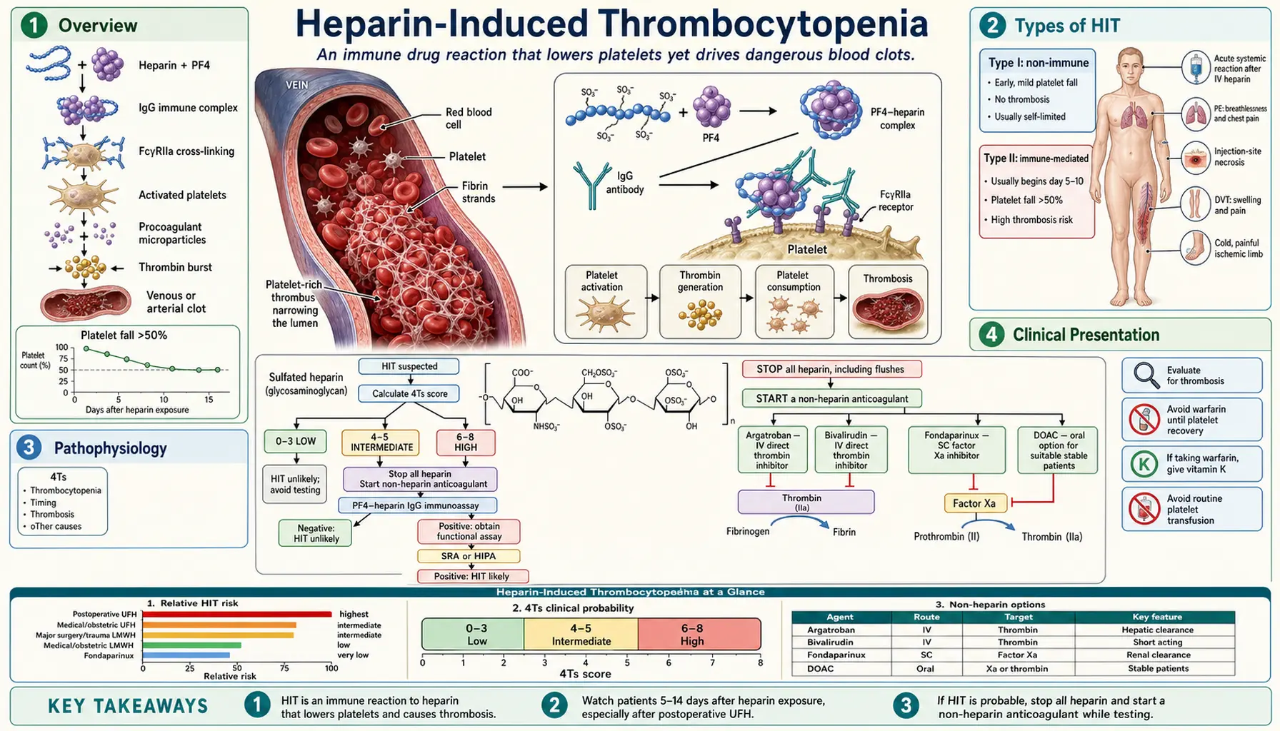
Table of Contents
Overview
Heparin-induced thrombocytopenia (HIT) is an immune-mediated adverse drug reaction to heparin that produces a dangerous and counterintuitive clinical picture: a falling platelet count accompanied not by bleeding but by paradoxical, life-threatening thrombosis. This makes HIT the most dangerous iatrogenic coagulopathy encountered in hospital practice.
The incidence of clinically significant (Type II) HIT ranges from approximately 0.5% to 5% of heparin-exposed patients, varying by heparin type, dose, duration, and patient population. Surgical patients — particularly those undergoing cardiac or orthopedic procedures — face the highest risk. Despite the low absolute incidence, the sheer volume of heparin use in hospitals worldwide means HIT affects tens of thousands of patients annually.
The condition was first clearly described in 1958, when Weismann and Tobin reported arterial embolism during heparin therapy; in 1964 Roberts and colleagues asked in print whether heparin was a cause of arterial emboli. For decades the mechanism remained unclear. In 1992 Amiral and colleagues reported that platelet factor 4 complexed with heparin is the target of the antibodies. The pivotal work of Theodore Warkentin, John Kelton, and Andreas Greinacher in the 1990s established that Type II HIT is driven by immunoglobulin G (IgG) antibodies targeting a complex of heparin bound to platelet factor 4 (PF4), a discovery that transformed clinical management.
The cardinal teaching point: HIT is a thrombotic disorder, not a bleeding disorder. The 2018 American Society of Hematology (ASH) guideline suggests against routine platelet transfusion — the instinctive response to a low platelet count — in people at average risk of bleeding (see Management). Early recognition, immediate heparin cessation, and prompt substitution with a non-heparin anticoagulant are the cornerstones of survival.
Types of HIT
Type I HIT (Non-Immune)
Type I HIT is a mild, benign, and self-limiting phenomenon that occurs within the first 1–2 days of heparin initiation. It results from a direct, non-immunologic interaction between heparin molecules and platelet membranes, causing transient platelet sequestration.
- Platelet count rarely falls below 100,000/µL and typically normalizes spontaneously even if heparin is continued.
- No anti-platelet factor 4/heparin antibodies are detectable.
- No association with thrombosis.
- No specific treatment is required; the diagnosis is essentially one of exclusion after ruling out the more dangerous Type II form.
Type II HIT (Immune-Mediated — The Dangerous Form)
Type II HIT is the clinically significant entity. It is an antibody-mediated immune reaction with a characteristic 5–10 day window from heparin initiation to platelet decline. Immunoglobulin G antibodies form against the heparin-platelet factor 4 neoantigen, activate platelets and the coagulation cascade, and drive a hypercoagulable state that can produce devastating venous and arterial thromboses. Hospitals treat it as urgent.
Rapid-Onset HIT
Patients who have received heparin within the preceding 100 days may carry pre-formed circulating anti-platelet factor 4/heparin antibodies. Upon re-exposure, the thrombocytopenia and thrombosis can begin within hours rather than days, because no time is needed to generate a new antibody response. Recent heparin exposure is therefore the key question in anyone who develops acute thrombocytopenia shortly after heparin administration.
Delayed HIT
A rare but underrecognized variant in which thrombocytopenia and thrombosis develop after heparin has already been discontinued — sometimes up to 3 weeks post-exposure. The antibodies persist after heparin is stopped, continue to activate platelets through low-level endogenous platelet factor 4, and can cause thrombosis even in the absence of ongoing heparin therapy. Delayed HIT is a recognized explanation for unexplained thrombosis within weeks of a heparin course.
The Phases of HIT and Why They Matter
The 2018 ASH guideline sorts confirmed HIT into phases by which blood results have returned to normal. These phases explain when heparin can be reconsidered.
- Acute HIT: the platelet count is low, and both the platelet-activation (functional) test and the antibody test (immunoassay) are positive. This is the highly clot-prone phase.
- Subacute HIT A: the platelet count is back to normal, but both tests are still positive.
- Subacute HIT B: the platelet count and the functional test are normal, but the antibody test is still positive.
- Remote HIT: the platelet count and both tests are normal.
Pathophysiology
The pathophysiology of Type II HIT is a precisely defined immunologic cascade that explains both the thrombocytopenia and — paradoxically — the thrombosis. Understanding each step is essential for rational management.
Step 1: Heparin-platelet factor 4 Complex Formation
Platelet factor 4 is a positively charged (+18 net charge) chemokine stored in platelet alpha-granules and released upon platelet activation. Heparin, a highly negatively charged glycosaminoglycan, electrostatically binds to platelet factor 4 and induces a conformational change, exposing a neoantigen not present on platelet factor 4 alone or heparin alone. This heparin-platelet factor 4 complex is immunogenic.
Step 2: Immunoglobulin G Antibody Formation
After approximately 5–7 days of antigen exposure, the immune system generates immunoglobulin G antibodies (predominantly IgG1 subclass) directed against the heparin-platelet factor 4 neoantigen. This explains the 5–10 day delay between heparin initiation and platelet count decline in most patients without prior exposure.
Step 3: Platelet Activation and Amplification
Immunoglobulin G-heparin-platelet factor 4 immune complexes bind simultaneously to the antigen-specific Fab region and, via the Fc portion of the immunoglobulin G, to FcγRIIa receptors on platelet surfaces. This crosslinking triggers explosive platelet activation, leading to:
- Platelet aggregation and consumption (explaining the thrombocytopenia).
- Release of procoagulant microparticles from platelet membranes — phosphatidylserine-rich surfaces that dramatically accelerate thrombin generation.
- Additional alpha-granule release, spilling more platelet factor 4 into the circulation — creating a self-amplifying cycle that perpetuates antibody formation and platelet activation.
Step 4: Monocyte and Endothelial Activation
HIT IgG complexes also bind FcγR on monocytes and vascular endothelial cells, triggering tissue factor expression. Tissue factor activates the extrinsic coagulation pathway, generating massive quantities of thrombin — the final common pathway to clot formation.
Result: The "White Clot Syndrome"
The net effect is paradoxical: platelet consumption drives the platelet count down (thrombocytopenia) while simultaneously the activated platelets, procoagulant microparticles, and tissue factor generate a profoundly prothrombotic state. The resulting thrombi are platelet-rich "white clots" — dense aggregates of activated platelets bound together by fibrin strands, morphologically distinct from the red fibrin-dominant thrombi of stasis or venous thromboembolism. This is why HIT is sometimes called white clot syndrome.
Clinical Presentation
Thrombocytopenia
The hallmark laboratory finding is a platelet count falling to less than 150,000/µL or by more than 50% from the pre-heparin baseline. In HIT the nadir typically ranges from 40,000 to 80,000/µL — low enough to be alarming but rarely low enough to cause spontaneous bleeding. A platelet nadir below 20,000/µL is unusual and points toward alternative diagnoses such as immune thrombocytopenic purpura (ITP) or thrombotic thrombocytopenic purpura (TTP).
Timing
In patients without recent prior heparin exposure, platelet decline begins on days 5–10 after heparin initiation. In patients with heparin exposure within the prior 100 days, onset may be within hours (rapid-onset HIT). In delayed HIT, thrombocytopenia manifests after heparin discontinuation.
Thrombosis
The 2018 ASH guideline states that thrombosis occurs in one-third to one-half of patients with HIT; it is the primary driver of morbidity and mortality. It may predate clinical recognition of thrombocytopenia: in a 14-year study of 127 patients, about half were recognized only after a clot had occurred, and 52.8% of those first recognized with a low platelet count alone clotted within 30 days.
- Venous thrombosis (most common): deep vein thrombosis (DVT) and pulmonary embolism (PE) are the most frequent manifestations.
- Arterial thrombosis (less common but more dramatic): acute limb ischemia from white clot occlusion of a major artery is a surgical emergency; stroke and myocardial infarction also occur. Arterial events are more limb- and life-threatening than venous events.
- Unusual sites: adrenal vein thrombosis causing hemorrhagic adrenal infarction and acute adrenal insufficiency; cerebral venous sinus thrombosis (can mimic other neurological emergencies).
Skin Necrosis at Heparin Injection Sites
Erythematous plaques or frank necrotic lesions at subcutaneous heparin injection sites are considered pathognomonic for HIT. They represent local microvascular thrombosis at sites of highest platelet factor 4-heparin concentration, and are treated as a reason for HIT workup even if the platelet count has not yet fallen significantly.
Acute Systemic Reactions
Some patients develop an acute inflammatory or anaphylactoid reaction within minutes of receiving an intravenous heparin bolus: fever, chills, flushing, hypertension, tachycardia, dyspnea, or in severe cases, cardiac arrest. This represents massive, near- instantaneous platelet activation by pre-formed antibodies and is a medical emergency. Such a reaction after intravenous heparin is treated as a sign of HIT until it is ruled out.
Diagnosis — 4Ts Scoring System
Diagnosis of HIT requires integrating clinical probability with laboratory testing. The validated 4Ts scoring system provides a standardized pretest probability assessment before sending laboratory tests.
The 4Ts Score
Each "T" is scored 0, 1, or 2 points; maximum score is 8.
T1 — Thrombocytopenia Severity
- 2 points: Platelet fall >50% AND nadir ≥20,000/µL.
- 1 point: Platelet fall 30–50%, OR nadir 10,000–19,000/µL.
- 0 points: Platelet fall <30%, OR nadir <10,000/µL.
T2 — Timing of Platelet Fall
- 2 points: Clear onset days 5–10, OR ≤1 day with prior heparin exposure within 30 days.
- 1 point: Consistent with days 5–10 but not clear; OR onset >10 days; OR onset ≤1 day with prior heparin exposure 30–100 days ago.
- 0 points: Platelet fall ≤4 days without recent heparin exposure.
T3 — Thrombosis or Other Sequelae
- 2 points: New confirmed thrombosis; skin necrosis at heparin injection sites; acute systemic reaction after intravenous heparin bolus.
- 1 point: Progressive or recurrent thrombosis while on heparin; erythematous (non-necrotic) skin lesions at injection sites; suspected thrombosis not yet proven.
- 0 points: None.
T4 — oTher Causes of Thrombocytopenia
- 2 points: No other cause evident.
- 1 point: Possible other cause present.
- 0 points: Definite other cause present (e.g., sepsis, recent surgery, drug-induced thrombocytopenia from another agent).
Score Interpretation
- 6–8 points: High probability — in a 2012 pooled analysis of 13 studies and 3068 patients, 64% of people with this score proved to have HIT. Heparin is typically stopped and a non-heparin anticoagulant started before the laboratory result returns.
- 4–5 points: Intermediate probability — 14% in the same analysis. Laboratory testing follows.
- 0–3 points: Low probability — a low score was correct 99.8% of the time in ruling HIT out. HIT is effectively excluded and other causes are looked for.
The numbers change with the setting. In a study at two centers, 28.6% of intermediate scores tested positive for clinically significant antibodies at one center and 7.9% at the other.
The 2018 ASH guideline strongly recommends against laboratory testing and against HIT treatment when the 4Ts score is low, because a low score rules HIT out so reliably. If key information is missing, it notes that it may be prudent to err on the side of a higher score.
Laboratory Testing
Two categories of tests are available:
- Anti-platelet factor 4/Heparin enzyme-linked immunosorbent assay (ELISA, an immunoassay): High sensitivity (~95%) but lower specificity — detects anti-platelet factor 4 antibodies regardless of whether they actually activate platelets. Many patients form antibodies without developing clinical HIT. The optical density (OD) value matters: an OD >2.0 correlates much more strongly with functional platelet-activating antibodies and clinical HIT. A negative immunoassay effectively rules out HIT. A positive immunoassay is read alongside the clinical picture or confirmed by functional testing.
- Serotonin Release Assay (SRA): The functional gold standard. Washed donor platelets are incubated with patient serum and therapeutic concentrations of heparin; release of [14C]-serotonin above 20% constitutes a positive result. Sensitivity ~95%, specificity ~99%. Complex methodology, radioactive substrate, and limited availability (few reference laboratories) restrict its routine use; results may take days.
- Heparin-Induced Platelet Activation (HIPA) assay: A European alternative functional assay using multiple donor platelet pools; comparable performance to SRA.
In practice, the immunoassay is sent first for its rapid turnaround; clinical management decisions (heparin cessation, alternative anticoagulation) are not held for laboratory confirmation in intermediate- or high-probability cases.
Management
Hospital management of suspected or confirmed HIT follows a sequential, time-critical protocol set out in the ASH 2018 and American College of Chest Physicians (ACCP) 2012 guidelines. Errors at any stage can be fatal.
Step 1 — All Heparin Is Stopped Immediately
The guidelines recommend stopping all forms of heparin — unfractionated heparin (UFH), low- molecular-weight heparin (LMWH), heparin flushes used to maintain catheter patency, and heparin-coated catheters or devices. LMWH cannot substitute for UFH in HIT: LMWH cross-reacts with HIT antibodies in approximately 85–95% of cases and is contraindicated.
Step 2 — Non-Heparin Anticoagulation Starts Immediately
The risk of thrombosis in untreated HIT is approximately 30–50% within 30 days, even after heparin cessation. The guidelines recommend a non-heparin anticoagulant whether or not thrombosis has occurred.
- Argatroban (direct thrombin inhibitor): Hepatically metabolized — preferred in renal failure. Given as a continuous intravenous infusion, adjusted to the activated partial thromboplastin time, a clotting test. Argatroban also prolongs the prothrombin time and its international normalized ratio (INR), which complicates the transition to warfarin. ASH 2018 states it is advisable to avoid argatroban or use a reduced dose in moderate or severe liver disease.
- Bivalirudin (direct thrombin inhibitor): Also used, particularly in the setting of cardiac surgery, percutaneous coronary intervention, or when argatroban is unavailable. Cleared mainly by enzymes in the blood (proteolytic cleavage), with a part filtered by the kidneys.
- Fondaparinux (synthetic pentasaccharide, indirect factor Xa inhibitor): Very low cross-reactivity with HIT antibodies. Widely used off-label for HIT management because of ease of administration (once-daily subcutaneous injection) and robust safety data. Renally cleared; the label lists it as contraindicated in severe kidney impairment (creatinine clearance below 30 mL/min).
- Danaparoid (anti-Xa mixture): Standard agent in Europe, limited availability elsewhere.
- Direct oral anticoagulants (rivaroxaban, apixaban, dabigatran): ASH 2018 lists them among the options for acute HIT, noting that an injected or intravenous non-heparin drug may be preferred for life- or limb-threatening clots because few such patients have been treated with one. For the phase after platelet recovery it suggests them over warfarin-type drugs. In a 2016 single-arm study of 22 people on rivaroxaban, 1 had a new clot (4.5%; 95% CI 0–23.5%), platelets recovered in 9 of 10 people with a low count, and 1 person needed a limb amputation; the authors note that the small size limits precision. They offer convenience for long-term outpatient management.
The evidence behind these choices is thin. ASH 2018 grades the choice among non-heparin anticoagulants as a conditional recommendation with very low certainty, so words such as "preferred" reflect expert judgement more than trial results.
Step 3 — Platelet Transfusions Are Not Routine
ASH 2018 suggests against routine platelet transfusion in acute HIT at average risk of bleeding (conditional recommendation, low certainty) and notes that a transfusion may be an option with active bleeding or a high risk of bleeding. ACCP 2012 suggests transfusions only for bleeding or during an invasive procedure with a high risk of bleeding. In the largest database study ASH reviewed, people given platelets had more arterial clots (adjusted odds ratio 3.4, 95% CI 1.2–9.5). Because HIT is a thrombotic — not hemorrhagic — disorder, transfused platelets are thought to become additional substrate for FcγRIIa-mediated activation, generating more procoagulant microparticles and amplifying thrombin generation. The clinical metaphor is "adding fuel to a fire."
Step 4 — Warfarin Is Delayed: Venous Limb Gangrene
ASH 2018 and ACCP 2012 recommend against starting warfarin until the platelet count has recovered (usually ≥150,000/µL), and the warfarin label states it is not for use as initial therapy in HIT. Warfarin in acute HIT depletes the natural anticoagulant protein C. Its half-life of about 8 hours is shorter than that of clotting factors II (60 hours), IX (24 hours) and X (48 to 72 hours), although factor VII is shorter still (4 to 6 hours) and protein S is longer (about 30 hours). A 1997 study of patients with limb gangrene attributed it to acquired failure of the protein C pathway, which creates a transient hypercoagulable window. This can precipitate venous limb gangrene: catastrophic ischemic necrosis of an extremity despite (or because of) anticoagulation. This counterintuitive complication is among the most devastating iatrogenic injuries in hematology.
If warfarin was already started before HIT was recognized, ASH 2018 describes stopping it and giving vitamin K, with therapeutic-dose non-heparin anticoagulation until the platelet count normalizes before warfarin is reintroduced with overlap.
Step 5 — Duration of Anticoagulation
With a clot (HIT with thrombosis), ASH 2018 notes that anticoagulation is typically given for 3 to 6 months when there is no other reason for it; the panel made no recommendation on this. Without a clot (isolated HIT), it suggests continuing at least until the platelet count recovers and suggests against continuing for 3 months or more unless HIT persists without platelet recovery (conditional recommendations, very low certainty). Long-term oral treatment uses warfarin (INR 2–3) or a direct oral anticoagulant once the acute phase resolves.
Special Situations
HIT and Cardiac Surgery
Cardiopulmonary bypass requires large volumes of heparin and cannot proceed with active HIT or high-titer antibodies. Management options include:
- Delay surgery 3 months: Anti-platelet factor 4 antibodies typically wane to undetectable levels within 100 days, after which brief heparin re-exposure is possible under close monitoring.
- Bivalirudin for bypass: Short-acting direct thrombin inhibitor that can be used as the sole anticoagulant during bypass and clears rapidly, permitting hemostasis at circuit removal.
- Non-heparin circuit coating and alternative pump priming solutions are also under investigation.
The ASH 2018 panel adds to this list. It agrees that surgery is delayed, if feasible, until the functional test is negative, and then suggests heparin during the operation itself (limited to the operation, very low certainty). If surgery cannot wait, it suggests one of three strategies (low certainty): bivalirudin during the operation, heparin after plasma exchange, or heparin with a strong antiplatelet drug. It notes that consensus protocols for the last two have not been established.
HIT in Renal Failure
Argatroban (hepatically metabolized) is the preferred agent. Fondaparinux is renally cleared and its label lists it as contraindicated below a creatinine clearance of 30 mL/min. Bivalirudin is partially renally cleared and requires dose adjustment. Danaparoid is also renally cleared and is used with caution.
HIT in Pregnancy
Both warfarin and direct oral anticoagulants are contraindicated in pregnancy. The ACCP 2012 guideline suggests danaparoid over other non-heparin anticoagulants in pregnant patients with acute or subacute HIT, and lepirudin or fondaparinux only if danaparoid is not available. Clinical guidance is based on case series rather than randomized trials.
Future Heparin Re-Exposure
Once HIT antibodies have cleared (typically after ~100 days), brief heparin re-exposure for an unavoidable indication such as cardiac bypass surgery is considered with extreme caution: antibody negativity is confirmed by immunoassay immediately before exposure, heparin is used only during the operation for the shortest effective duration, and non-heparin anticoagulation restarts immediately afterward.
The same guideline suggests carrying or wearing an emergency identifier (such as a pendant or bracelet naming heparin, the reaction and the date of diagnosis) for 3 months after HIT, and recommends a non-heparin blood thinner, rather than heparin, for anyone with past HIT who later needs treatment or prevention of clots.
Vaccine-Induced Immune Thrombocytopenia and Thrombosis (VITT)
Following the rollout of adenovirus-vector COVID-19 vaccines, a rare but severe syndrome emerged characterized by thrombocytopenia and thrombosis — particularly in unusual venous sites such as cerebral venous sinuses, splanchnic veins, and pulmonary arteries. The 2021 study that described it says it clinically mimics HIT: spontaneously activating anti-platelet factor 4 antibodies that do not require heparin as a cofactor.
Management of VITT parallels HIT management: heparin is avoided (contraindicated), non-heparin anticoagulation is used, and intravenous immunoglobulin is given to compete with the pathogenic antibodies for FcγRIIa receptor binding. The HIT framework — developed over three decades — proved directly applicable to this vaccine-associated emergency, demonstrating the clinical importance of understanding the underlying pathophysiology.
Prognosis
With appropriate, timely management the prognosis of HIT has improved markedly over the past three decades, though the condition remains serious.
- Mortality: With appropriate treatment, overall mortality from HIT is approximately 5–10%, largely driven by thromboembolic complications — PE, stroke, and myocardial infarction — and by delays in diagnosis.
- Thrombosis risk: Without anticoagulation, 30–50% of HIT patients develop new or progressive thrombosis within 30 days of diagnosis, even after heparin has been stopped. This thrombotic risk — not the thrombocytopenia itself — defines the urgency of treatment.
- Limb amputation: ASH 2018 puts the rate of amputation in HIT at about 1% to 3%.
- Key message: Early recognition and rapid sequential action — heparin stopped, non-heparin anticoagulant started, no routine platelet transfusions, warfarin deferred — dramatically improves outcomes. The mnemonic holds true: thinking about HIT saves limbs and lives.
The Danger With Modern Care
A 2025 study followed 1393 people with suspected HIT at 11 centers and confirmed it in 119 (8.5%). Among those, 23% later had a clot in a vein, 9% a clot in an artery, 12.6% major bleeding, and 18% died, so the danger remains large with modern care. Treatment with argatroban, bivalirudin or a direct oral anticoagulant was linked to a lower risk of arterial clots.
Monitoring Recommendations
The guidelines tie platelet count monitoring to the risk of HIT. ACCP 2012 suggests counts every 2 or 3 days from day 4 to day 14 when the risk is above 1%, and suggests no monitoring when it is below 1%. ASH 2018 suggests none at low risk (below 0.1%), every 2 to 3 days at intermediate risk (0.1% to 1.0%) and at least every other day at high risk (above 1.0%), from day 4 to day 14, or from the first day of heparin if heparin was also given in the previous 30 days. This is the window within which Type II HIT characteristically manifests. Postoperative cardiac and orthopedic patients are described as high-risk populations.
Awareness Is the Best Prevention
HIT is paradoxical in almost every respect: a drug given to prevent clots causes clots; a falling platelet count signals thrombotic, not hemorrhagic, danger; the intuitive treatment (platelets for low platelets; warfarin for thrombosis) can be catastrophic. Understanding this paradox is the foundation of prevention and survival.
Key Research Papers
- Warkentin TE et al. "Heparin-induced thrombocytopenia in patients treated with low-molecular-weight heparin or unfractionated heparin." N Engl J Med. 1995;332(20):1330–5. — PubMed PMID: 7715641
- Greinacher A, Amiral J, Dummel V, et al. "Laboratory diagnosis of heparin-associated thrombocytopenia and comparison of platelet aggregation test, heparin-induced platelet activation test, and platelet factor 4/heparin enzyme-linked immunosorbent assay." Transfusion. 1994;34(5):381–5. — PubMed PMID: 8191560
- Warkentin TE et al. "The pathogenesis of venous limb gangrene associated with heparin-induced thrombocytopenia." Ann Intern Med. 1997;127(9):804–12. — PubMed PMID: 9382401
- Cuker A et al. "Predictive value of the 4Ts scoring system for heparin-induced thrombocytopenia: a systematic review and meta-analysis." Blood. 2012;120(20):4160–7. — PubMed PMID: 22990018
- Linkins LA et al. "Treatment and prevention of heparin-induced thrombocytopenia: Antithrombotic Therapy and Prevention of Thrombosis, 9th ed: ACCP guidelines." Chest. 2012;141(2 Suppl):e495S–e530S. — PubMed PMID: 22315270
- Arepally GM, Ortel TL. "Clinical practice: heparin-induced thrombocytopenia." N Engl J Med. 2006;355(8):809–17. — PubMed PMID: 16928996
- Lobo B, Finch C, Howard A, Minhas S. "Fondaparinux for the treatment of patients with acute heparin-induced thrombocytopenia." Thromb Haemost. 2008;99(1):208–14. — PubMed PMID: 18217156
- Linkins LA, Warkentin TE, Pai M, et al. "Rivaroxaban for treatment of suspected or confirmed heparin-induced thrombocytopenia study." J Thromb Haemost. 2016;14(6):1206–10. — PubMed PMID: 27061271
- Greinacher A, Thiele T, Warkentin TE, et al. "Thrombotic thrombocytopenia after ChAdOx1 nCov-19 vaccination." N Engl J Med. 2021;384(22):2092–2101. — PubMed PMID: 33835769
- Warkentin TE. "Heparin-induced thrombocytopenia: diagnosis and management." Circulation. 2004;110(18):e454–8. — PubMed PMID: 15520327
- Cuker A, Arepally GM, Chong BH, et al. "American Society of Hematology 2018 guidelines for management of venous thromboembolism: heparin-induced thrombocytopenia." Blood Adv. 2018;2(22):3360–3392. — PubMed PMID: 30482768
- Lo GK, Juhl D, Warkentin TE, et al. "Evaluation of pretest clinical score (4 T's) for the diagnosis of heparin-induced thrombocytopenia in two clinical settings." J Thromb Haemost. 2006;4(4):759–65. — PubMed PMID: 16634744
- Warkentin TE, Kelton JG. "A 14-year study of heparin-induced thrombocytopenia." Am J Med. 1996;101(5):502–7. — PubMed PMID: 8948273
- Weismann RE, Tobin RW. "Arterial embolism occurring during systemic heparin therapy." AMA Arch Surg. 1958;76(2):219–25. — PubMed PMID: 13497418
- Roberts B, Rosato FE, Rosato EF. "Heparin--a cause of arterial emboli?" Surgery. 1964;55:803–8. — PubMed PMID: 14168000
- Amiral J, Bridey F, Dreyfus M, et al. "Platelet factor 4 complexed to heparin is the target for antibodies generated in heparin-induced thrombocytopenia." Thromb Haemost. 1992;68(1):95–6. — PubMed PMID: 1514184
- Nilius H, Sinitsa E, Studt JD, et al. "Outcomes of patients with suspected heparin-induced thrombocytopenia in a contemporary multicenter cohort." Blood Adv. 2025;9(21):5546–5555. — PubMed PMID: 40758946
Connections
- Hematology
- Thrombotic Thrombocytopenic Purpura
- Deep Vein Thrombosis
- Pulmonary Embolism
- Antiphospholipid Syndrome
- Cardiology Diseases
- Thrombocytopenia — HIT is one of the drug-induced immune causes of a low platelet count.