Essential Thrombocythemia
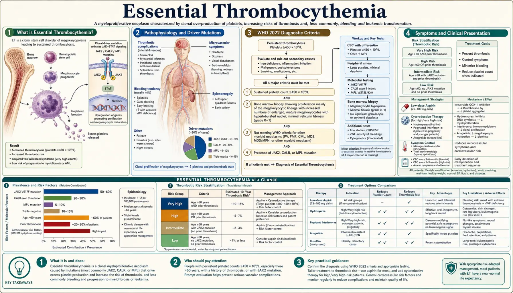
Table of Contents
- What Essential Thrombocythemia Is
- Pathophysiology and Driver Mutations
- WHO 2022 Diagnostic Criteria
- Symptoms and Clinical Presentation
- Thrombosis and Bleeding Risk
- Risk Stratification (IPSET-Thrombosis Revised)
- Treatment Approach
- Prognosis and Disease Progression
- Key Research Papers
- Connections
- Featured Videos
What Essential Thrombocythemia Is
Essential Thrombocythemia (ET) is a clonal hematopoietic stem cell disorder belonging to the Philadelphia chromosome-negative myeloproliferative neoplasms (MPNs), characterized by sustained thrombocytosis with a platelet count persistently at or above 450 × 109/L. It is the second most common MPN after polycythemia vera, and carries a relatively benign course compared to other MPNs — yet the major risks of thrombosis and hemorrhage can significantly affect quality of life and longevity.
The annual incidence is approximately 1–2.5 per 100,000 persons per year. ET shows a bimodal age distribution: a younger peak in women during their 30s (possibly reflecting hormonal influences on clonal expansion) and a larger peak in both sexes during their 50s and 60s. A 2024 review reports that life expectancy is shorter than in the general population: median survival is about 18 years, and more than 35 years in younger patients. The principal long-term risks are thrombosis, hemorrhage, progression to post-ET myelofibrosis (10–20% at 15–20 years), and — less commonly — transformation to acute myeloid leukemia (AML, fewer than 5%).
Pathophysiology and Driver Mutations
ET arises from an acquired somatic mutation in a multipotent hematopoietic stem cell that drives constitutive activation of the JAK-STAT signaling pathway, resulting in excessive megakaryocyte proliferation and platelet production. Three principal driver mutations account for approximately 83% of ET cases:
JAK2 V617F (approximately 55% of ET)
A point mutation (valine-to-phenylalanine substitution at codon 617) in the pseudo-kinase domain of Janus kinase 2. This mutation disables the autoinhibitory JH2 domain, causing constitutive JAK2 kinase activity and continuous downstream STAT3/STAT5 signaling independent of thrombopoietin (TPO) or erythropoietin binding. JAK2-mutated ET carries a higher risk of arterial thrombosis, higher red blood cell and white blood cell counts, and greater polycythemia vera-like features than other mutation subtypes.
CALR Exon 9 Mutations (approximately 25% of ET)
Insertions or deletions in exon 9 of the calreticulin gene create a frameshift producing a novel mutant C-terminus. This abnormal calreticulin protein binds and activates the thrombopoietin receptor MPL (myeloproliferative leukemia protein) in a ligand-independent manner, driving megakaryocyte expansion. Two main types exist: Type 1 (52-bp deletion) and Type 2 (5-bp insertion). CALR-mutated ET has a lower thrombosis risk compared to JAK2-mutated ET but a somewhat higher long-term risk of transformation to post-ET myelofibrosis. Prognosis hierarchy for thrombosis risk: JAK2 V617F > CALR type 1 > CALR type 2 > MPL.
MPL W515L/K Mutations (approximately 3% of ET)
Activating mutations in codon 515 of the thrombopoietin receptor gene (MPL) directly activate the receptor without ligand binding, producing constitutive JAK-STAT pathway signaling and megakaryocytic hyperplasia similar to JAK2 V617F.
Triple-Negative ET (approximately 17%)
Patients lacking JAK2, CALR, and MPL mutations. Some harbor mutations in other signaling genes (e.g., SH2B3). Diagnosis requires strict exclusion of reactive thrombocytosis and other myeloid neoplasms; bone marrow biopsy is particularly important in this group.
Driver mutations alone are insufficient to cause ET — the defining bone marrow finding is megakaryocyte hyperplasia with characteristic large, mature megakaryocytes featuring hyperlobated ("staghorn") nuclei and loose clustering, without significant granulocyte or erythrocyte proliferation.
WHO 2022 Diagnostic Criteria
The 2022 WHO Classification of Haematolymphoid Tumours requires all four major criteria, or the first three major criteria plus the one minor criterion, for a diagnosis of ET:
Major Criteria
- Platelet count ≥450 × 109/L on two measurements at least 4 weeks apart.
- Bone marrow biopsy showing megakaryocyte hyperplasia with large, mature megakaryocytes with hyperlobated (staghorn) nuclei; no significant increase in neutrophil granulopoiesis or erythropoiesis; very rarely increased reticulin fibrosis (grade 1).
- Not meeting WHO criteria for BCR-ABL1-positive chronic myeloid leukemia (CML), polycythemia vera (PV), primary myelofibrosis (PMF), myelodysplastic syndrome (MDS), or other myeloid neoplasm.
- Presence of a JAK2 V617F, CALR, or MPL mutation.
Minor Criterion
- Presence of a clonal marker (e.g., abnormal karyotype by conventional cytogenetics) OR absence of evidence of reactive thrombocytosis.
Reactive Thrombocytosis: Mimics to Exclude
Before ET is diagnosed, the WHO criteria require common causes of secondary (reactive) thrombocytosis to be ruled out, as reactive platelet elevation rarely causes thrombotic complications:
- Iron deficiency anemia (most common cause of thrombocytosis in young women)
- Acute or chronic infection and inflammation (interleukin-6 (IL-6) drives hepatic TPO production)
- Post-splenectomy state (loss of platelet sequestration)
- Tissue damage, surgery, or trauma (acute phase response)
- Rebound thrombocytosis following recovery from thrombocytopenia
- Certain malignancies producing inflammatory cytokines
Symptoms and Clinical Presentation
Approximately 50% of ET patients are asymptomatic at diagnosis, with the condition discovered incidentally on a routine complete blood count. When symptoms are present, they generally fall into two categories: microvascular disturbances and major thrombohemorrhagic events.
Microvascular Symptoms
- Erythromelalgia: Burning, aching pain with redness and warmth of the hands and feet, caused by platelet-mediated arteriolar occlusion of small vessels. Dramatically and rapidly responsive to aspirin — one of the most specific features of ET among MPNs.
- Headaches and dizziness: Due to impaired microvascular circulation in cerebral vessels; often relieved by aspirin.
- Visual disturbances: Transient scotomas, blurred vision, or amaurosis fugax from ocular microvascular platelet plugging.
- Paresthesias: Tingling or numbness in hands and feet from peripheral microvascular platelet-mediated ischemia.
- Aquagenic pruritus: Itching triggered by water contact; less common in ET than in polycythemia vera.
Splenomegaly
Present in approximately 40% of ET patients. Usually mild to moderate; massive splenomegaly suggests progression toward post-ET myelofibrosis.
Thrombotic Events
Arterial and venous thromboses are the leading causes of morbidity and mortality in ET. Arterial events are more common than in other MPNs and include stroke and transient ischemic attack (TIA), myocardial infarction, and peripheral arterial occlusion. Venous events include deep vein thrombosis, pulmonary embolism, and splanchnic vein thrombosis (Budd-Chiari syndrome, portal vein thrombosis, mesenteric vein thrombosis).
Hemorrhagic Manifestations
Paradoxically, marked thrombocytosis (platelets >1,000–1,500 × 109/L) can cause bleeding rather than clotting, because very high platelet numbers lead to excessive binding and proteolytic degradation of large von Willebrand factor (vWF) multimers by ADAMTS13 — a condition called acquired von Willebrand syndrome. Loss of these high-molecular-weight vWF multimers impairs platelet adhesion, and bleeding ensues despite the very high platelet count. Manifestations include gastrointestinal bleeding, epistaxis, easy bruising, and mucosal hemorrhage.
Pregnancy Complications
ET poses specific risks during pregnancy, including first-trimester miscarriage (placental microthrombi), placental insufficiency, fetal growth restriction, preeclampsia, and postpartum thrombosis.
Thrombosis and Bleeding Risk
Understanding the dual thrombotic and hemorrhagic risk in ET is essential for guiding therapy. The relationship between platelet count and clinical events is not strictly linear, and individual mutation profile strongly modulates risk.
Arterial vs. Venous Thrombosis
Arterial thrombosis predominates in ET (particularly in JAK2-mutated patients), in contrast to venous predominance in PV. The excess of reactive oxygen species from JAK2 V617F platelets activates endothelial cells and promotes platelet-leukocyte aggregates. Major arterial events include ischemic stroke and transient ischemic attack, myocardial infarction, and peripheral arterial occlusion. Major venous events include DVT/PE and splanchnic vein thrombosis — the latter can be the initial manifestation of an underlying MPN in young patients presenting with Budd-Chiari syndrome or portal vein thrombosis.
Acquired von Willebrand Syndrome
When platelet counts exceed 1,000 × 109/L, reviews describe checking for acquired vWF deficiency before aspirin is started. If vWF ristocetin cofactor activity falls below 30% of normal, aspirin is contraindicated due to bleeding risk. Cytoreductive therapy normalizing the platelet count reverses the acquired vWD and restores high-molecular-weight vWF multimers — eliminating the hemorrhagic risk.
Cardiovascular Risk Factors
Traditional cardiovascular risk factors (hypertension, diabetes, hyperlipidemia, smoking, obesity) synergize powerfully with ET-related platelet activation to increase arterial thrombosis risk. Reviews describe the management of modifiable cardiovascular risk factors as a core part of ET care, independent of cytoreductive therapy decisions. In a 433-patient study of low-risk ET, having both a JAK2 V617F mutation and cardiovascular risk factors raised the rate of clots (incidence rate ratio 9.8), even after aspirin use was taken into account.
Risk Stratification (IPSET-Thrombosis Revised)
The Revised International Prognostic Score for ET Thrombosis (IPSET-Thrombosis Revised), built from 1,019 patients with WHO-defined ET, sorts patients into four clot-risk groups using age, any past clot (thrombosis) and JAK2 mutation status. As summarized in the 2024 review by Tefferi, Vannucchi and Barbui:
Very Low Risk
- Age 60 years or younger AND
- No past thrombosis AND
- No JAK2 mutation (JAK2 wild-type)
Low Risk
- Same as very low risk, but a JAK2 mutation is present
Intermediate Risk
- Same as low risk, but age over 60 years (no past thrombosis, no JAK2 mutation)
High Risk
- Past thrombosis at any age, OR
- Age over 60 years with a JAK2 mutation
How the groups are used: the 2024 review states that once-daily low-dose aspirin is advised for all patients and twice daily for low-risk disease, that cytoreductive (count-lowering) therapy is advised for high-risk disease, and that it is optional for intermediate-risk disease.
What Is Not Settled
Aspirin for low-risk patients with a CALR mutation is not settled. In a study of 433 low-risk patients (271 with a CALR mutation, 162 with a JAK2 V617F mutation), aspirin did not lower the risk of clots in the CALR group but was linked to more bleeding (12.9 versus 1.8 bleeding episodes per 1,000 patient-years). In the JAK2 group, aspirin was linked to fewer vein clots with no rise in bleeding. The authors concluded that in low-risk, CALR-mutated ET, low-dose aspirin does not reduce the risk of thrombosis and may increase the risk of bleeding.
Treatment Approach
Treatment goals in ET are to prevent thrombotic and hemorrhagic complications, control microvascular symptoms, and minimize the small risk of disease transformation. Cure is not achievable with current therapies except potentially with allogeneic stem cell transplantation, which is rarely indicated in ET given its relatively indolent course.
Aspirin in All Risk Groups
A 2024 review states that once-daily low-dose aspirin (81–100 mg/day) is advised for all ET patients and twice daily for low-risk disease; the exception for low-risk patients with a CALR mutation is described above under Risk Stratification. Aspirin inhibits thromboxane A2-mediated platelet activation and reliably resolves erythromelalgia and microvascular symptoms. Critical exception: reviews describe checking vWF activity before aspirin is started when the platelet count exceeds 1,000 × 109/L. If vWF ristocetin cofactor activity is below 30%, aspirin is contraindicated and cytoreduction to normalize the platelet count takes precedence.
Cytoreductive Therapy: First-Line Options
Hydroxyurea (Hydroxycarbamide)
First-line cytoreductive therapy for high-risk ET. In the landmark PT-1 trial (Harrison et al., 2005; 809 high-risk patients, all on low-dose aspirin, median follow-up 39 months), patients on anagrelide were more likely than those on hydroxyurea to reach the combined end point of arterial clots, vein clots, serious bleeding or death from clotting or bleeding (odds ratio 1.57). Anagrelide was linked to more arterial clots, more serious bleeding and more change to myelofibrosis, but to fewer vein clots (venous thromboembolism). Both drugs controlled the platelet count equally well over the long term. Hydroxyurea suppresses DNA synthesis in all rapidly dividing cells, including megakaryocyte progenitors. Typical dosing: 500–1,000 mg/day titrated to platelet count target (typically <400 × 109/L). Long-term use in young patients is generally limited because of a theoretical leukemia risk and harm to an unborn baby (teratogenicity).
Pegylated Interferon-Alpha (pegIFN-α2a)
Preferred cytoreductive agent for young patients (<40 years), patients of childbearing potential, or those who are pregnant or planning pregnancy (hydroxyurea is teratogenic and contraindicated). PegIFN-α2a directly suppresses clonal hematopoiesis at the stem cell level. A unique advantage is the potential for molecular remission — reduction or elimination of the JAK2 V617F or CALR allele burden — which is not achievable with hydroxyurea. In the MPD-RC 112 trial (168 untreated high-risk patients with ET or polycythemia vera), complete response at 12 months was similar with hydroxyurea and pegylated interferon (37% vs 35%); at 24 to 36 months it was 20% to 17% with hydroxyurea and 29% to 33% with pegylated interferon. Pegylated interferon lowered the JAK2 V617F mutation load more by 24 months, hydroxyurea gave more bone-marrow responses, clots and disease progression were uncommon in both groups, and severe (grade 3/4) side effects were more frequent with pegylated interferon (46% vs 28%). Tolerability is improved versus standard interferon, but side effects (fatigue, depression, autoimmune phenomena) still limit use.
Anagrelide
Reduces platelet count by inhibiting megakaryocyte maturation (platelet production is selectively impaired). However, the PT-1 trial demonstrated anagrelide is inferior to hydroxyurea for high-risk ET: the anagrelide arm had significantly more arterial thrombotic events, more episodes of myelofibrotic transformation, and more bleeding complications, though fewer vein clots. A 2024 review lists hydroxyurea and pegylated interferon-α as the first-line cytoreductive drugs and busulfan as second-line; the 2025 SURPASS ET trial describes anagrelide as a standard second-line agent for patients who cannot take hydroxyurea or whose disease does not respond to it. Cardiovascular side effects (palpitations, fluid retention) due to its vasodilatory/inotropic properties require attention in older patients.
Second-Line and Special Situations
- Busulfan: An alkylating agent that provides durable platelet control in elderly patients; not used in younger patients due to leukemogenicity. Well-tolerated in patients over 70.
- Ruxolitinib (JAK1/JAK2 inhibitor): Used for hydroxyurea-resistant or hydroxyurea-intolerant ET; reduces splenomegaly and constitutional symptoms via JAK pathway blockade. Not curative; disease rebounds on discontinuation. In the randomized MAJIC-ET trial (110 patients resistant to or unable to take hydroxyurea), complete response within 1 year was 46.6% with ruxolitinib versus 44.2% with best available therapy, and clots, bleeding and disease change at 2 years were not significantly different; some disease symptoms improved with ruxolitinib. The authors concluded that ruxolitinib is not superior to current second-line treatments for ET.
- Ropeginterferon alfa-2b: In the SURPASS ET trial (2025; 174 high-risk patients with a raised white cell count who could not take hydroxyurea or whose disease did not respond to it; 96% of participants were Asian), 43% on ropeginterferon alfa-2b had a lasting blood-count response at months 9 and 12, versus 6% on anagrelide. Severe (grade 3 or worse) side effects occurred in 23% on ropeginterferon alfa-2b and 34% on anagrelide.
- Pregnancy management: Low-dose aspirin throughout pregnancy; cytoreduction with pegIFN-α2a if cytoreduction is required (hydroxyurea, busulfan, and anagrelide are all contraindicated during pregnancy). Low molecular weight heparin added peripartum for high-risk patients. Close obstetric monitoring for placental complications.
Prognosis and Disease Progression
The natural history of ET is generally favorable compared to other MPNs. Many low-risk patients have a near-normal life expectancy. The 10-year overall survival is approximately 80%, with most deaths related to cardiovascular complications, thrombosis, or transformation rather than to ET-specific disease burden.
Disease Transformation
- Post-ET Myelofibrosis: Occurs in approximately 10–20% of patients at 15–20 years of follow-up (fibrotic progression, splenomegaly, constitutional symptoms, cytopenias). Post-ET MF has worse prognosis than ET but better prognosis than primary myelofibrosis. CALR mutations, particularly type 1, carry a somewhat higher transformation risk than JAK2.
- Acute Myeloid Leukemia (AML): Blastic transformation occurs in fewer than 5% of ET patients and is significantly more likely in those who receive alkylating agents (busulfan, pipobroman) or radioactive phosphorus — now avoided in younger patients for this reason. Spontaneous AML transformation is rare in ET, lower than in MDS or PV.
Prognostic Factors
- Mutation type: JAK2 V617F confers higher thrombosis risk; CALR type 1 has the longest median survival and lowest thrombosis risk. Triple-negative ET carries intermediate prognosis.
- Age and prior thrombosis: The two strongest independent predictors of future thrombotic events and overall survival.
- White blood cell count: Leukocytosis (WBC >11 × 109/L) is an emerging independent risk factor for thrombosis and overall survival in several cohort studies.
- IPSS-ET score: Incorporates age, WBC count, and prior thrombosis to stratify overall survival risk separate from thrombosis risk.
- Triple A survival model: The 2024 review describes a model built on Age, Absolute neutrophil count and Absolute lymphocyte count that separates four survival groups (high, intermediate-1, intermediate-2 and low risk) with median survival of 8, 14, 21 and 47 years.
- Extra mutations: About 50% of patients carry mutations beyond the driver genes, most often TET2 (9%–11%), ASXL1 (7%–20%), DNMT3A (7%) and SF3B1 (5%). MPL and CALR type 1 mutations have been linked to a higher risk of change to myelofibrosis, and TP53 to change to leukemia. The 2024 review gives the 10-year rate of change to leukemia as under 1%.
Key Research Papers
Pivotal peer-reviewed studies on essential thrombocythemia, covering clinical trials, molecular pathogenesis, diagnostic criteria, and risk stratification.
- Harrison CN, Campbell PJ, Buck G, Wheatley K, East CL, Bareford D, et al. Hydroxyurea compared with anagrelide in high-risk essential thrombocythemia. N Engl J Med. 2005;353(1):33–45. — PubMed PMID: 16000354
- Tefferi A, Vannucchi AM, Barbui T. Essential thrombocythemia: 2024 update on diagnosis, risk stratification, and management. Am J Hematol. 2024;99(4):697–718. — PubMed PMID: 38269572
- Klampfl T, Gisslinger H, Harutyunyan AS, Nivarthi H, Rumi E, Milosevic JD, et al. Somatic mutations of calreticulin in myeloproliferative neoplasms. N Engl J Med. 2013;369(25):2379–2390. — PubMed PMID: 24325356
- Nangalia J, Massie CE, Baxter EJ, Nice FL, Gundem G, Wedge DC, et al. Somatic CALR mutations in myeloproliferative neoplasms with nonmutated JAK2. N Engl J Med. 2013;369(25):2391–2405. — PubMed PMID: 24325359
- Kralovics R, Passamonti F, Buser AS, Teo SS, Tiedt R, Passweg JR, et al. A gain-of-function mutation of JAK2 in myeloproliferative disorders. N Engl J Med. 2005;352(17):1779–1790. — PubMed PMID: 15858187
- Barbui T, Finazzi G, Carobbio A, Thiele J, Passamonti F, Rumi E, et al. Development and validation of an International Prognostic Score of thrombosis in World Health Organization-essential thrombocythemia (IPSET-thrombosis). Blood. 2012;120(26):5128–5133. — PubMed PMID: 23033268
- Barbui T, Vannucchi AM, Buxhofer-Ausch V, De Stefano V, Betti S, Rambaldi A, et al. Practice-relevant revision of IPSET-thrombosis based on 1019 patients with WHO-defined essential thrombocythemia. Blood Cancer J. 2015;5(11):e369. — PubMed PMID: 26617062
- Passamonti F, Thiele J, Girodon F, Rumi E, Carobbio A, Gisslinger H, et al. A prognostic model to predict survival in 867 World Health Organization-defined essential thrombocythemia at diagnosis: a study by the International Working Group on Myelofibrosis Research and Treatment. Blood. 2012;120(6):1197–1201. — PubMed PMID: 22740446
- Gisslinger H, Gotic M, Holowiecki J, Penka M, Thiele J, Kvasnicka HM, et al. Anagrelide compared with hydroxyurea in WHO-classified essential thrombocythemia: the ANAHYDRET Study, a randomized controlled trial. Blood. 2013;121(10):1720–1728. — PubMed PMID: 23315161
- Rumi E, Pietra D, Ferretti V, Klampfl T, Harutyunyan AS, Milosevic JD, et al. JAK2 or CALR mutation status defines subtypes of essential thrombocythemia with substantially different clinical course and outcomes. Blood. 2014;123(10):1544–1551. — PubMed PMID: 24366362
- Alvarez-Larrán A, Pereira A, Guglielmelli P, Hernández-Boluda JC, Arellano-Rodrigo E, Ferrer-Marín F, et al. Antiplatelet therapy versus observation in low-risk essential thrombocythemia with a CALR mutation. Haematologica. 2016;101(8):926–931. — PubMed PMID: 27175028
- Harrison CN, Mead AJ, Panchal A, Fox S, Yap C, Gbandi E, et al. Ruxolitinib vs best available therapy for ET intolerant or resistant to hydroxycarbamide. Blood. 2017;130(17):1889–1897. — PubMed PMID: 29074595
- Mascarenhas J, Kosiorek HE, Prchal JT, Rambaldi A, Berenzon D, Yacoub A, et al. A randomized phase 3 trial of interferon-α vs hydroxyurea in polycythemia vera and essential thrombocythemia. Blood. 2022;139(19):2931–2941. — PubMed PMID: 35007321
- Mesa R, Gill H, Zhang L, Jin J, Kirito K, Komatsu N, et al. Ropeginterferon alfa-2b in hydroxyurea-intolerant or hydroxyurea-refractory essential thrombocythaemia (SURPASS ET): a multicentre, open-label, randomised, active-controlled, phase 3 study. Lancet Haematol. 2025;12(11):e862–e875. — PubMed PMID: 41193116