⚠ Evidence warning: Clinical trials or other evidence do not support the claim that a negative diffusion-weighted scan 18 months after surgery has a negative predictive value of about 95% for residual cholesteatoma over 3 mm. This page keeps it as a report of what people, websites or other media say.
Cholesteatoma
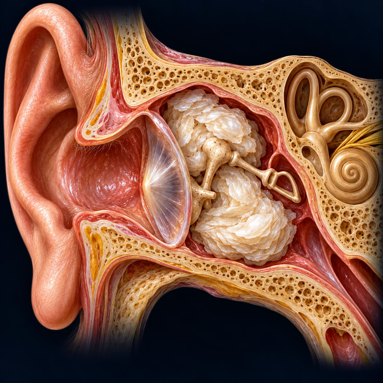
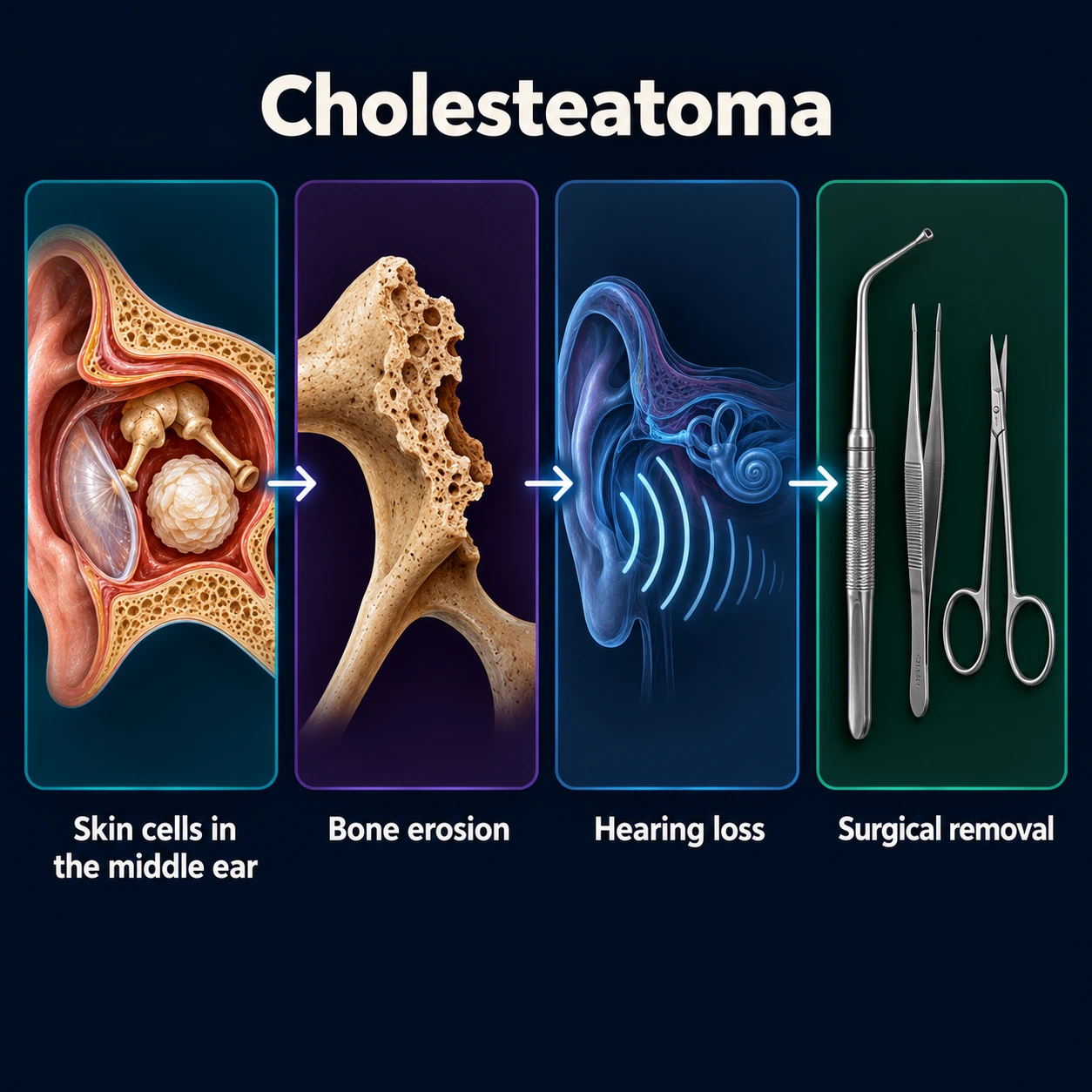
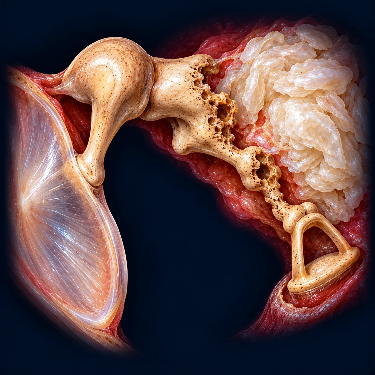
Cholesteatoma is an abnormal, destructive accumulation of keratinizing squamous epithelium in the middle ear and mastoid. Despite the name, it is not a tumor and it contains no cholesterol crystals in the modern pathological sense — the term is a historical misnomer that has persisted in clinical usage. Left untreated, cholesteatoma slowly but relentlessly erodes the ossicles, mastoid air cells, and surrounding bone through enzymatic and pressure mechanisms, leading to progressive hearing loss and potentially life-threatening intracranial complications.
Table of Contents
- What Cholesteatoma Is
- Types: Acquired vs. Congenital
- Pathophysiology and Bone Erosion
- Symptoms and Clinical Presentation
- Diagnosis and Imaging
- Ossicular Chain Destruction
- Complications
- Surgical Treatment
- Canal Wall Up vs. Canal Wall Down
- Second-Look Surgery and Residual Disease
- Recidivism and Recurrence
- How Common Cholesteatoma Is
- Key Research Papers
- Featured Videos
What Cholesteatoma Is
A cholesteatoma is a cystic structure lined by stratified squamous keratinizing epithelium — essentially skin that has migrated or grown into a location where it does not belong. Normally, the skin of the ear canal migrates outward (a process called epithelial migration), carrying debris away from the eardrum. In cholesteatoma, this migration is disrupted: keratinized squames accumulate in the middle ear or mastoid, forming an expanding mass of concentric keratin layers surrounding a central core of desquamated debris.
The mass appears as a pearlescent white lesion on otoscopic examination when visible. Its margins may produce a foul-smelling, cheesy discharge when infected. The term "cholesteatoma" was coined by Johannes Müller in 1838 based on the lesion's gross appearance, which he thought resembled fatty (cholesterol-containing) material.
Cholesteatoma is not a malignancy, but its behavior is locally aggressive. The epithelium stimulates the production of collagenase, matrix metalloproteinases (MMPs), and interleukins that dissolve the surrounding bone through both enzymatic digestion and osteoclast activation. This destructive capacity means that the goal of treatment is always surgical eradication.
Types: Acquired vs. Congenital
Acquired Cholesteatoma (90%)
Acquired cholesteatoma forms in the setting of chronic otitis media with middle ear and Eustachian tube dysfunction. Two subtypes are recognized:
Primary Acquired (Pars Flaccida / Attic Cholesteatoma)
The most common form. Chronic negative pressure in the middle ear — caused by Eustachian tube dysfunction — draws the pars flaccida portion of the tympanic membrane (the superior, less-taut portion) inward, forming a retraction pocket. Over years, the pocket deepens into the epitympanum (attic), accumulating keratin debris that cannot self-clean. This retraction pocket theory is the dominant mechanistic explanation for primary acquired disease. Early otoscopy reveals a subtle attic retraction pocket that can be mistaken for a simple perforation.
Secondary Acquired
Squamous epithelium migrates through a pre-existing perforation of the tympanic membrane — typically a marginal perforation at the posterosuperior quadrant — into the middle ear space. This is less common than the retraction pocket mechanism.
Congenital Cholesteatoma (10%)
Congenital cholesteatoma presents as a white, pearlescent mass visible through an intact tympanic membrane in children, typically discovered incidentally. It arises from remnant embryonic epithelial rests (Michaels' bodies) that normally involute during fetal development. Typical location is the anterosuperior quadrant of the middle ear, distinguishing it from the posterosuperior location of acquired disease. There is no history of otitis media, ear infections, or tympanic membrane perforation. Mean age at diagnosis is 4–7 years.
Pathophysiology and Bone Erosion
The destructive mechanism of cholesteatoma involves two interacting processes:
Enzymatic Bone Destruction
Cholesteatoma epithelium and the inflammatory cells it attracts (macrophages, mast cells) secrete elevated levels of matrix metalloproteinases (MMP-2, MMP-9), cathepsins, and various interleukins (interleukin-1 alpha (IL-1α), interleukin-6 (IL-6), tumor necrosis factor alpha (TNF-α)). These enzymes directly dissolve collagen in the ossicular ligaments, periosteium, and surrounding bone matrix.
Pressure-Mediated Resorption
The expanding keratin mass exerts continuous pressure on adjacent bony structures, activating osteoclasts at the bone-lesion interface. This combination of enzymatic and pressure-driven resorption explains why cholesteatoma erodes bone at a rate disproportionate to its size.
The incus long process is the most vulnerable ossicle because of its tenuous blood supply and central position in the erosion pathway. It is the ossicle most commonly destroyed by cholesteatoma, followed by the stapes superstructure. The malleus handle is relatively protected and often remains intact until disease is extensive.
Symptoms and Clinical Presentation
Cholesteatoma is often slowly progressive and may be surprisingly asymptomatic until ossicular destruction or complications occur.
Recurrent or Chronic Ear Discharge
The hallmark presenting symptom is chronic, foul-smelling otorrhea that fails to clear completely with antibiotic eardrops. The discharge has a characteristic fetid odor from the bacteria colonizing the keratin debris. These are usually a mix: air-tolerant (aerobic) bacteria, particularly Pseudomonas aeruginosa and Proteus mirabilis, together with bacteria that grow without oxygen (anaerobes). A 1978 study of 114 chronically discharging middle ears found anaerobes in 33% (mostly Bacteroides), always mixed with aerobic bacteria such as staphylococci, gut-type gram-negative rods and Pseudomonas; ears with suspected cholesteatoma grew anaerobes significantly more often (Karma P et al., 1978). Patients often report multiple courses of antibiotics with only partial improvement.
Conductive Hearing Loss
Progressive conductive hearing loss (CHL) results from ossicular discontinuity or the mass effect of the cholesteatoma filling the middle ear space and damping tympanic membrane vibration. A large cholesteatoma may paradoxically improve sound transmission by bridging an ossicular gap (the "shell-through" effect) — a discovery that can delay the diagnosis.
Ear Fullness and Otalgia
A sensation of fullness or pressure in the affected ear is common. Pain is unusual in uncomplicated cholesteatoma but may indicate active infection or impending complications such as mastoiditis.
Otoscopic Findings
The classic appearance is a white, cheese-like mass visible in the attic (pars flaccida region) through an otoscope. Squamous debris may be seen extending from a retraction pocket. A marginal perforation in the posterosuperior quadrant with white pearlescent material is highly suggestive. A systematic examination covers the entire tympanic membrane, including the attic and posterior annulus, because early lesions can be subtle and obscured by otorrhea.
Diagnosis and Imaging
Otoscopy and Microscopy
The definitive diagnosis is clinical: direct visualization of the cholesteatoma sac on otoscopy or microscopy (operating microscope or otoendoscope). The standard workup for suspected cholesteatoma includes formal hearing tests: pure-tone audiometry and tympanometry.
High-Resolution Computed Tomography of the Temporal Bone (HRCT)
HRCT is essential for surgical planning. It defines:
- Extent of disease (attic, antrum, mastoid, hypotympanum, petrous apex)
- Degree of ossicular erosion
- Tegmen integrity (roof of the middle ear — if eroded, intracranial extension is possible)
- Sigmoid sinus position and dehiscence risk
- Facial nerve canal integrity (especially the horizontal segment)
- Labyrinthine fistula (erosion of the lateral semicircular canal)
Computed tomography (CT) cannot reliably distinguish cholesteatoma from granulation tissue or middle ear effusion based on density alone, but its anatomical detail is indispensable for the surgeon.
Diffusion-Weighted MRI (DWI)
Non-echo-planar DWI (non-EPI DWI) has emerged as the most accurate non-invasive tool for detecting residual or recurrent cholesteatoma after surgery, with sensitivity and specificity exceeding 90%. Cholesteatoma keratin shows restricted diffusion (bright on DWI, dark on the apparent diffusion coefficient (ADC) map), distinguishing it from scar, effusion, and granulation tissue. DWI has largely replaced planned second-look surgery at many centers for post-operative surveillance.
A 2017 meta-analysis pooled 26 studies (1,152 patient episodes) in which the scan result was checked against what surgeons actually found. Non-echo-planar DWI had a pooled sensitivity of 91% (it picked up about 91 of every 100 cholesteatomas present) and a specificity of 92% (about 92 of every 100 ears without cholesteatoma were correctly read as clear). The figures stayed high when first-time cases, post-surgery cases, children and adults were looked at separately (sensitivity 86–93%, specificity 88–97%). The authors called for a large trial comparing the scan with second-look surgery (Lingam RK, Bassett P, 2017).
Ossicular Chain Destruction
Cholesteatoma destroys ossicles in a predictable sequence determined by tumor extension from the epitympanum:
- Incus long process: most commonly eroded first (80–90% of cases with ossicular involvement); its distal segment has a poor blood supply from the stapes end
- Stapes superstructure (crura): second most common; erosion here produces a significant air-bone gap and increases the risk of perilymphatic fistula and sensorineural hearing loss during surgery
- Malleus handle: more resistant; erosion here suggests extensive cholesteatoma
Ossicular reconstruction (ossiculoplasty) is performed at the time of cholesteatoma removal or staged at a second procedure. Partial ossicular replacement prostheses (PORPs) replace the incus-stapes connection; total ossicular replacement prostheses (TORPs) replace the entire ossicular chain when the stapes arch is absent. Success rates (post-operative air-bone gap <20 dB) are approximately 60–80% for PORPs and 50–70% for TORPs at experienced centers.
Complications
Untreated or inadequately managed cholesteatoma can produce potentially life-threatening complications from erosion into adjacent structures:
Labyrinthine Fistula
Erosion of the bony labyrinthine capsule — most commonly the lateral semicircular canal — creates a perilymph fistula. This is the most common intratemporal complication (incidence 5–10%). Clinically, it produces sudden sensorineural hearing loss and episodic vertigo provoked by changes in middle ear pressure (positive fistula test / Hennebert sign). Intraoperative management is challenging because opening the fistula risks irreversible sensorineural hearing loss.
Facial Nerve Palsy
Erosion of the bony fallopian canal — typically at the horizontal (tympanic) segment or second genu — exposes and compresses the facial nerve. The incidence is 2–5% in untreated cases. Presenting as progressive ipsilateral peripheral facial weakness, it represents a surgical emergency requiring prompt decompression. In an Italian series of 13 patients with facial paralysis from acquired cholesteatoma, all those operated on less than 7 days after the paralysis began had normal facial function at long-term follow-up, while those operated on 7 days or more after onset had variable results (Quaranta N et al., 2007).
Mastoiditis and Coalescent Mastoiditis
Secondary infection of mastoid air cells produces postauricular swelling, erythema, and anterior displacement of the auricle. Coalescent mastoiditis (breakdown of septa between air cells) may require urgent mastoidectomy.
Intracranial Complications
These are rare (1–2%) but potentially fatal:
- Meningitis: from tegmen erosion with direct bacterial seeding of the subarachnoid space
- Epidural and subdural abscess
- Brain abscess (typically temporal lobe or cerebellar)
- Sigmoid sinus thrombosis: from adjacent infection with thrombophlebitis; presents with fever, headache, and septic emboli
- Otitic hydrocephalus (rare)
Surgical Treatment
Surgery is the only definitive treatment for cholesteatoma. The goals, in order of priority, are:
- Eradication of disease: complete removal of all cholesteatoma matrix to prevent recurrence
- A safe, dry ear: anatomically stable middle ear that resists future infection and recolonization
- Hearing reconstruction: ossiculoplasty to restore the ossicular chain
Tympanomastoidectomy — surgical exploration and exenteration of the mastoid air cell system — is the standard operative framework, with the specific technique chosen based on disease extent, mastoid pneumatization, and patient factors.
Canal Wall Up vs. Canal Wall Down
The central surgical controversy in cholesteatoma management is whether to preserve the posterior external auditory canal wall (canal wall up, CWU) or remove it (canal wall down, CWD / modified radical mastoidectomy).
Canal Wall Up (CWU) / Intact Canal Wall Mastoidectomy
The posterior bony ear canal wall is preserved, maintaining near-normal anatomy. Advantages: better hearing reconstruction outcomes, no open mastoid cavity requiring lifelong maintenance, can use bone-anchored hearing aids if needed. Disadvantages: higher recidivism (residual or recurrent cholesteatoma) rate of 10–40%; requires planned second-look surgery or serial DWI surveillance to detect residual disease hidden behind the intact wall.
Canal Wall Down (CWD) / Modified Radical Mastoidectomy
The posterior canal wall is removed, creating a large, open mastoid cavity (mastoid bowl) that is exteriorized through the ear canal. The Eustachian tube is obliterated. Advantages: lower recidivism rate (5–15%); the entire cavity can be inspected at clinic visits; suitable for extensive disease or in patients unlikely to attend follow-up. Disadvantages: the open cavity requires regular clinic debridement and lifelong water precautions; hearing reconstruction is less reliable; cavity problems (chronic discharge, erosion of the mastoid tip, inadequate meatoplasty) occur in 10–30% of cases.
Mastoid Obliteration
Obliteration of the mastoid cavity with bone pâté, hydroxyapatite, or cartilage after CWD reduces the burden of cavity management and can improve hearing outcomes. Various obliteration techniques are used at different centers; outcomes depend heavily on surgeon experience.
Second-Look Surgery and Residual Disease
In CWU tympanomastoidectomy, planned second-look surgery at 9–18 months was historically standard to detect and remove residual cholesteatoma not visible at the primary operation. Reported residual rates at second look range from 15–40% in adult series and up to 50% in pediatric cases.
Non-echo-planar DWI MRI has largely replaced routine second-look surgery at centers where it is available and where the surgeon is confident in intraoperative technique. A negative DWI scan at 18 months post-operatively has a negative predictive value of approximately 95% for residual cholesteatoma >3 mm. In the 2017 meta-analysis described under Diagnosis, its accuracy for post-surgery cases was as high as for first-time cases. DWI is less reliable for small (<3 mm) residuals and for lesions near metallic ossicular prostheses. The older echo-planar form of the scan was much weaker here: in a 2006 Belgian study of 45 patients scanned 8–18 months after surgery, it correctly showed only 1 of 7 residual cholesteatomas that surgery later confirmed (Vercruysse JP et al., 2006).
When to scan after surgery is still debated. A 2026 meta-analysis of 16 studies (1,187 patients) pooled the share of post-surgery MRI scans that showed cholesteatoma at each point in time: 12% at 1 month, 19% at 6 months, 15% at 12 months, 22% at 24 months, 12% at 36 months, a second peak of 21% at 48 months and 13% at 60 months. Ears that had a canal wall up operation had more positive scans than ears that had mastoid obliteration (22% v 7% at 12 months; 18% v 9% at 36 months). The authors concluded that their findings support a first routine scan 12 to 24 months after surgery, then scans every 2 years for people whose first scan is clear, continued for at least 5 years (Wolfovitz A et al., 2026).
Recidivism and Recurrence
It is important to distinguish:
- Residual disease: incompletely removed cholesteatoma matrix left at surgery, discovered on second-look or surveillance imaging
- Recurrent disease: new cholesteatoma formation from a retraction pocket or re-migration of epithelium after apparent complete removal
Recurrence rates are higher in children (immature Eustachian tube function, longer time horizon for disease) and in patients with extensive primary disease. Surgical practice describes long-term follow-up of at least 5 years for CWU cases, and the 2026 imaging meta-analysis above likewise concluded that follow-up scans continue for at least 5 years. In a Finnish series of 349 patients followed for a mean of 7.3 years, 43 (12.3%) had a recurrence; recurrence was more frequent in children than in adults and in ears that were discharging before surgery, and the type of surgical technique had no significant effect on the recurrence rate (Vartiainen E, 1995). Some surgeons advocate lifetime annual surveillance given the lifelong risk of recurrence, particularly in the pediatric population.
How Common Cholesteatoma Is
A 2026 systematic review and meta-analysis of 46 studies estimated the worldwide prevalence of cholesteatoma at about 4 per 1,000 people (4.02 per 1,000; 95% confidence interval 1.79–7.10). The figure differed widely between World Health Organization regions — for example 2.32 per 1,000 in the European Region and 7.32 per 1,000 in the African Region. Hearing loss was present in about 76 of every 100 people with cholesteatoma (Melariri H et al., 2026).
Key Research Papers
- Olszewska E, Wagner M, Bernal-Sprekelsen M, et al. Etiopathogenesis of cholesteatoma. Eur Arch Otorhinolaryngol. 2004;261(1):6–24. — PubMed PMID: 12835944
- Tos M. A new pathogenesis of mesotympanic (congenital) cholesteatoma. Laryngoscope. 2000;110(11):1890–1897. — PubMed PMID: 11081605
- Persaud R, Hajioff D, Thevasagayam MS, et al. Keratosis obturans and external ear canal cholesteatoma: how and why we should distinguish between these conditions. Clin Otolaryngol Allied Sci. 2004;29(6):577–581. — PubMed PMID: 15533140
- Kuo CL, Shiao AS, Yung M, et al. Updates and knowledge gaps in cholesteatoma research. Biomed Res Int. 2015;2015:854024. — PubMed PMID: 25866816
- Vartiainen E. Factors associated with recurrence of cholesteatoma. J Laryngol Otol. 1995;109(7):590–592. — PubMed PMID: 7561462
- Yung M, Tono T, Olszewska E, et al. EAONO/JOS Joint Consensus Statements on the Definitions, Classification and Staging of Middle Ear Cholesteatoma. J Int Adv Otol. 2017;13(1):1–8. — PubMed PMID: 28059056
- Vercruysse JP, De Foer B, Pouillon M, Somers T, Casselman J, Offeciers E. The value of diffusion-weighted MR imaging in the diagnosis of primary acquired and residual cholesteatoma: a surgical verified study of 100 patients. Eur Radiol. 2006;16(7):1461–1467. — PubMed PMID: 16514469
- Saleh HA, Mills RP. Classification and staging of cholesteatoma. Clin Otolaryngol Allied Sci. 1999;24(4):355–359. — PubMed PMID: 10472474
- Quaranta N, Cassano M, Quaranta A. Facial paralysis associated with cholesteatoma: a review of 13 cases. Otol Neurotol. 2007;28(3):405–407. — PubMed PMID: 17414046
- Karma P, Jokippi L, Ojala K, Jokipii AM. Bacteriology of the chronically discharging middle ear. Acta Otolaryngol. 1978;86(1-2):110–114. — PubMed PMID: 358732
- Lingam RK, Bassett P. A Meta-Analysis on the Diagnostic Performance of Non-Echoplanar Diffusion-Weighted Imaging in Detecting Middle Ear Cholesteatoma: 10 Years On. Otol Neurotol. 2017;38(4):521–528. — PubMed PMID: 28195998
- Wolfovitz A, Segal E, Murad H, Agay N, Tessler I. Defining the Timing of Radiological Follow-Up for Detection of Cholesteatoma Recidivism: A Meta-analysis. Otolaryngol Head Neck Surg. 2026;174(6):1410–1420. — PubMed PMID: 41904979
- Melariri H, Balakrishna Y, Mukhtar A, et al. The Global Burden of Cholesteatoma: A Systematic Review and Meta-analysis. Otolaryngol Head Neck Surg. 2026;174(1):45–56. — PubMed PMID: 41147202
PubMed Topic Searches
The following PubMed topic searches retrieve current peer-reviewed literature on Cholesteatoma.
- PubMed: Cholesteatoma surgery outcomes
- PubMed: Cholesteatoma DWI MRI detection
- PubMed: Cholesteatoma canal wall recurrence
- PubMed: Congenital cholesteatoma pediatric
- PubMed: Cholesteatoma bone erosion mechanisms
- PubMed: Cholesteatoma ossiculoplasty hearing reconstruction
- PubMed: Cholesteatoma intracranial complications
- PubMed: Cholesteatoma second-look surgery residual