⚠ Evidence warning: Clinical trials or other evidence do not support the claims that the average delay to diagnosis exceeds seven years, that aspirin plus dipyridamole is among the best-established regimens, and that complete remission is uncommon. This page keeps it as a report of what people, websites or other media say.
Livedoid Vasculopathy (Atrophie Blanche)
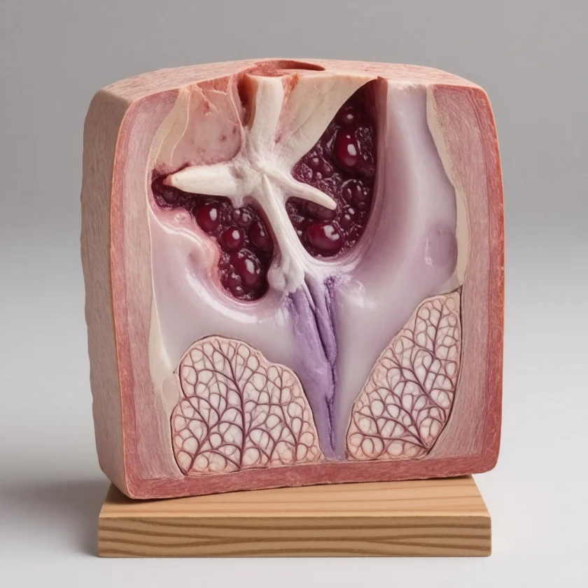
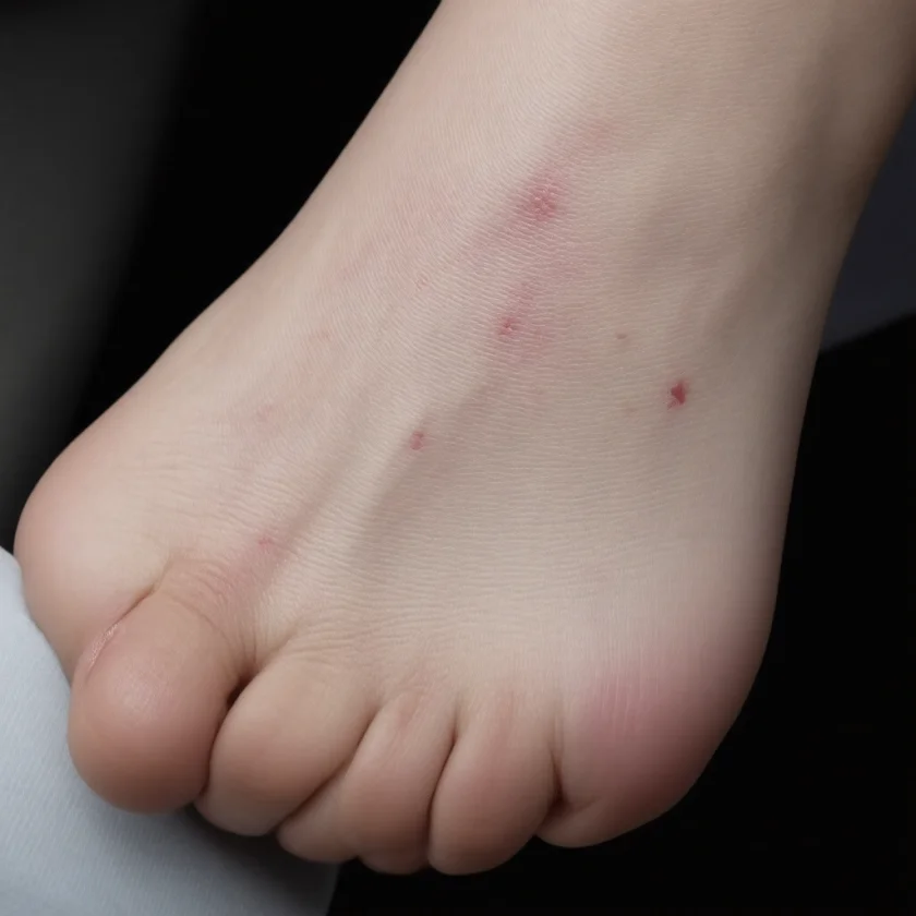
Table of Contents
- Overview
- Pathophysiology
- Clinical Presentation
- Diagnosis
- Treatment: Antiplatelet and Antithrombotic Therapy
- Treatment: Wound Care and Adjunctive Therapies
- Differential Diagnosis
- Prognosis and Quality of Life
- Practical Facts for People With Livedoid Vasculopathy
- Key Research Papers
- Featured Videos
- Connections
Overview
Livedoid vasculopathy (LV) — also known as segmental hyalinizing vasculitis or livedoid vasculitis, and originally called atrophie blanche — is a rare, chronic skin condition characterized by recurrent, extremely painful ulcerations on the lower legs and feet that heal with distinctive porcelain-white stellate scars. The term atrophie blanche (French: "white atrophy") describes the hallmark healed scar: a star-shaped (stellate), hypopigmented ivory-white area rimmed by dilated capillaries (telangiectasias) and brown hemosiderin deposits. These scars are permanent. A 2013 international consensus notes that "atrophie blanche" was originally used as a synonym, but that the scars seen with LV and those seen with chronic venous insufficiency are two unrelated forms that need separate diagnostic and treatment approaches.
Despite the older name "livedoid vasculitis," LV is not primarily an inflammatory condition. The fundamental pathological event is intravascular thrombosis — small blood clots blocking the superficial dermal blood vessels — leading to ischemia (lack of blood flow) and ultimately painful ulceration. This distinction is critical because it means anti-inflammatory treatments alone are often ineffective, while antiplatelet and anticoagulant strategies are the therapeutic mainstay. In the treatment histories of a German cohort of 27 patients, anti-inflammatory regimens were used in 17 of 24 and worked in 0 of 17, while heparin worked in 12 of 17.
LV disproportionately affects women (approximately 3:1 female-to-male ratio), typically in young to middle adulthood. A review of 339 patients counted 230 women and 69 men (sex not stated for 40), the German cohort had a ratio of 2.1:1 and a median age of 53, and a Mayo Clinic series of 45 patients had a mean age of 45 (range 10 to 85). Both lower legs are usually involved (bilateral). Many patients experience summer exacerbations, when heat causes vasodilation and may worsen local circulatory dynamics. The condition is frequently underdiagnosed or misdiagnosed for years. Some accounts give an average diagnostic delay exceeding seven years from symptom onset; one measured figure is 4.61 years (± 0.69) from onset to diagnosis at a Beijing hospital, where 85% of patients were first given another diagnosis.
Pathophysiology
The central mechanism in livedoid vasculopathy is occlusive thrombosis of small superficial dermal blood vessels. Unlike true vasculitis — where the vessel wall itself is attacked by immune cells — in LV the primary event is the formation of fibrin thrombi (blood clots composed largely of fibrin) within the lumen (interior) of small dermal vessels. These clots obstruct blood flow, depriving the overlying skin of oxygen and nutrients, leading to ischemic ulceration.
On skin biopsy, the pathological findings reflect this thrombotic process:
- Fibrin thrombi occluding small superficial dermal vessels (highlighted by PAS staining)
- Hyalinization of vessel walls — a glassy, pink thickening of the vessel wall due to fibrin and protein deposition
- Endothelial cell proliferation within vessel lumens
- Minimal or absent inflammatory infiltrate — the key histological feature distinguishing LV from true vasculitis
- Hemosiderin deposits in surrounding tissue from red blood cell extravasation
A significant proportion of LV patients have an underlying prothrombotic (hypercoagulable) state that drives this abnormal clotting. Identified thrombophilias in LV patients include:
- Antiphospholipid antibodies (anticardiolipin antibodies, anti-β2 glycoprotein I, lupus anticoagulant) — present in approximately 25% of LV patients
- Factor V Leiden mutation — the most common inherited thrombophilia
- Prothrombin G20210A mutation
- Protein C or Protein S deficiency — natural anticoagulant proteins
- Antithrombin III deficiency
- Hyperhomocysteinemia — elevated homocysteine damages endothelium and promotes clotting
- Elevated lipoprotein(a) — an independent prothrombotic risk factor
How often a clotting disorder is found varies by study and by the tests done. In a prospective study of 34 patients, 18 (52%) had laboratory abnormalities of this kind. At the Mayo Clinic, anticardiolipin antibodies were present in 8 of 28 tested and lupus anticoagulant in 5 of 28; in the German cohort, homocysteine was raised in 10 of 12 tested and lipoprotein(a) in 5 of 12. Many LV cases occur without a clearly identifiable systemic thrombophilia, suggesting that local vascular factors — such as sluggish venous flow in the dependent lower leg, reduced local fibrinolysis, or endothelial dysfunction — contribute to the localized thrombotic tendency. The overlapping background of livedo racemosa (a purplish, irregular, non-blanchable reticulated skin mottling) reflects chronic local vascular insufficiency in the affected areas.
Clinical Presentation
Livedoid vasculopathy follows a chronic, relapsing, episodic course. Most patients experience recurrent flares interspersed with partial or complete healing, though the scars from previous ulcerations are permanent. The full clinical picture unfolds in three overlapping phases:
Phase 1: Active Ulceration
Flares begin with the sudden appearance of painful, punched-out ulcers on the lower legs, ankles, and dorsal (top) surface of the feet. Key characteristics:
- Size: typically 1–5 cm, though lesions can be larger
- Shape: irregular, stellate (star-shaped), with undermined or sharply demarcated edges
- Pain: excruciating and markedly out of proportion to the wound size — a hallmark feature that raises suspicion of LV; often described as burning, stabbing, or throbbing
- Distribution: bilateral lower legs (both sides), particularly around the ankles and malleoli
- Background livedo racemosa: a purple-red, irregular (non-geometric) net-like mottling of the surrounding skin; non-blanchable; represents local vascular insufficiency
- Seasonal pattern: flares are more common and severe in summer months
Phase 2: Slow, Painful Healing
Unlike many wounds, LV ulcers heal extremely slowly — over weeks to months — and are notoriously resistant to standard wound care. The prothrombotic state in the dermal vessels impairs the normal healing process. Infection can complicate open wounds and delay healing further.
Phase 3: Atrophie Blanche (Healed Scar)
As ulcers finally heal, they leave behind the characteristic scar of livedoid vasculopathy:
- Atrophie blanche: stellate (star-shaped), ivory-white, porcelain-smooth scarred areas
- Margins: bordered by dilated superficial capillaries (telangiectasias) giving a red or pink rim
- Hemosiderin deposits: brown pigmentation surrounding the white scar from iron-containing hemoglobin breakdown products
- Permanent: these scars do not resolve; they accumulate over years of recurrent ulceration
Important note: atrophie blanche scars are characteristic of the healed phase of LV but can also be seen in other conditions causing lower leg ischemia, including chronic venous insufficiency and antiphospholipid syndrome. The clinical context and history differentiate these.
The severe, chronic pain of LV has profound quality-of-life implications. Many patients are unable to walk normally during flares, are kept from work, and develop sleep disturbance and psychological distress. The long diagnostic delay (more than seven years in some accounts, 4.61 years at the Beijing hospital) means many patients suffer for years without appropriate treatment.
Diagnosis
Diagnosis of livedoid vasculopathy rests on three pillars: clinical recognition, skin biopsy, and a systematic thrombophilia workup. Because LV is rare and its presentation overlaps with more common conditions, a high index of suspicion is required.
Clinical Diagnosis
The clinical picture — recurrent extremely painful lower leg ulcers healing with stellate ivory-white atrophie blanche scars, bilateral distribution, and background livedo racemosa — is highly characteristic. Clinicians consider LV in people with:
- Recurring lower leg/ankle ulcers with disproportionate pain
- Slow healing despite standard wound care
- White stellate scars on the lower legs
- Young to middle-aged female patient (though men are affected too)
Skin Biopsy
An incisional biopsy from the edge of an active ulcer (not the center, which may show only necrosis) is the most informative. Findings diagnostic of LV:
- Fibrin thrombi in the lumens of superficial dermal blood vessels (PAS stain highlights these)
- Hyalinization of vessel walls (pink, glassy thickening)
- Endothelial proliferation
- Minimal or no inflammatory cell infiltrate — this key feature distinguishes LV from true vasculitis, where a brisk inflammatory infiltrate surrounds and invades vessel walls
- Extravasated red blood cells and hemosiderin in surrounding dermis
Thrombophilia Workup
The authors of a 2010 prospective study suggest that all patients diagnosed with LV be investigated for an underlying prothrombotic state, as identifying a specific thrombophilia guides treatment decisions (particularly the use of anticoagulation). The workup includes:
- Factor V Leiden mutation (polymerase chain reaction-based genetic test)
- Prothrombin G20210A mutation
- Protein C activity and Protein S activity (functional assays; note: levels may be falsely low during acute illness or anticoagulation)
- Antithrombin III activity
- Antiphospholipid antibody panel: anticardiolipin immunoglobulin G (IgG)/immunoglobulin M (IgM), anti-β2 glycoprotein I immunoglobulin G/immunoglobulin M, lupus anticoagulant
- Serum homocysteine (fasting)
- Lipoprotein(a)
- Complete blood count, coagulation studies (PT, PTT)
- Antinuclear antibody (ANA) / rheumatological screen to exclude associated connective tissue disease
An identified thrombophilia was found in 52% (18 of 34) in that prospective study; other series give figures for single tests (see Pathophysiology). Even when no systemic thrombophilia is found, the treatment approach remains antithrombotic.
Vascular Studies
An ankle-brachial index (ABI) is used to exclude significant arterial insufficiency, which would influence wound care and compression decisions. Duplex ultrasound of the venous system may reveal underlying chronic venous insufficiency, which frequently co-exists.
Treatment: Antiplatelet and Antithrombotic Therapy
Because LV is driven by intravascular thrombosis rather than inflammation, antiplatelet and antithrombotic agents are the cornerstone of treatment. The goal is to prevent new clot formation in the superficial dermal vessels, allowing existing ulcers to heal and reducing the frequency of new flares.
No treatment is approved for LV. A 2025 report from Germany states there are "no approved treatments", so every drug below is used off-label. A 2018 systematic review found "no established treatment": its 339 patients came from 29 case series, and its authors asked for randomized trials. The German S1 guideline recommends anticoagulation (low-molecular-weight heparins, rivaroxaban and other direct oral anticoagulants) as first-line treatment. In the 2018 review, anticoagulants used alone gave a favorable response in 62 of 63 patients.
Aspirin and Dipyridamole (A Long-Used Combination)
Practitioners have described the combination of aspirin 100 mg/day plus dipyridamole 225 mg/day as among the best-established regimens for LV; a 1982 report describes excellent results in two patients on the pair. The authors of a 2019 German multicentre analysis concluded instead that anticoagulants belong first, used alone. Aspirin irreversibly inhibits platelet thromboxane A2 production, reducing platelet aggregation. Dipyridamole inhibits phosphodiesterase and adenosine uptake, further impairing platelet aggregation and causing vasodilation. Together they provide complementary antiplatelet effects relevant to the small-vessel thrombosis of LV.
Aspirin Monotherapy
Low-dose aspirin alone (75–100 mg/day) is used as an initial step, particularly in patients who cannot tolerate dipyridamole (which can cause headaches). Practitioners move to combination therapy or anticoagulation if aspirin monotherapy fails to control ulcers.
Warfarin (Vitamin K Antagonist)
Warfarin (target international normalized ratio (INR) 2.0–3.0) is used when:
- A specific thrombophilia (especially antiphospholipid syndrome) is identified
- Antiplatelet therapy has failed to control the disease
- The patient has particularly severe or rapidly recurring ulcerations
Warfarin is effective for LV, particularly in the antiphospholipid syndrome-associated subgroup, but requires regular international normalized ratio monitoring and has significant drug and food interactions. A 2006 case report describes a 50-year-old man whose lesions, after failing several other medicines, responded dramatically to oral warfarin. In a 2010 study, 11 of 13 patients responded to warfarin or heparin; pain eased in about 1.8 weeks, lesions healed in about 2.3 months, and remission lasted an average of 7.8 months after treatment stopped.
Direct Oral Anticoagulants
Rivaroxaban and apixaban have emerging evidence supporting their use in LV. A 2021 systematic review reports rivaroxaban at 10 to 20 mg per day (20 mg/day is described as taken with the evening meal). The key trial, RILIVA, was single-arm and open-label, not randomized: 25 people received rivaroxaban 10 mg twice daily, cut to once daily once pain had halved. Median pain fell from 65 to 6 on a visual pain scale at 12 weeks, and 6 of 20 needed added enoxaparin. In the 2021 review, 60 of 73 patients (82.2%) reached remission of both pain and ulceration, a result its authors say still needs confirming in large studies. Direct oral anticoagulants offer practical advantages over warfarin: no routine international normalized ratio monitoring and fewer food interactions. They are increasingly favored, especially in patients without antiphospholipid antibodies (where these drugs may be equivalent or superior to warfarin).
Side effects in the trial: 8 treatment-related events occurred in 6 of the 25 people, 5 of them heavy menstrual bleeding (one serious, in a woman with endometriosis, who left the study). This matters for a condition that mostly affects women.
Safety in high-risk antiphospholipid syndrome: a randomized trial in people who tested positive on all three antiphospholipid antibody tests (not a study of LV) was stopped early because of excess events on rivaroxaban: 11 (19%) of 59 had an event, against 2 (3%) of 61 on warfarin, including 4 strokes and 3 heart attacks on rivaroxaban and none on warfarin.
Low-Molecular-Weight Heparin
Low-molecular-weight heparin (e.g., enoxaparin; a 2003 report describes 2 treated cases) is used in two specific scenarios:
- Acute flares: rapid anticoagulation during severe active ulceration while bridging to oral therapy
- Pregnancy: the preferred anticoagulant when LV flares during pregnancy (warfarin is teratogenic; direct oral anticoagulants are contraindicated)
Danazol
Danazol, a synthetic androgen, was used in older LV literature based on its ability to reduce fibrinogen levels and increase fibrinolytic activity. While some patients responded, its use has fallen out of favor due to significant adverse effects including hepatotoxicity, virilization in women (acne, hirsuitism, voice deepening), and lipid abnormalities. It may still be considered in refractory cases where other options have failed.
Treatment: Wound Care and Adjunctive Therapies
Antithrombotic therapy forms the backbone of LV management, but adjunctive treatments address wound healing, microcirculation, and pain — all critical aspects of comprehensive care.
Pentoxifylline
Pentoxifylline 400 mg three times daily is a hemorheological agent that improves the flow properties of blood by increasing red blood cell deformability and reducing blood viscosity. It also has mild anti-inflammatory and anti-platelet effects. Multiple studies support its use as an adjunct in LV; it is generally well-tolerated. Side effects include nausea and headache.
Iloprost (Prostacyclin Analog)
Iloprost, a synthetic prostacyclin analog given by intravenous infusion, potently vasodilates microvessels, inhibits platelet aggregation, and improves microvascular perfusion. It is reserved for severe or refractory LV and is administered in specialist centers. Treatment courses typically involve daily infusions over several days. It is not available orally for this indication in many countries.
Compression Stockings
Graduated compression stockings (20–30 mmHg) reduce venous hypertension in the lower legs and improve venous return, which benefits many LV patients — particularly those with co-existing chronic venous insufficiency. Compression is generally not applied to actively ulcerated skin until wounds are healing, and arterial insufficiency is excluded (ABI check) before it is prescribed.
Hydroxychloroquine
Hydroxychloroquine 200–400 mg/day has a dual mechanism relevant to LV: it has antiplatelet properties (inhibits platelet aggregation) and mild anti-inflammatory effects. It is safe for long-term use with appropriate ophthalmological monitoring and is a useful adjunct, particularly in patients with connective tissue disease associations or those who need a low-risk add-on therapy.
Wound Care
LV wounds require meticulous care:
- Moist wound healing: non-adherent dressings (e.g., silicone-coated, hydrocolloid, or foam dressings) maintain a moist wound environment and prevent trauma on dressing removal
- Trauma: aggressive debridement is not used; LV skin is friable
- Infection management: signs of secondary infection are cellulitis, increased erythema and purulence; antibiotics are the treatment
- Elevation: elevating the legs reduces edema and improves venous return
- Heat: warm water baths and prolonged heat exposure can exacerbate LV
Intravenous Immunoglobulin
A 2022 systematic review of 80 patients with resistant LV, from 17 articles, reported that 76 (95%) responded to intravenous immunoglobulin at 1 to 2.1 g/kg every 4 weeks, with no severe adverse events. These were uncontrolled reports, and the authors say large randomized trials are still needed.
Biologic Drugs and Janus Kinase Inhibitors
A 2025 systematic review of 15 articles covered 41 patients with resistant LV treated with biologic drugs or Janus kinase inhibitors; 36 (87.8%) improved, including in pain. Tumor necrosis factor inhibitors were the drugs used most often alone. These findings come from small reports, and the authors describe them as a foundation for future studies.
Pain Management
The severe pain of LV — often neuropathic in character — requires a proactive, multimodal approach:
- Gabapentin or pregabalin (neuropathic pain agents) — often the most effective oral options
- Topical anesthetics (e.g., lidocaine gel) for wound dressing changes
- Nonsteroidal anti-inflammatory drugs (NSAIDs) — limited utility and potential adverse effects on renal function and platelet function
- Opioids — may be required for severe pain, but long-term opioid dependence is a recognized complication of undertreated chronic LV pain; practitioners describe formal pain management consultation for this
- Psychological support: chronic pain and visible scarring have significant psychological impact; mental health referral is part of the care described
Differential Diagnosis
Because LV is rare and its features overlap with several more common conditions, a broad differential is considered and systematically excluded:
- True cutaneous vasculitis (e.g., leukocytoclastic vasculitis, polyarteritis nodosa): biopsy shows vessel wall inflammation and neutrophil infiltration — absent in LV. Antineutrophil cytoplasmic antibody (ANCA) testing helps distinguish.
- Chronic venous insufficiency (CVI) ulcers: the most common lower leg ulcer in adults. Atrophie blanche can occur in CVI. Distinguishing features of LV: younger patient, extreme pain (CVI ulcers are often less painful), biopsy showing thrombi rather than venous changes.
- Arterial insufficiency ulcers: occur over pressure points; ABI typically reduced. LV can co-exist with mild arterial disease.
- Antiphospholipid syndrome: LV can be a cutaneous manifestation of antiphospholipid syndrome; the syndrome can cause livedo racemosa and lower leg ulcers indistinguishable from primary LV. Positive antiphospholipid antibodies on two separate occasions define it.
- Cryoglobulinemia: causes purpuric lower leg skin lesions and ulcers; cryoglobulin levels and hepatitis C serology distinguish this.
- Pyoderma gangrenosum: neutrophilic ulceration; violaceous undermined edges; often associated with inflammatory bowel disease or hematological malignancy; biopsy shows dense neutrophilic infiltrate.
- Calciphylaxis: painful skin necrosis in renal failure patients; calcium deposits in vessel walls on biopsy; different patient population.
- Sickle cell disease: chronic lower leg ulcers; hemoglobin electrophoresis diagnostic.
- Cholesterol emboli: painful lower leg ischemia/livedo following vascular procedures or in severe atherosclerosis; eosinophilia + needle-shaped clefts in vessel lumens on biopsy.
Prognosis and Quality of Life
Livedoid vasculopathy is a chronic, relapsing condition. It is not curable, but it is manageable — many patients achieve significant reduction in ulcer frequency and severity with appropriate antithrombotic therapy. Key prognostic considerations include:
- Chronic relapsing course: most patients experience recurrent flares over many years. With treatment, the frequency and severity of ulcers can be reduced. Complete remission has been described as uncommon, but the low-level evidence available shows more: 60 of 73 people on rivaroxaban reached remission of both pain and ulceration in a 2021 review, and remission lasted an average of 7.8 months after warfarin or heparin stopped in a 2010 study.
- Permanent scarring: atrophie blanche scars accumulate over time and do not resolve. In longstanding LV, patients may have extensive areas of scarring on both lower legs.
- Diagnostic delay: some accounts say the average delay from symptom onset to correct diagnosis exceeds seven years; a Beijing hospital measured 4.61 years. During this time, patients suffer without targeted treatment, accumulating scars and often developing opioid dependence from inadequate pain management.
- Underlying thrombophilia: when an identifiable prothrombotic state (especially antiphospholipid syndrome) is present, there is a risk of systemic thrombotic events (deep vein thrombosis, pulmonary embolism, stroke). Long-term anticoagulation is typically required in this subgroup.
- Pain and opioid dependence: the severe chronic pain of LV, combined with the long diagnostic delay, places patients at significant risk of long-term opioid use. Proactive neuropathic pain management and pain specialist involvement are essential.
- Psychological impact: chronic pain, visible scarring, and recurrent disability contribute to anxiety, depression, and reduced social function. Mental health support is an underutilized but important component of comprehensive care.
- Lifestyle triggers: heat, prolonged standing, trauma to the lower legs, and dehydration can provoke flares. Patient education about these triggers is important for self-management.
The overall prognosis for life expectancy is generally not reduced by LV itself, unless there is associated antiphospholipid syndrome with systemic thrombotic events. The primary burden is the chronic, debilitating nature of the pain and scarring.
Practical Facts for People With Livedoid Vasculopathy
Living with livedoid vasculopathy involves a collaborative, proactive approach. These are the practical facts most often described for patients:
- Specialists involved: LV care is described as co-management between a dermatologist (skilled in skin ulcer diagnosis and wound care) and a hematologist or vascular medicine specialist (for thrombophilia workup and anticoagulation management). Practitioners describe bringing in a pain management specialist early when pain is severe or opioids are being considered.
- Thrombophilia workup: identifying an underlying blood clotting disorder changes the treatment plan and may prevent serious systemic clots. The tests cover Factor V Leiden, protein C and S, antiphospholipid antibodies, and the other factors listed in the Diagnosis section.
- The treatment goal: antithrombotic therapy (aspirin, warfarin, or a direct oral anticoagulant like rivaroxaban) is the most important medication for LV — not antibiotics or steroids. Anti-inflammatory regimens worked in 0 of 17 people in the German cohort; antiplatelet and anticoagulant drugs are the options described when steroids have been the only treatment.
- Compression stockings: once ulcers are healing, graduated compression stockings (20–30 mmHg) reduce venous pressure in the lower leg and are described as helping prevent new ulcers. Arterial disease is excluded first.
- Triggers in the lower legs: heat (hot baths, prolonged sun exposure to legs), standing for long periods without movement, and trauma to the shins or ankles are the triggers described. Even minor injuries can trigger new ulcers.
- Wound care: moist non-adherent dressings, changed gently, are the dressings described for these ulcers. Increasing redness, warmth, pus and fever are signs of infection, which is treated with antibiotics.
- Uncontrolled pain: severe pain in LV is real and is treated. Gabapentin or pregabalin for neuropathic pain, topical anesthetics for dressing changes, and formal pain management consultation are the options described, alongside or instead of long-term opioids.
- Patient support: connecting with others who have LV through patient advocacy groups and online communities can be valuable for coping with a rare, chronic, and often misunderstood condition.
- Summer: flares are more common in summer, and having compression stockings, wound dressings and medications on hand before warmer months is a practice described for that reason.
Key Research Papers
- Alavi A, Hafner J, Dutz JP, et al. Livedoid vasculopathy: an in-depth analysis using a modified Delphi approach. J Am Acad Dermatol. 2013;69(6):1033–1042.e1. [ PubMed PMID: 24028907]
- Criado PR, Rivitti EA, Sotto MN, et al. Livedoid vasculopathy: an intringuing cutaneous disease. An Bras Dermatol. 2011;86(5):961–977. [ PubMed PMID: 22147037]
- Kern AB. Atrophie blanche. Report of two patients treated with aspirin and dipyridamole. J Am Acad Dermatol. 1982;6(6):1048–1053. [ PubMed PMID: 7096668]
- Vasudevan B, Neema S, Verma R. Livedoid vasculopathy: a review of pathogenesis and principles of management. Indian J Dermatol Venereol Leprol. 2016;82(5):478–488. [ PubMed PMID: 27297279]
- Weishaupt C, Strölin A, Kahle B, et al. Characteristics, risk factors and treatment reality in livedoid vasculopathy - a multicentre analysis. J Eur Acad Dermatol Venereol. 2019;33(9):1784–1791. [ PubMed PMID: 31009111]
- Di Giacomo TB, Hussein TP, Souza DG, Criado PR. Frequency of thrombophilia determinant factors in patients with livedoid vasculopathy and treatment with anticoagulant drugs--a prospective study. J Eur Acad Dermatol Venereol. 2010;24(11):1340–1346. [ PubMed PMID: 20337814]
- Hairston BR, Davis MD, Pittelkow MR, Ahmed I. Livedoid vasculopathy: further evidence for procoagulant pathogenesis. Arch Dermatol. 2006;142(11):1413–1418. [ PubMed PMID: 17116831]
- Schroeter AL, Diaz-Perez JL, Winkelmann RK, Jordan RE. Livedo vasculitis (the vasculitis of atrophie blanche). Immunohistopathologic study. Arch Dermatol. 1975;111(2):188–193. [ PubMed PMID: 1090260]
- Browning CE, Callen JP. Warfarin therapy for livedoid vasculopathy associated with cryofibrinogenemia and hyperhomocysteinemia. Arch Dermatol. 2006;142(1):75–78. [ PubMed PMID: 16415390]
- Weishaupt C, Strölin A, Kahle B, et al. Anticoagulation with rivaroxaban for livedoid vasculopathy (RILIVA): a multicentre, single-arm, open-label, phase 2a, proof-of-concept trial. Lancet Haematol. 2016;3(2):e72–e79. [ PubMed PMID: 26853646]
- Micieli R, Alavi A. Treatment for Livedoid Vasculopathy: A Systematic Review. JAMA Dermatol. 2018;154(2):193–202. [ PubMed PMID: 29141075]
- Gao Y, Jin H. Rivaroxaban for treatment of livedoid vasculopathy: A systematic review. Dermatol Ther. 2021;34(5):e15051. [ PubMed PMID: 34197012]
- Hillgruber C, Mitschang C, Eveslage M, et al. Long-Term Efficacy of Guideline-Followed Treatment in Patients with Livedoid Vasculopathy: A Single-Center Study. Adv Wound Care (New Rochelle). 2026;15(12):898–906. [ PubMed PMID: 41264385]
- Qi F, Gao Y, Jin H. Identification of Challenging Diagnostic Factors in Livedoid Vasculopathy: A Retrospective Study. Clin Cosmet Investig Dermatol. 2024;17:1747–1756. [ PubMed PMID: 39109220]
- Pengo V, Denas G, Zoppellaro G, et al. Rivaroxaban vs warfarin in high-risk patients with antiphospholipid syndrome. Blood. 2018;132(13):1365–1371. [ PubMed PMID: 30002145]
- Gao Y, Jin H. Efficacy and safety of intravenous immunoglobulin for treating refractory livedoid vasculopathy: a systematic review. Ther Adv Chronic Dis. 2022;13:20406223221097331. [ PubMed PMID: 35634570]
- Liu Y, Li T, Shi W. Janus kinase inhibitors and biologics for treatment of livedoid vasculopathy: a systematic review. J Dermatolog Treat. 2025;36(1):2451804. [ PubMed PMID: 39828272]
- Hairston BR, Davis MD, Gibson LE, Drage LA. Treatment of livedoid vasculopathy with low-molecular-weight heparin: report of 2 cases. Arch Dermatol. 2003;139(8):987–990. [ PubMed PMID: 12925383]