Linear Immunoglobulin A (IgA) Bullous Dermatosis
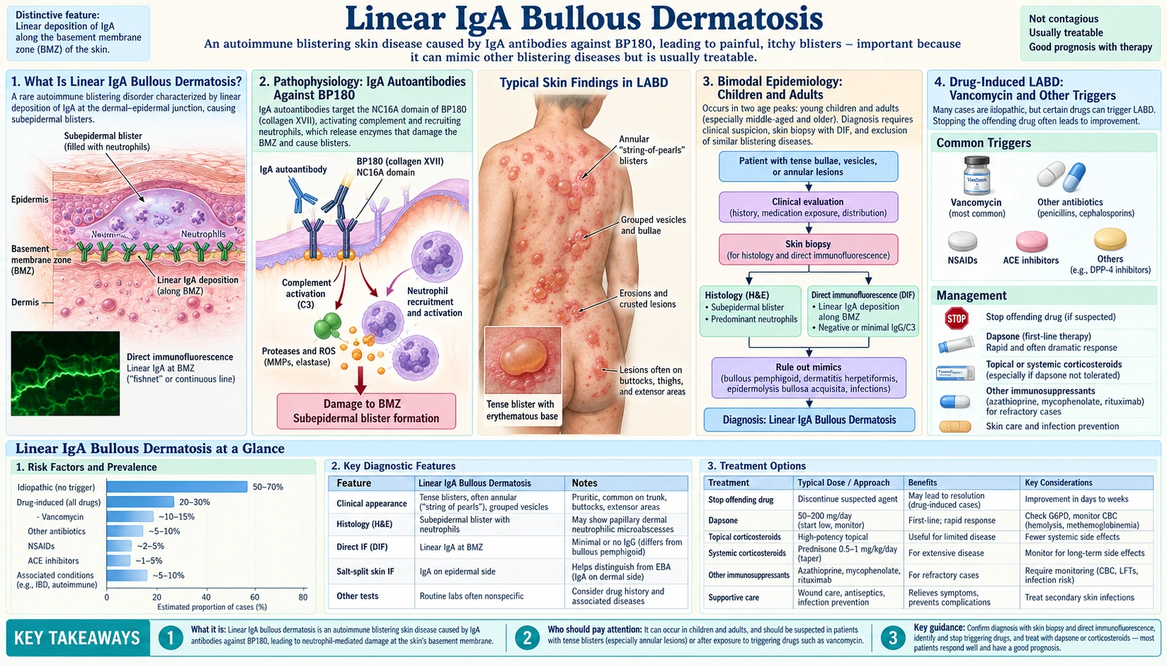
Linear immunoglobulin A (IgA) bullous dermatosis (LABD) is a rare autoimmune blistering disease defined by a single diagnostic hallmark: linear deposits of immunoglobulin A along the basement membrane zone on direct immunofluorescence. It affects two distinct age groups — young children and older adults — and has an important drug-induced form triggered most classically by vancomycin. Understanding LABD means understanding how immunoglobulin A autoantibodies attack the structural protein BP180, and why the pattern of that attack is the key to diagnosis.
Table of Contents
- Linear Immunoglobulin A Bullous Dermatosis Explained
- Pathophysiology: Immunoglobulin A Autoantibodies Against BP180
- Bimodal Epidemiology: Children and Adults
- Drug-Induced LABD: Vancomycin and Other Triggers
- Clinical Features and "Cluster of Jewels" Pattern
- Diagnosis: Linear Immunoglobulin A on Direct Immunofluorescence
- Treatment Approach
- Mucosal and Ocular Complications
- Key Research Papers
- Related PubMed Searches
- Connections
- Featured Videos
Linear Immunoglobulin A Bullous Dermatosis Explained
Linear immunoglobulin A bullous dermatosis is an autoimmune subepidermal blistering disease in which the immune system produces immunoglobulin A class antibodies directed against proteins at the dermoepidermal junction. The disease gets its name from its defining immunofluorescence pattern: a smooth, linear band of immunoglobulin A running along the basement membrane zone — the structural boundary between the outer layer of skin (epidermis) and the deeper connective tissue (dermis).
The disease exists under several names that reflect its history and the populations it affects. In children it is often called linear immunoglobulin A disease of childhood (LAD) or chronic bullous disease of childhood (CBDC). In adults the term linear immunoglobulin A bullous dermatosis is standard. Both are the same fundamental condition: immunoglobulin A-mediated destruction of the basement membrane zone leading to separation of the epidermis from the dermis and the formation of fluid-filled blisters.
What sets LABD apart from other autoimmune blistering diseases is the immunoglobulin class involved. Most subepidermal blistering diseases — including bullous pemphigoid and mucous membrane pemphigoid — are driven by immunoglobulin G (IgG) autoantibodies. LABD is defined by immunoglobulin A. This distinction matters clinically because immunoglobulin A activates different inflammatory pathways, recruits neutrophils rather than eosinophils as the dominant effector cell, and responds to different treatments, particularly dapsone.
Pathophysiology: Immunoglobulin A Autoantibodies Against BP180
The primary autoantigen in LABD is BP180 (also called BPAG2 or collagen XVII), a transmembrane glycoprotein that is a critical structural component of hemidesmosomes — the attachment complexes that anchor the basal keratinocytes of the epidermis to the underlying basement membrane. BP180 spans from inside the basal keratinocyte through the basement membrane into the dermis, making it a keystone of the epidermal-dermal connection.
In LABD, IgA autoantibodies target several proteins of the basement membrane zone. A 2020 Italian study of 54 patients lists the main ones as LAD-1, LABD97, BP180, BP230, type VII collagen and laminin 332. Two of the best-studied targets are:
- LAD285 — a 285 kDa antigen that is separate from BP180. In a 2003 immunoblot study of serum from 63 adults and 34 children, LAD285 and a 180 kDa protein (probably BP180) were the major dermal antigens identified, and a 2001 report of vancomycin-induced cases found immunoglobulin A antibodies to both LAD285 and BP180.
- LAD97 (also written LABD97) — a 97 kDa proteolytic fragment shed from the extracellular portion of BP180, studied together with the related fragment LAD-1. In the 2020 Italian study, immunoglobulin A to LAD-1 was found in 52% of sera, and LAD-1 was a major antigen of the lamina lucida subtype; in a 2017 French series of 72 adults, the positive immunoblots were mostly for LAD-1/LABD97 (44%) and full-length BP180 (33%).
The sequence of tissue damage follows a well-characterized pathway. Immunoglobulin A autoantibodies bind to these BP180 epitopes along the basement membrane zone, creating immune complexes. These complexes activate complement via the alternative or lectin pathways, generating chemoattractant fragments (C3a, C5a) that recruit neutrophils to the dermoepidermal junction. Neutrophil degranulation releases proteases and reactive oxygen species that degrade the extracellular matrix and structural proteins of the basement membrane zone, ultimately severing the epidermal-dermal bond and allowing fluid to accumulate in the subepidermal space as a blister.
The resulting blisters are subepidermal — they form beneath the entire epidermis rather than within it. This explains why LABD blisters are tense and intact (the full thickness of epidermis forms the blister roof), unlike the fragile flaccid blisters of pemphigus diseases, which split within the epidermis and collapse easily.
The linear distribution of immunoglobulin A deposits along the basement membrane zone — as opposed to the granular pattern seen in dermatitis herpetiformis — reflects the continuous nature of BP180 distribution along hemidesmosomes rather than a localized accumulation in dermal papillae.
Bimodal Epidemiology: Children and Adults
LABD has a distinctive bimodal age distribution that is one of its epidemiological signatures. It is not a disease that smoothly affects all ages — it clusters in two peaks separated by decades of relative rarity in between.
The childhood peak occurs between ages 2 and 5 years. This form — often called linear immunoglobulin A disease of childhood or chronic bullous disease of childhood — tends to be self-limited. The 2026 European S2k guideline on autoimmune blistering diseases in children and adolescents reports that linear IgA disease is the most common of these diseases in children (42–83% of cases) and that the way it is diagnosed does not differ from adults. Most affected children achieve spontaneous remission by puberty, though the disease can persist longer in some individuals. The childhood form has a characteristic distribution involving the perineal and genital area, which helps distinguish it from other childhood blistering conditions.
The adult peak occurs in the sixth and seventh decades of life (ages 60–70 years). Adult LABD tends to be more chronic and relapsing than the childhood form. It affects both sexes and all ethnicities, without a strong sex predilection (unlike many other autoimmune diseases which disproportionately affect women).
In adults, LABD is associated with several systemic conditions:
- Inflammatory bowel disease — both Crohn's disease and ulcerative colitis have been reported in association with adult LABD. The shared mucosal immunoglobulin A immune system may explain this link.
- Hematologic malignancies — chronic lymphocytic leukemia (CLL) and lymphoma have been found more frequently in adult LABD patients than in the general population. In a 2024 study of 81 adults at one large US referral centre, 12.3% had a cancer and 11.1% had inflammatory bowel disease; the authors write that screening for an underlying cancer, a triggering medicine and possibly inflammatory bowel disease may be advisable, particularly when LABD is newly diagnosed.
- Other autoimmune diseases — occasional associations with rheumatoid arthritis, thyroid disease, and other autoimmune conditions have been reported.
In the same 2024 study, drug-induced and cancer-associated cases lasted far shorter (a median of 1.2 and 1.7 months) than idiopathic LABD (48.8 months), and idiopathic disease came back more often (76.1% of cases, against 11.5% of drug-induced and 33.3% of cancer-associated cases).
LABD is rare overall, with estimates in the range of 0.5–1 per million population per year in Western countries, though exact incidence figures vary by geography and whether drug-induced cases are included. A 2024 Korean study of the nationwide health insurance database identified 670 cases between 2010 and 2022 and an annual incidence of 1.3 per 100,000 people, higher than in other studies; the disease was more common at age 60 and over, and use of the antibiotics vancomycin and third-generation cephalosporins was a risk factor.
Drug-Induced LABD: Vancomycin and Other Triggers
A clinically important subset of LABD is triggered by drugs. Drug-induced LABD can be clinically and histologically indistinguishable from idiopathic LABD — the same linear immunoglobulin A deposits on immunofluorescence, the same tense blisters, even similar mucosal involvement in some cases. The key diagnostic clue is the temporal relationship between drug initiation and blister onset.
Vancomycin is the classic and most common trigger — this is a high-yield teaching point in dermatology and internal medicine. Hospitalized patients receiving vancomycin for serious infections can develop LABD within days to weeks of starting the antibiotic. The blistering can be severe and widespread, sometimes mimicking Stevens-Johnson syndrome or toxic epidermal necrolysis in its extent. Stopping vancomycin typically leads to resolution within weeks to months.
In a 2019 French pharmacovigilance study of 69 drug-induced cases, vancomycin was suspected in 39 (57%), 20% of patients had lesions mimicking toxic epidermal necrolysis, and of the 60 patients with follow-up, 52 reached remission and 4 died. A 2025 analysis of the US Food and Drug Administration (FDA) Adverse Event Reporting System (2004–2024) found 1,394 reports of LABD, with vancomycin (559 reports) and amoxicillin (58 reports) the most frequently reported drugs.
Other documented drug triggers include:
- Amoxicillin-clavulanate — the most common antibiotic trigger after vancomycin
- Nonsteroidal anti-inflammatory drugs (NSAIDs) — particularly diclofenac and naproxen
- Captopril — angiotensin-converting enzyme inhibitor (ACE inhibitor)
- Lithium — mood stabilizer
- Phenytoin — anticonvulsant
- Furosemide — loop diuretic
- Atorvastatin, cephalosporins, trimethoprim-sulfamethoxazole — less commonly reported
The mechanism of drug-induced LABD is not fully established. Proposed mechanisms include drug-induced hapten formation on BP180 or related proteins, alteration of BP180 antigenicity, or direct stimulation of autoreactive immunoglobulin A-producing B cells.
When evaluating a patient with new-onset LABD, a thorough medication review is essential. The first and most important step in drug-induced LABD is identifying and stopping the causative drug. In many cases this alone is sufficient treatment, though systemic immunosuppression may be needed while waiting for resolution if the blistering is extensive or mucosal involvement is present.
Clinical Features and "Cluster of Jewels" Pattern
The primary lesions of LABD are tense vesicles and bullae — fluid-filled blisters that arise on skin that may appear normal, erythematous, or urticarial. Because the blister is subepidermal (the full epidermal thickness forms the roof), the blisters are tense and resistant to rupture, unlike the fragile, easily broken blisters of pemphigus diseases where the split occurs within the epidermis.
A characteristic but not universally present arrangement is the "cluster of jewels" or rosette pattern: new small vesicles or bullae arrange themselves in a ring or arc around a central older bulla or healing crust. This peripheral extension of blistering around a central lesion gives the cluster-of-jewels appearance that dermatologists recognize as suggestive of LABD, though it can also be seen in dermatitis herpetiformis and bullous pemphigoid.
The distribution of lesions varies by age:
- Adults: trunk, buttocks, inner thighs, and periumbilical area are most commonly affected. Lesions may also appear on the scalp, face, and extremities. The distribution can resemble bullous pemphigoid.
- Children: the genital and perineal area is classically involved, which is an important distinguishing feature from other childhood blistering diseases. Buttocks, inner thighs, and lower abdomen are also common sites.
- Drug-induced LABD: often presents with widespread, rapidly developing bullae that may cover large body surface areas. A 2019 French study of 69 drug-induced cases found no major clinical difference from idiopathic LABD, except that more drug-induced cases (20%) had lesions mimicking toxic epidermal necrolysis.
Lesions heal with postinflammatory hyperpigmentation or milia (small keratin cysts) but typically without scarring on non-mucosal skin. However, mucosal surfaces — particularly the conjunctiva — can develop permanent scarring that progresses even after the skin disease is controlled.
Pruritus (itching) is variable. Some patients have significant itch, similar to dermatitis herpetiformis; others have minimal pruritus despite extensive blistering.
Diagnosis: Linear Immunoglobulin A on Direct Immunofluorescence
The diagnosis of LABD rests on a combination of clinical presentation and immunopathological testing. No single clinical finding is pathognomonic, but the immunofluorescence pattern is.
Skin biopsy for direct immunofluorescence (DIF) is the cornerstone of diagnosis. The biopsy is taken from perilesional skin — normal-appearing skin immediately adjacent to a blister, not from within the blister itself. The DIF finding that defines LABD is:
- Linear immunoglobulin A deposits along the basement membrane zone — smooth, continuous, linear fluorescence running along the dermoepidermal junction
- Immunoglobulin G and C3 may be present but do not predominate
- On direct immunofluorescence microscopy the line is n-serrated, the pattern found in all pemphigoid diseases except epidermolysis bullosa acquisita (which shows a u-serrated pattern). The exact layer varies: immunoelectron microscopy of biopsies from 31 patients in the 2017 French series placed the immunoglobulin A in the lamina lucida (26%), lamina densa (23%), anchoring-fibril zone (19%) or both lamina lucida and anchoring-fibril zone (23%).
Understanding how LABD differs from the other immunoglobulin A-mediated blistering disease — dermatitis herpetiformis (DH) — is essential:
- LABD: LINEAR IgA along the basement membrane zone
- Dermatitis herpetiformis: GRANULAR IgA in the tips of the dermal papillae — a completely different pattern reflecting immune complex deposition in skin away from the dermoepidermal junction
This distinction is pathognomonic and diagnostic. The pattern cannot be mimicked by any other disease and directly defines which condition you are dealing with.
Comparing LABD with bullous pemphigoid (BP):
- Bullous pemphigoid: linear immunoglobulin G + C3 along the basement membrane zone (not immunoglobulin A)
- LABD: linear immunoglobulin A along the basement membrane zone (not immunoglobulin G as the dominant immunoglobulin)
Additional diagnostic tests include:
- Indirect immunofluorescence (IIF) using salt-split skin: serum from LABD patients contains circulating immunoglobulin A antibodies that bind to the roof (epidermal side) of the artificial split created by incubating skin in sodium chloride solution. This epidermal-side binding pattern, with immunoglobulin A as the immunoglobulin class, is characteristic of LABD.
- Enzyme-linked immunosorbent assay (ELISA) for anti-BP180 immunoglobulin A antibodies: commercially available; useful for monitoring disease activity and confirming the diagnosis.
- Histology (hematoxylin and eosin, H&E, staining): shows a subepidermal blister with a predominantly neutrophilic infiltrate at the dermoepidermal junction. Eosinophils may also be present, sometimes making histology overlap with bullous pemphigoid. The neutrophil predominance aligns with the immunoglobulin A-driven mechanism (immunoglobulin A is a potent neutrophil activator via FcαRI).
Treatment Approach
Treatment of LABD is guided by whether the disease is drug-induced or idiopathic, its severity, and the presence or absence of mucosal involvement.
Step 1: Removing the causative drug — In drug-induced LABD, identifying and stopping the offending medication is the single most important intervention. Most drug-induced cases resolve within weeks to months after drug withdrawal, though systemic therapy may be needed to manage severe or widespread blistering in the interim.
Dapsone (first-line for idiopathic LABD): Dapsone, a sulfone antibiotic with potent anti-inflammatory and anti-neutrophil properties, is the mainstay of treatment for idiopathic LABD. Responses can be dramatic and rapid — often within days — resembling the dapsone response in dermatitis herpetiformis. Typical doses range from 50 to 200 mg daily. A 2012 review names dapsone and sulfapyridine as the two common therapies, which reduce the inflammatory response and bring remission over a variable period of time.
- Testing before dapsone: people are tested for glucose-6-phosphate dehydrogenase (G6PD) deficiency before dapsone is started. Patients with glucose-6-phosphate dehydrogenase deficiency cannot tolerate dapsone because the drug causes oxidative hemolysis in red blood cells lacking this enzyme. Glucose-6-phosphate dehydrogenase-deficient patients can develop life-threatening hemolytic anemia on dapsone.
- Known side effects: hemolytic anemia (even in glucose-6-phosphate dehydrogenase-normal patients at higher doses), methemoglobinemia, peripheral neuropathy (rare, with long-term use), agranulocytosis (rare but serious), and dapsone hypersensitivity syndrome.
- Dapsone hypersensitivity syndrome develops in about 0.5 to 3.6% of people treated with dapsone and has a reported mortality of 9.9%. A 2013 genetic study of people given dapsone for leprosy found that the gene variant HLA-B*13:01 was a strong risk factor (odds ratio 20.53). This variant is present in about 2 to 20% of Chinese people, 1.5% of Japanese, 1 to 12% of Indians and 2 to 4% of Southeast Asians, but is largely absent in Europeans and Africans.
Systemic corticosteroids (prednisone 0.5–1 mg/kg/day): used when dapsone is contraindicated (glucose-6-phosphate dehydrogenase deficiency), when the response to dapsone is incomplete, or when mucosal involvement requires rapid control. Often used in combination with dapsone for severe disease.
Colchicine: Used as an alternative to dapsone and valued by some clinicians for mucosal disease, although a 2025 systematic review of 650 articles covering 1,627 cases found that colchicine and amoxicillin-clavulanate had lower response rates overall. Colchicine inhibits neutrophil chemotaxis and degranulation, targeting the downstream mechanism of LABD. It may be better tolerated than dapsone in some patients.
Trimethoprim-sulfamethoxazole (TMP-SMX): Effective for mild cases and particularly useful in childhood LABD. The mechanism may relate to its anti-inflammatory properties or the sulfamethoxazole component (structurally related to dapsone).
Refractory or severe disease:
- Mycophenolate mofetil: steroid-sparing immunosuppressant; well-tolerated long-term
- Azathioprine: traditional second-line immunosuppressant; thiopurine methyltransferase (TPMT) enzyme levels are checked before it is started
- Intravenous immunoglobulin (IVIg): useful for rapid disease control in severe or refractory LABD; expensive and requires intravenous (IV) access
- Rituximab: anti-CD20 monoclonal antibody that depletes B cells; increasingly used for refractory autoimmune blistering diseases including LABD; growing evidence base
Childhood LAD often responds well to dapsone or TMP-SMX and frequently enters spontaneous remission by puberty, allowing treatment to be tapered and discontinued.
The same 2025 systematic review found that dapsone was the primary therapy, while biologic drugs were reported with higher complete-response rates; intravenous immunoglobulin and mycophenolate mofetil had comparable responses in childhood cases but lower response rates in adult LABD.
Over the long term, a 2017 French series of 72 adults with idiopathic LABD, followed for a median of 39 months, found that 36% reached lasting complete remission, 29% relapsed and 35% had chronic disease. People under 70 years of age with mucous-membrane involvement were the most likely to have a chronic course.
Mucosal and Ocular Complications
Mucosal involvement is common — 60% of 72 adults with idiopathic LABD in the 2017 French series had mucous-membrane involvement, as did 42% of 69 drug-induced cases in the 2019 French study — and represents the most clinically significant aspect of the disease beyond the skin blisters. Unlike skin blistering, which heals without permanent scarring on non-mucosal surfaces, mucosal involvement can lead to irreversible damage.
Oral mucosal involvement is the most common form of mucosal disease. Patients develop:
- Desquamative gingivitis — erythematous, painful, peeling gums
- Erosions and blisters of the buccal mucosa, palate, and tongue
- Difficulty eating, speaking, and maintaining oral hygiene
Genital mucosal involvement occurs in some patients, causing erosions, scarring, and significant discomfort.
Ocular involvement is the most dangerous complication of LABD. Conjunctival inflammation leads to:
- Conjunctival scarring — fibrous bands form in the conjunctival tissue
- Symblepharon — adhesion between the eyelid conjunctiva and the bulbar conjunctiva covering the eyeball
- Fornix shortening — loss of the conjunctival fold depth due to progressive scarring
- Ankyloblepharon — in advanced cases, complete fusion of eyelids
- Corneal damage — scarring and vascularization of the cornea leading to visual impairment
- Blindness — the end-stage of uncontrolled ocular LABD
This progressive ocular scarring places LABD in the same risk category as mucous membrane pemphigoid (ocular cicatricial pemphigoid), which is well-recognized as a blinding disease. Unlike skin lesions, ocular scarring in LABD can progress even when systemic disease appears controlled. Cicatricial (scarring) changes in the conjunctiva are irreversible once established.
Management of ocular LABD:
- Eye symptoms in a person with LABD — redness, irritation, foreign-body sensation, visual changes — are signs that eye specialists (ophthalmologists) treat as urgent
- Topical corticosteroid eye drops and lubricants provide symptomatic relief but do not stop the underlying immune damage
- Systemic immunosuppression is essential: dapsone alone is often insufficient for ocular disease; systemic corticosteroids plus a steroid-sparing agent (mycophenolate mofetil, azathioprine) are standard
- IVIg or rituximab may be required for progressive ocular disease despite conventional immunosuppression
- Scarring of the eye can progress without symptoms, even in apparently stable patients, which is why regular eye examinations are part of follow-up
Key Research Papers
- Amber KT, Murrell DF, Schmidt E, et al. "Autoimmune Subepidermal Bullous Diseases of the Skin and Mucosae: Clinical Features, Diagnosis, and Management." Clin Rev Allergy Immunol. 2018;54(1):26–51. — PubMed PMID: 28779299
- Fortuna G, Marinkovich MP. "Linear immunoglobulin A bullous dermatosis." Clin Dermatol. 2012;30(1):38–50. — PubMed PMID: 22137225
- Egan CA, Zone JJ. "Linear IgA bullous dermatosis." Int J Dermatol. 1999;38(11):818–827. — PubMed PMID: 10583613
- Palmer RA, Ogg G, Allen J, Banerjee A, Ryatt KS, Ratnavel R, Wojnarowska F. "Vancomycin-induced linear IgA disease with autoantibodies to BP180 and LAD285." Br J Dermatol. 2001;145(5):816–820. — PubMed PMID: 11736908
- Guide SV, Marinkovich MP. "Linear IgA bullous dermatosis." Clin Dermatol. 2001;19(6):719–727. — PubMed PMID: 11705681
- Nousari HC, Anhalt GJ. "Pemphigus and bullous pemphigoid." Lancet. 1999;354(9179):667–672. — PubMed PMID: 10466686
- Chorzelski TP, Jabłońska S, Maciejowska E. "Linear IgA bullous dermatosis of adults." Clin Dermatol. 1991;9(3):383–392. — PubMed PMID: 1806226
- Armstrong AW, Fazeli A, Yeh SW, et al. "Vancomycin-induced linear IgA disease manifesting as bullous erythema multiforme." J Cutan Pathol. 2004;31(5):393–397. — PubMed PMID: 15059226
- Wojnarowska F, Marsden RA, Bhogal B, Black MM. "Chronic bullous disease of childhood, childhood cicatricial pemphigoid, and linear IgA disease of adults. A comparative study demonstrating clinical and immunopathologic overlap." J Am Acad Dermatol. 1988;19(5 Pt 1):792–805. — PubMed PMID: 3056993
- Allen J, Wojnarowska F. "Linear IgA disease: the IgA and IgG response to dermal antigens demonstrates a chiefly IgA response to LAD285 and a dermal 180-kDa protein." Br J Dermatol. 2003;149(5):1055–1058. — PubMed PMID: 14632815
- Cozzani E, Di Zenzo G, Gasparini G, et al. "Autoantibody Profile of a Cohort of 54 Italian Patients with Linear IgA Bullous Dermatosis: LAD-1 Denoted as a Major Auto-antigen of the Lamina Lucida Subtype." Acta Derm Venereol. 2020;100(4):adv00070. — PubMed PMID: 32011724
- Gottlieb J, Ingen-Housz-Oro S, Alexandre M, et al. "Idiopathic linear IgA bullous dermatosis: prognostic factors based on a case series of 72 adults." Br J Dermatol. 2017;177(1):212–222. — PubMed PMID: 27995619
- Holtsche MM, van Beek N, Künstner A, et al. "Diagnostic Value and Practicability of Serration Pattern Analysis by Direct Immunofluorescence Microscopy in Pemphigoid Diseases." Acta Derm Venereol. 2021;101(3):adv00410. — PubMed PMID: 33491096
- Garel B, Ingen-Housz-Oro S, Afriat D, et al. "Drug-induced linear immunoglobulin A bullous dermatosis: A French retrospective pharmacovigilance study of 69 cases." Br J Clin Pharmacol. 2019;85(3):570–579. — PubMed PMID: 30511379
- Wang KL, Lehman JS, Todd A, Davis DMR. "Adult-onset linear IgA bullous dermatosis: a retrospective single-center cohort study of 81 patients and literature review." Int J Dermatol. 2024;63(7):936–941. — PubMed PMID: 38251749
- Kim YR, Kim JH, Kim SW, Lee JM, Bae JS. "Linear IgA Bullous Dermatosis in Korea Using the Nationwide Health Insurance Database." J Clin Med. 2024;13(4):1159. — PubMed PMID: 38398470
- Nanda A, Tedbirt B, Bodemer C, et al. "European S2k guidelines on management of autoimmune blistering diseases in children and adolescents." J Eur Acad Dermatol Venereol. 2026;40(7):1137–1161. — PubMed PMID: 41678328
- Mar K, Landells F, Khalid B, et al. "Clinical characteristics and treatment outcomes of linear IgA bullous dermatosis." J Dtsch Dermatol Ges. 2025;23(5):576–585. — PubMed PMID: 40018881
- Zhang FR, Liu H, Irwanto A, et al. "HLA-B*13:01 and the dapsone hypersensitivity syndrome." N Engl J Med. 2013;369(17):1620–1628. — PubMed PMID: 24152261
- Yang Y, Xie H, Liu S, et al. "Linear IgA bullous dermatosis secondary to drugs: a real-world pharmacovigilance study of the FDA adverse event reporting system." Front Med (Lausanne). 2025;12:1521697. — PubMed PMID: 39917062
Related PubMed Searches
- Linear immunoglobulin A bullous dermatosis treatment — PubMed
- Vancomycin-induced linear immunoglobulin A disease — PubMed
- Chronic bullous disease of childhood linear immunoglobulin A — PubMed
- BP180 LAD97 autoantibody subepidermal blistering — PubMed
- Linear immunoglobulin A bullous dermatosis ocular complications — PubMed
Connections
- Dermatology
- Dermatitis Herpetiformis — the granular immunoglobulin A counterpart: distinguish by DIF pattern (granular papillary vs linear BMZ)
- Epidermolysis Bullosa — inherited subepidermal blistering from structural protein mutations vs autoimmune in LABD
- Pemphigoid Gestationis — pregnancy-associated subepidermal blistering, immunoglobulin G-mediated, same target antigen BP180
- Celiac Disease — gluten-sensitive enteropathy with immunoglobulin A autoimmunity; associated with dermatitis herpetiformis
- Gastroenterology Conditions — inflammatory bowel disease (IBD) associations including Crohn's disease and ulcerative colitis in adult LABD