Peripheral Arterial Disease
Interactive Visualization Nitric Oxide — make an artery relax Watch the endothelium turn L-arginine into nitric oxide and widen the artery — then let oxidative stress uncouple eNOS and stiffen it, or pour in beet nitrate to rescue the flow. Launch →
Table of Contents
- Overview
- Epidemiology
- Pathophysiology
- Risk Factors
- Clinical Spectrum
- Ankle-Brachial Index (ABI)
- Classification Systems
- Diagnosis
- Medical Management
- Supervised Exercise and Lifestyle
- Revascularization and Critical Limb Ischemia
- Key Research Papers
- Connections
- Featured Videos
1. Overview
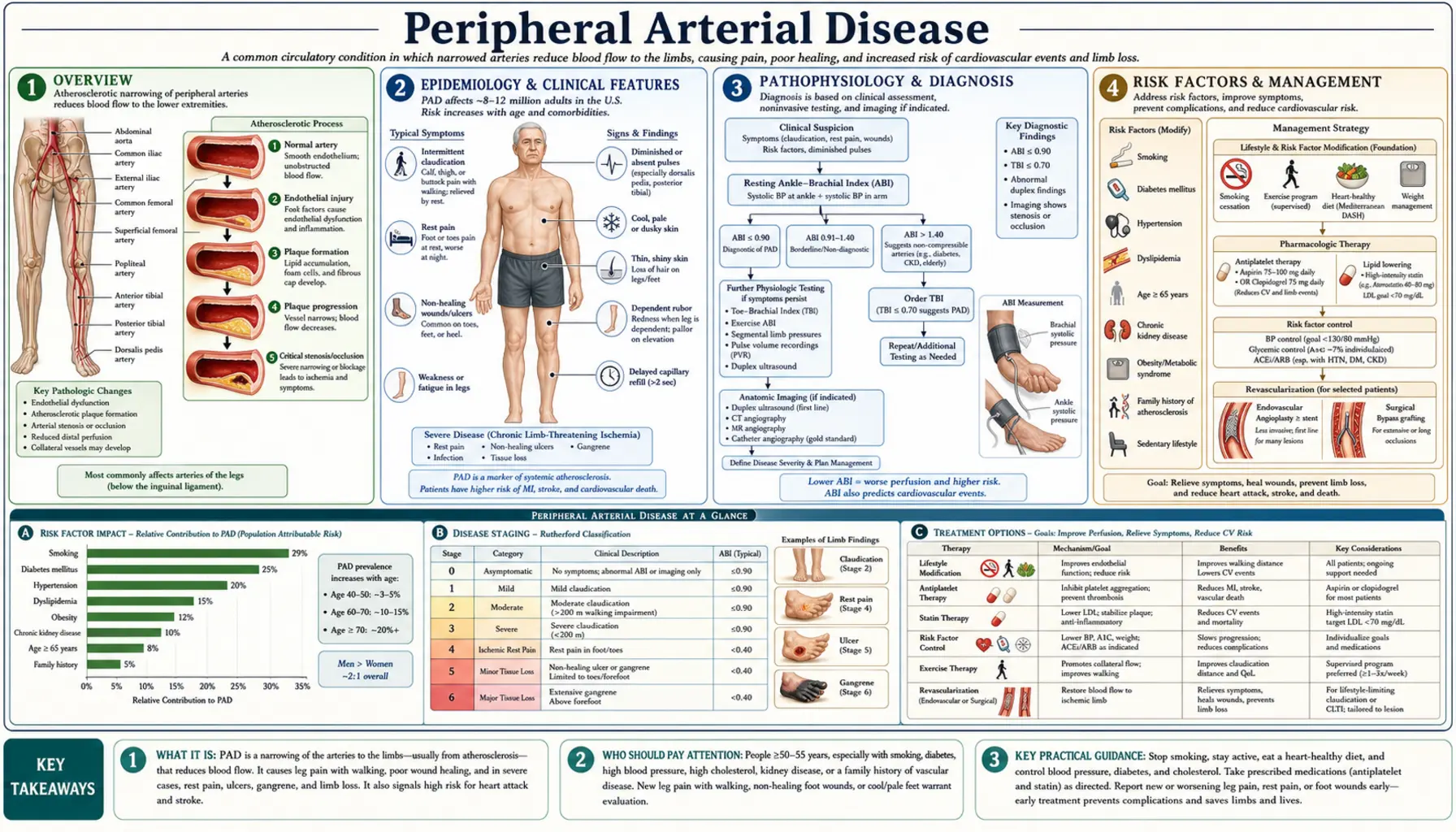
Peripheral arterial disease (peripheral artery disease, PAD) is a manifestation of systemic atherosclerosis in which plaque accumulation narrows the lumen of arteries supplying the extremities — predominantly the lower limbs — reducing blood flow and oxygen delivery to the muscles and tissues downstream. Peripheral artery disease is the third leading cause of atherosclerotic cardiovascular morbidity worldwide, after coronary artery disease and stroke, yet it remains substantially underdiagnosed and undertreated relative to those better-known conditions.
The defining pathological process is atherosclerotic stenosis or occlusion of the aortoiliac, femoropopliteal, or tibial/peroneal arterial segments. As plaques enlarge and calcify over decades, the cross-sectional lumen narrows until blood flow can no longer meet the metabolic demands of exercising muscle — producing the hallmark symptom of intermittent claudication. In its most severe form, peripheral artery disease reduces flow so critically that even resting tissues cannot receive adequate oxygen, causing critical limb-threatening ischemia (CLTI) — rest pain, non-healing wounds, and gangrene that may necessitate limb amputation.
What makes peripheral artery disease particularly important beyond the limb itself is its role as a systemic vascular risk marker. Patients with peripheral artery disease have a 3- to 6-fold increased risk of myocardial infarction and a 2- to 4-fold increased risk of stroke compared with people without peripheral artery disease, because the same atherosclerotic process that narrows leg arteries is simultaneously advancing in the coronary and carotid circulations. Approximately 60% of symptomatic peripheral artery disease patients have significant coronary artery disease (CAD), and a similar fraction have associated cerebrovascular disease. The 5-year cardiovascular mortality in patients with peripheral artery disease exceeds that of many cancers — roughly 20–30% in symptomatic patients.
Despite this burden, peripheral artery disease is often diagnosed late, partially because the majority of patients — an estimated 50–80% — are asymptomatic or have atypical symptoms misattributed to arthritis, lumbar radiculopathy, or normal aging. Systematic use of the ankle-brachial index (ABI), a simple, inexpensive, non-invasive hemodynamic test, can identify peripheral artery disease long before limb-threatening complications develop. Earlier recognition enables aggressive cardiovascular risk-factor modification, supervised exercise therapy, antiplatelet and statin therapy, and — when required — timely revascularization to preserve the limb.
2. Epidemiology
Peripheral artery disease affects an estimated 8–12 million Americans and approximately 236 million people worldwide — making it one of the most prevalent cardiovascular conditions globally. A landmark 2013 analysis by Fowkes et al. ( PubMed PMID: 23915883) counted 202 million people living with peripheral artery disease worldwide in 2010, 69.7% of them in low- and middle-income countries. Over the preceding decade the number of people with the disease rose by 28.7% in low- and middle-income countries and by 13.1% in high-income countries. Prevalence climbed with age: in high-income countries it was about 5.3–5.4% at ages 45–49 and about 18.4–18.8% at ages 85–89. Current smoking carried an odds ratio of 2.72 in high-income countries and 1.42 in low- and middle-income countries.
The newest worldwide estimate, a 2019 update by Song et al. ( PubMed PMID: 31303293) using 2015 data, counts 236.62 million people aged 25 and older with peripheral artery disease — a global prevalence of 5.56% (7.37% in high-income countries and 5.09% in low- and middle-income countries), with 72.91% of all cases in low- and middle-income countries. At ages 80–84, prevalence reached 21.24% in high-income countries and 12.04% in low- and middle-income countries.
Age is the dominant demographic predictor. Peripheral artery disease prevalence is under 3% in individuals below age 50, rises to 8–10% in those aged 60–69, and reaches 15–20% in adults over age 70. The dramatic age-dependence reflects the decades-long accumulation of atherosclerotic plaque — peripheral artery disease is largely a disease of cumulative vascular injury. Men have a modestly higher overall prevalence than women in most high-income population studies, though sex differences attenuate at older ages. Notably, women with peripheral artery disease are more likely to have atypical or absent claudication, leading to delayed diagnosis.
Racial and ethnic disparities are pronounced. Non-Hispanic Black Americans have a peripheral artery disease prevalence approximately twice that of non-Hispanic White Americans — a disparity driven by higher rates of hypertension, diabetes, smoking, and chronic kidney disease, as well as possible genetic and social determinants of health. Hispanic Americans have intermediate prevalence. These disparities are compounded by differential access to vascular specialty care; Black patients with CLTI have substantially higher rates of major limb amputation and lower rates of revascularization compared with White patients, even after adjusting for disease severity.
Geographic variation is substantial. Prevalence is highest in South and Southeast Asia and sub-Saharan Africa in absolute numbers due to population size and rising diabetes epidemics. In the United States, the Southeast — the "Stroke Belt" — has disproportionately high peripheral artery disease burden, correlating with regional patterns of smoking, obesity, and diabetes. Urban-rural disparities also exist, with rural populations having higher prevalence and lower access to vascular imaging and intervention.
Economic burden: peripheral artery disease imposes enormous healthcare costs. In the US alone, peripheral artery disease-related hospitalizations exceed 1 million annually, and the estimated direct and indirect costs exceed $21 billion per year. Patients with CLTI requiring hospitalization, endovascular or surgical revascularization, wound care, and amputation rehabilitation represent a disproportionate share of this cost. Amputation — the endpoint that peripheral artery disease management aims to prevent — carries a 1-year mortality of 50–70% in CLTI patients, comparable to many advanced cancers.
3. Pathophysiology
The pathophysiology of peripheral artery disease is fundamentally that of atherosclerosis in the peripheral circulation, with several features distinguishing it from coronary or cerebrovascular disease: the predominance of calcification, the central role of diabetes in accelerating tibial disease, and the unique physiology of skeletal muscle ischemia under exercise versus rest conditions.
Atherosclerotic Plaque Development
The process begins with endothelial dysfunction — a disruption of the normal anti-inflammatory, antithrombotic, and vasodilatory properties of the arterial endothelium. Shear stress disturbances at arterial bifurcations and curvatures (particularly the aortoiliac bifurcation, the femoral bifurcation in the groin, and the popliteal trifurcation behind the knee) promote the expression of adhesion molecules and chemokines, attracting monocytes into the subintimal space. There, monocytes differentiate into macrophages and engulf oxidized low-density lipoprotein (LDL) particles to form lipid-laden foam cells — the cellular hallmark of early atherosclerosis.
Over decades, foam cell accumulation, smooth muscle cell migration from the media, extracellular matrix deposition, and neovascularization build a progressively enlarging fibrous plaque that protrudes into the arterial lumen. The plaque's fibrous cap separates its lipid-rich, necrotic core from the flowing bloodstream. Plaque rupture or erosion exposes the prothrombotic core, triggering platelet aggregation and acute thrombus formation — a mechanism that, in the coronary circulation, produces myocardial infarction, and in the peripheral circulation, can produce acute limb ischemia.
Calcification and Medial Sclerosis
In the peripheral vasculature — particularly the tibial arteries of diabetic and chronic kidney disease (CKD) patients — medial calcification (Monckeberg's sclerosis) occurs alongside or instead of intimal atherosclerosis. In medial sclerosis, calcium deposits accumulate within the arterial media, hardening the wall without necessarily obstructing the lumen. This calcification renders vessels non-compressible and falsely elevates ankle pressures, producing spuriously normal or high ABI readings (>1.40) that underestimate true hemodynamic impairment. Toe-brachial index (TBI) measurement — using photoplethysmography at the toe, where digital arteries are less affected by medial calcification — is essential in these patients.
Exercise Physiology and Claudication
At rest, even significantly stenosed arteries may provide adequate blood flow to meet the low oxygen demands of resting muscle. During exercise, however, skeletal muscle oxygen demand increases 10- to 20-fold — a demand that requires a proportional increase in blood flow. Stenotic or occluded arteries cannot deliver this increment. Downstream, anaerobic metabolism increases, lactate and other metabolites accumulate in the interstitium, and pain receptors in the muscle are activated, producing the characteristic claudication pain — cramping or aching in the calf (most common), thigh, or buttock depending on the location of the occlusion.
Stopping exercise immediately relieves the ischemic demand; metabolites are cleared and pain resolves within 2–5 minutes of standing rest — a temporal pattern that distinguishes claudication from musculoskeletal or neurogenic causes of exertional leg pain. Walking uphill or faster shortens the distance to claudication onset; walking on flat ground more slowly extends it.
Collateral Circulation
The body responds to progressive arterial occlusion by arteriogenesis — the remodeling and enlargement of pre-existing collateral vessels stimulated by increased shear stress and local growth factors (VEGF, FGF). Effective collateral development can partially compensate for main-channel occlusion, explaining why some patients with complete femoral artery occlusion on imaging have only mild claudication while others with lesser stenoses have severe symptoms. Supervised exercise training is a potent stimulus for collateral development — it is the most effective intervention for claudication.
Critical Limb Ischemia
When stenoses progress to multiple-level disease with inadequate collateral compensation, resting blood flow falls below the threshold for tissue viability. The skin and subcutaneous tissues — especially of the toes and distal foot — begin to receive insufficient oxygen even without the added metabolic demand of exercise. The result is ischemic rest pain (typically burning, constant pain in the forefoot and toes, relieved partially by hanging the foot dependent to increase perfusion by gravity), ischemic ulceration at pressure points or minor trauma sites, and ultimately gangrene of digits or the foot. This stage — now termed CLTI (chronic limb-threatening ischemia) — represents end-stage peripheral artery disease and carries the highest risk of amputation and death.
4. Risk Factors
Peripheral artery disease shares the classic atherosclerotic risk factor profile with coronary artery disease and cerebrovascular disease, but the relative magnitudes differ importantly. Tobacco use and diabetes exert particularly strong effects in the peripheral circulation.
Tobacco Use (Strongest Modifiable Risk Factor)
Smoking is the single most powerful modifiable risk factor for peripheral artery disease, conferring a 2- to 4-fold increased risk of developing the disease. The risk is dose-dependent — heavier and longer-duration smokers have greater risk — and persists for years after cessation, though quitting substantially reduces progression and improves outcomes. Smoking accelerates atherosclerosis through endothelial dysfunction, oxidative stress, platelet activation, reduced prostacyclin production, and impaired fibrinolysis. Approximately 80% of patients with symptomatic peripheral artery disease are current or former smokers. In patients who continue to smoke after peripheral artery disease diagnosis, the risk of progressing to CLTI and requiring amputation is dramatically higher compared with those who quit.
Diabetes Mellitus (Strongest Risk Factor for Amputation)
Diabetes confers a 3- to 4-fold increased risk of peripheral artery disease and is the most important risk factor for major limb amputation once peripheral artery disease is established. Several mechanisms converge in diabetes to produce especially severe peripheral artery disease: accelerated tibial artery disease (the below-knee vessels are preferentially affected, making revascularization more technically challenging), peripheral neuropathy (which impairs pain perception and delays presentation), impaired wound healing from reduced neutrophil function and growth factor signaling, and medial calcification that renders vessels non-compressible. In one hospital series of 136 patients with peripheral arterial disease (Jude 2001), diabetic patients were five times more likely to have an amputation than non-diabetic patients (41.4% vs 11.5%; odds ratio 5.4) and their mortality was higher (51.7% vs 25.6%) — a stark inequality.
Hypertension
Hypertension is strongly associated with peripheral artery disease, with relative risk estimates of 1.5- to 2-fold in most population studies. The mechanism involves sustained wall stress, endothelial dysfunction from oxidative stress, and accelerated intimal plaque deposition. The association is particularly strong in African Americans, in whom hypertension is more prevalent, more severe, and more likely to be undertreated. Blood pressure control with a target below 130/80 mmHg reduces cardiovascular events in peripheral artery disease patients, though aggressive lowering below 120 mmHg in patients with CLTI may paradoxically reduce distal perfusion pressure and worsen ischemia.
Dyslipidemia
Elevated low-density lipoprotein cholesterol promotes plaque formation in peripheral arteries through the same mechanisms as in the coronary circulation. High-intensity statin therapy reduces peripheral artery disease progression, the rate of major adverse limb events (MALE), and systemic cardiovascular events. Low high-density lipoprotein (HDL) cholesterol and elevated triglycerides (features of the metabolic syndrome) are independently associated with peripheral artery disease risk. Beyond lipids, elevated Lp(a) — a genetically determined lipoprotein particle — is an emerging independent risk factor for peripheral artery disease, and emerging targeted therapies (inclisiran, PCSK9 inhibitors) may specifically reduce Lp(a)-associated vascular risk.
Chronic Kidney Disease
Chronic kidney disease amplifies peripheral artery disease risk substantially. Patients with an estimated glomerular filtration rate (eGFR) below 60 mL/min/1.73m² have a 2- to 3-fold higher peripheral artery disease prevalence than those with normal renal function, and this risk rises steeply with worsening kidney function. Chronic kidney disease promotes vascular calcification (through phosphate retention and disordered mineral metabolism), endothelial dysfunction, and a chronically inflamed, prothrombotic state. The combination of chronic kidney disease and diabetes is particularly devastating — such patients develop diffuse, heavily calcified tibial disease that is resistant to both endovascular and surgical revascularization.
Other Risk Factors
Additional established risk factors include age over 65 (or over 50 with one additional risk factor), male sex (though sex differences narrow at older age), elevated C-reactive protein (reflecting systemic inflammation), hyperhomocysteinemia (which impairs endothelial nitric oxide production), hypercoagulable states (antiphospholipid syndrome, factor V Leiden — relevant particularly for acute limb ischemia), and a personal or family history of coronary or cerebrovascular disease.
5. Clinical Spectrum
Peripheral artery disease presents across a wide clinical spectrum — from completely asymptomatic disease discovered incidentally, to classic intermittent claudication, to limb-threatening ischemia. Understanding this spectrum is essential for appropriate triage, counseling, and intervention decisions.
Asymptomatic Peripheral Artery Disease (50–80% of Cases)
The majority of individuals with peripheral artery disease — estimated at 50–80% in population studies using ABI-based definitions — have no classic claudication symptoms. Many are truly asymptomatic, while others have reduced their activity level to below the ischemic threshold without recognizing the cause. Still others have peripheral neuropathy (as in diabetes) that blunts the pain signal of ischemia. Asymptomatic peripheral artery disease still carries the full systemic cardiovascular risk of symptomatic disease — such patients have the same elevated rates of myocardial infarction (MI) and stroke — and the same risk-factor modification and antiplatelet/statin therapy used for symptomatic patients are applied to them.
Intermittent Claudication
Intermittent claudication (IC) is the hallmark symptomatic presentation of peripheral artery disease. The term derives from the Latin claudicare — to limp — reflecting the limping gait that develops as patients protect the ischemic limb during walking. Patients describe reproducible, predictable cramping, aching, tightening, or fatigue in specific muscle groups during walking, which resolves completely with 2–5 minutes of standing rest (not sitting, which is important diagnostically — true claudication does not require sitting).
The location of claudication symptoms maps to the level of arterial occlusion:
- Calf claudication (most common): occlusion in the superficial femoral or popliteal arteries.
- Thigh claudication: occlusion at the common femoral artery or proximal superficial femoral artery.
- Buttock and hip claudication (Leriche syndrome): aortoiliac occlusive disease; in men, this is classically accompanied by erectile dysfunction from reduced internal iliac artery flow.
- Foot claudication: tibial/peroneal disease, most common in diabetics.
The absolute claudication distance (ACD) — the distance walked before onset of pain — and the pain-free walking distance are key clinical metrics for monitoring symptom severity over time and assessing response to therapy. A standardized treadmill claudication test (typically 3.2 km/h at 12% grade) provides objective quantification.
Critical Limb-Threatening Ischemia (CLTI)
CLTI represents the severe end of peripheral artery disease and is defined by the triad of ischemic rest pain, non-healing wounds (ischemic ulcers or gangrene), and hemodynamic evidence of severe arterial insufficiency (ABI <0.40, ankle pressure <50 mmHg, or toe pressure <30 mmHg). Approximately 1–3% of symptomatic peripheral artery disease patients per year progress to CLTI; the annual incidence in the US is estimated at 150,000–200,000 patients.
Ischemic rest pain is a constant, burning, or gnawing pain in the toes and forefoot, typically worse at night when the foot is horizontal (gravity can no longer assist perfusion). Patients characteristically hang the foot dependently over the bed edge or sleep in a chair — this dependent position slightly increases hydrostatic pressure and marginally improves distal flow, providing partial relief.
Ischemic ulcers are a distinguishing feature of CLTI. They differ fundamentally from venous or neuropathic ulcers:
- Location: toes, toe web spaces, heel (pressure points), or sites of minor trauma. Venous ulcers occur at the medial malleolus; neuropathic ulcers at pressure points on the plantar surface.
- Appearance: pale, necrotic base with minimal or absent granulation tissue (reflecting absent perfusion); punched-out edges; minimal exudate. Venous ulcers have a moist, sloughy base with surrounding venous stasis changes (lipodermatosclerosis, hemosiderin staining).
- Pain: ischemic ulcers are typically very painful (unless neuropathy is present); venous ulcers are less painful and often relieved by leg elevation.
Without revascularization, CLTI carries a 1-year amputation rate of 25–30% and a 1-year mortality of 25% — a prognosis that rivals many advanced malignancies and underscores the urgency of expert vascular evaluation.
Acute Limb Ischemia
Acute limb ischemia (ALI) is a sudden reduction in limb perfusion that threatens tissue viability, classified as a separate syndrome from chronic peripheral artery disease though often occurring on a background of pre-existing atherosclerotic disease. The causes include acute thrombosis of a stenotic native artery, embolism (usually from the heart — AF, LV thrombus — or proximal arterial aneurysm), or bypass graft occlusion. The clinical hallmarks are the "6 Ps": Pain, Pallor, Pulselessness, Paresthesias, Paralysis, and Poikilothermia (coldness). Paresthesias and paralysis signal impending irreversible ischemia and represent a vascular emergency requiring revascularization within hours to avoid permanent limb loss.
6. Ankle-Brachial Index (ABI)
The ankle-brachial index (ABI) is the gold-standard diagnostic and screening test for peripheral artery disease — inexpensive, non-invasive, reproducible, and strongly predictive of both peripheral artery disease severity and systemic cardiovascular prognosis. It is the single most valuable tool in the initial evaluation of any patient suspected to have peripheral artery disease.
Measurement Technique
ABI is measured with the patient supine at rest for at least 5 minutes. A handheld Doppler probe (typically 8 MHz) is used to detect audible arterial flow signals while a standard blood pressure cuff is inflated and deflated above each measurement site. Systolic pressures are recorded at:
- Both brachial arteries (right and left)
- Both dorsalis pedis arteries (dorsum of foot)
- Both posterior tibial arteries (behind the medial malleolus)
The ABI for each leg is calculated as:
ABI = Highest ankle systolic pressure (dorsalis pedis or posterior tibial) / Highest brachial systolic pressure
The higher brachial pressure is used in the denominator to avoid falsely elevating ABI if one arm has a subclavian stenosis. Each leg is calculated separately; the lower of the two leg values is the diagnostic ABI for that patient.
Interpretation
- Normal: 1.00–1.40 — no significant arterial obstruction.
- Borderline: 0.91–0.99 — possible early peripheral artery disease; usually followed by repeat measurement or further evaluation, especially with symptoms or risk factors.
- Peripheral artery disease: <0.90 — diagnostic of significant arterial obstruction. This is the accepted diagnostic threshold in all major guidelines (AHA/ACC 2016, updated by the 2024 ACC/AHA multisociety guideline).
- Moderate peripheral artery disease: 0.50–0.89 — corresponds clinically to most patients with claudication.
- Severe peripheral artery disease: <0.50 — high risk of CLTI; rest pain and tissue loss are common at this level.
- Non-compressible vessels: >1.40 — indicates medial arterial calcification (common in diabetes and chronic kidney disease); ankle pressure is falsely elevated and does not reflect true hemodynamics. Requires alternative testing.
ABI as a Cardiovascular Risk Marker
Even mildly reduced ABI values in the range of 0.70–0.89 are associated with substantially increased cardiovascular mortality. A 2008 individual-participant meta-analysis by the Ankle Brachial Index Collaboration ( PubMed PMID: 18612117) of 24,955 men and 23,339 women across 16 population cohorts found that a low ABI (0.90 or below) predicts cardiovascular events beyond the Framingham risk score: it was associated with approximately twice the 10-year total mortality, cardiovascular mortality and major coronary event rate compared with the overall rate in each Framingham risk category. Adding the ABI would change the risk category, and the treatment recommendations, of about 19% of men and 36% of women.
Toe-Brachial Index (TBI)
When ankle vessels are non-compressible (ABI >1.40), the toe-brachial index (TBI) provides an accurate hemodynamic assessment; a 2005 study in people with and without diabetes found the TBI and qualitative waveform analysis more effective screening methods than the ABI and foot pulses, particularly in limbs with peripheral neuropathy ( PubMed PMID: 16123491). Digital arteries of the toes are rarely affected by medial calcification. TBI is measured with small pneumatic cuffs at the base of the first or second toe and photoplethysmography (PPG) to detect digital pulse waveforms. A TBI below 0.70 is diagnostic of hemodynamically significant peripheral artery disease. A toe pressure below 30 mmHg is a key threshold for CLTI.
Post-Exercise ABI
Patients with equivocal resting ABI (borderline range) may have normal resting pressures that fall significantly after exercise-induced ischemia. Post-exercise ABI (measured immediately after a standardized treadmill test) can unmask hemodynamically significant peripheral artery disease in symptomatic patients with otherwise normal resting values — a drop of >20 mmHg or >15% after exercise confirms the diagnosis.
7. Classification Systems
Two major classification systems are used clinically and in research to describe peripheral artery disease severity: the Fontaine classification (older, widely used in Europe) and the Rutherford classification (more granular, preferred in North America and most contemporary clinical trials).
Rutherford Classification
The Rutherford system grades peripheral artery disease from Category 0 to Category 6, combining subjective symptoms with objective hemodynamic findings from treadmill testing and segmental pressure measurements:
- Category 0: Asymptomatic. ABI normal at rest; normal treadmill performance.
- Category 1 (Mild claudication): Completes standard treadmill; ankle pressure after exercise >50 mmHg but at least 20 mmHg below brachial pressure.
- Category 2 (Moderate claudication): Between categories 1 and 3.
- Category 3 (Severe claudication): Cannot complete standard treadmill; ankle pressure after exercise <50 mmHg.
- Category 4 (Ischemic rest pain): Resting ankle pressure <40 mmHg; flat or barely pulsatile ankle or metatarsal pulse volume recording.
- Category 5 (Minor tissue loss): Resting ankle pressure <60 mmHg; non-healing ulcer or focal gangrene with diffuse pedal ischemia.
- Category 6 (Major tissue loss): Major tissue loss extending above the transmetatarsal level; functional foot no longer salvageable.
Fontaine Classification
- Stage I: Asymptomatic.
- Stage IIa: Mild claudication (walking distance >200 m).
- Stage IIb: Moderate to severe claudication (walking distance <200 m).
- Stage III: Ischemic rest pain.
- Stage IV: Ulceration or gangrene.
WIfI Classification for CLTI
The WIfI (Wound, Ischemia, foot Infection) classification, developed by the Society for Vascular Surgery, provides a more nuanced three-dimensional staging system specifically for CLTI that better predicts 1-year amputation risk and guides revascularization decision-making. It grades each dimension (Wound severity 0–3, Ischemia severity 0–3, Infection severity 0–3) independently, producing a composite clinical stage (1–4) that guides management intensity — from medical optimization alone in Stage 1 to urgent revascularization in Stage 4.
8. Diagnosis
The diagnostic workup for peripheral artery disease serves three goals: confirming the diagnosis, assessing anatomical disease extent and severity for revascularization planning, and evaluating systemic cardiovascular disease burden. Guidelines recommend a stepwise approach beginning with non-invasive testing.
History and Physical Examination
A thorough vascular history documents the presence, location, onset, and progression of claudication symptoms; risk factors (tobacco use, diabetes duration and control, hypertension, dyslipidemia, prior cardiovascular events); and functional status (maximum walking distance, activity limitations). Physical examination includes:
- Pulse examination: palpation of femoral, popliteal, dorsalis pedis, and posterior tibial pulses bilaterally. Absent or diminished pulses indicate significant proximal disease. However, pulse examination is insensitive and operator-dependent — normal pulses do not exclude peripheral artery disease.
- Bruit auscultation: femoral bruits at the groin suggest aortoiliac or proximal femoral stenosis. Their absence does not exclude disease.
- Skin and tissue assessment: skin temperature (cooler in ischemic limbs), hair loss on the dorsum of the foot and toes, thickened dystrophic nails, muscle wasting, skin atrophy, pallor on elevation, and dependent rubor (a sign of advanced ischemia — the foot turns deep red when dependent as maximally dilated arterioles fill from gravity).
ABI and Duplex Ultrasound
After ABI confirmation of peripheral artery disease, duplex ultrasound provides detailed anatomical mapping of disease extent and severity without radiation or contrast exposure. Duplex combines B-mode imaging with Doppler velocity measurements: a focal velocity ratio >2.0 at a stenosis corresponds to >50% diameter reduction. Duplex is highly accurate for femoropopliteal disease and is frequently sufficient for endovascular intervention planning, though its resolution decreases in heavily calcified vessels and in evaluating the aortoiliac segment in obese patients.
Computed Tomography Angiography (CTA)
CTA of the aorta and lower extremity arteries provides high-resolution three-dimensional anatomical mapping across the full aortoiliac-tibial circuit in a single study. It is the preferred pre-operative imaging modality for complex CLTI cases requiring bypass surgery planning. Limitations include iodinated contrast (nephrotoxic — relevant in the chronic kidney disease patients who constitute a large fraction of CLTI), radiation exposure, and artifact from heavy calcification that obscures lumen assessment in tibial vessels.
Magnetic Resonance Angiography (MRA)
MRA — particularly contrast-enhanced MRA with gadolinium — provides excellent anatomical resolution without radiation and with superior soft-tissue contrast. It is preferred in patients with contrast allergy or chronic kidney disease (where gadolinium is used at lower doses than iodinated contrast). Limitations include lower availability, longer acquisition times, and inability to image patients with certain metallic implants (pacemakers, some stents).
Digital Subtraction Angiography (DSA)
Invasive angiography remains the gold standard for pre-intervention anatomical assessment, particularly when percutaneous revascularization is planned in the same session ("diagnostic and therapeutic" procedure). It provides real-time visualization of flow, collateral circulation, and distal runoff, and allows immediate pressure-gradient measurements across stenoses. Its limitation is procedural risk (access site complications, contrast nephropathy, radiation) and cost, restricting its use to cases where revascularization is being planned.
Systemic Cardiovascular Assessment
Given the high prevalence of concurrent coronary artery disease and cerebrovascular disease in peripheral artery disease patients, diagnostic evaluation commonly includes an electrocardiogram (ECG), echocardiography if cardiac symptoms are present, and assessment for carotid disease (duplex ultrasound of carotids in patients with peripheral artery disease and bruit or neurological symptoms). Serum biomarkers — fasting lipid panel, hemoglobin A1c (HbA1c), renal function (creatinine, estimated glomerular filtration rate), complete blood count (CBC), and high-sensitivity C-reactive protein (hsCRP) — characterize the systemic vascular risk profile.
9. Medical Management
Medical management forms the foundation of peripheral artery disease care for all patients, regardless of symptom severity. Its goals are to reduce systemic cardiovascular event risk (myocardial infarction, stroke, vascular death — the dominant causes of mortality in peripheral artery disease), prevent peripheral artery disease progression to CLTI, and improve walking function. Medical therapy has evolved substantially, with the addition of low-dose rivaroxaban to aspirin (the COMPASS regimen) representing the most significant advance in peripheral artery disease pharmacotherapy in two decades.
Antiplatelet Therapy
Single antiplatelet therapy with aspirin (75–100 mg/day) or clopidogrel (75 mg/day) is recommended for all symptomatic peripheral artery disease patients to reduce myocardial infarction and stroke risk (2016 AHA/ACC PAD guideline; PubMed PMID: 28494710). The EUCLID trial ( PubMed PMID: 27959717) compared clopidogrel directly with ticagrelor in 13,885 peripheral artery disease patients and found no significant difference in the composite of cardiovascular death, myocardial infarction, or ischemic stroke (10.8% with ticagrelor vs 10.6% with clopidogrel). Ticagrelor was not shown to be superior, and major bleeding occurred at the same rate in both groups (1.6% each).
The landmark COMPASS trial ( PubMed PMID: 29132880) demonstrated that the combination of low-dose rivaroxaban 2.5 mg twice daily plus aspirin 100 mg daily reduced major adverse cardiovascular events (MACE) and major adverse limb events (MALE) significantly more than aspirin alone in peripheral artery disease patients, at the cost of increased non-fatal bleeding. The 2024 updated guidelines now give Class IIa recommendation to the rivaroxaban-aspirin combination for symptomatic peripheral artery disease patients at low bleeding risk — this "vascular dose" rivaroxaban is the most important pharmacological advance in peripheral artery disease in recent years.
Statin Therapy
High-intensity statin therapy (atorvastatin 40–80 mg or rosuvastatin 20–40 mg) is indicated for all peripheral artery disease patients to reduce cardiovascular events and slow peripheral artery disease progression. Statins reduce atherosclerotic plaque progression, stabilize existing plaques, improve endothelial function, and reduce systemic inflammation. In peripheral artery disease-specific analyses, statin therapy reduces the risk of MALE (amputation, bypass graft failure, revascularization) by 25–35% and all-cause mortality. Low-density lipoprotein cholesterol (LDL-C) targets are aggressive: below 55 mg/dL in very-high-risk peripheral artery disease patients (those with CLTI or concurrent coronary artery disease) per current ACC/AHA guidelines.
Blood Pressure Control
A blood pressure target of <130/80 mmHg is recommended in peripheral artery disease patients. Angiotensin-converting enzyme inhibitors (ACE inhibitors) and angiotensin receptor blockers (ARBs) are first-line for patients with peripheral artery disease and diabetes or chronic kidney disease. The concern that antihypertensives might reduce distal perfusion pressure has been largely refuted — multiple trials show no significant worsening of peripheral artery disease symptoms with beta-blockers or angiotensin-converting enzyme inhibitors in claudication, and the cardiovascular risk reduction benefit clearly outweighs any marginal hemodynamic concern. The one exception is patients with CLTI and severely compromised ankle pressures — in this group, excessive blood pressure lowering may precipitate critical ischemia.
Glycemic Management
For diabetic peripheral artery disease patients, hemoglobin A1c target below 7.0% is the standard goal, with individualized targets (7.5–8.0%) in elderly or complex patients. Beyond glycemic control, the choice of glucose-lowering agent matters: sodium-glucose cotransporter-2 (SGLT2) inhibitors (empagliflozin, canagliflozin) and glucagon-like peptide-1 (GLP-1) receptor agonists (liraglutide, semaglutide) reduce MACE independently of glucose lowering and are preferred in peripheral artery disease patients with atherosclerotic cardiovascular disease. Some sodium-glucose cotransporter-2 inhibitors have shown adverse signals for lower extremity amputation in high-risk peripheral artery disease patients — canagliflozin in CANVAS — though subsequent data and meta-analyses have clarified this risk to be specific to already-severe peripheral artery disease and largely offset by overall cardiovascular benefit.
In the STRIDE trial ( PubMed PMID: 40169145), 792 people with type 2 diabetes and symptomatic peripheral artery disease received weekly semaglutide 1.0 mg or placebo for 52 weeks. Maximum treadmill walking distance rose more with semaglutide (median ratio to baseline 1.21 vs 1.08; treatment ratio 1.13, p=0.0004). Serious side effects judged possibly or probably treatment related were rare in both groups (1% vs 2%). The trial included only people with type 2 diabetes; the authors call for studies in people with peripheral artery disease who do not have diabetes.
Smoking Cessation
Smoking cessation is the single intervention with the greatest impact on peripheral artery disease progression and limb salvage. Structured cessation counseling at clinical visits is part of standard peripheral artery disease care. Pharmacological aids — varenicline (most effective), bupropion, and nicotine replacement therapy — are all appropriate. Even partial reduction in smoking improves peripheral artery disease outcomes. Patients who quit have significantly better post-revascularization patency rates, fewer amputations, and better overall cardiovascular survival compared with those who continue to smoke.
Cilostazol for Claudication
Cilostazol (50–100 mg twice daily) is one of two drugs with a US label for claudication: the current label for pentoxifylline extended-release tablets ( DailyMed label) states that it is “indicated for the treatment of patients with intermittent claudication on the basis of chronic occlusive arterial disease of the limbs.” It is a phosphodiesterase-3 (PDE3) inhibitor that increases intracellular cAMP in platelets and vascular smooth muscle, producing vasodilation, antiplatelet effects, and reduced smooth muscle cell proliferation. A 2012 systematic review and network meta-analysis by Stevens et al. ( PubMed PMID: 23034699) found that, compared with placebo, cilostazol increased maximum walking distance by 25% (95% credible interval 11–40%) and pain-free walking distance by 13%. In the same analysis pentoxifylline increased them by 11% and 9%, and naftidrofuryl oxalate by 60% and 49%, which ranked it first. Cilostazol is contraindicated in heart failure of any severity — a critical prescribing caution, as peripheral artery disease and HF co-occur frequently. Side effects include headache, diarrhea, and palpitations.
10. Supervised Exercise and Lifestyle
Supervised exercise therapy (SET) is the most effective treatment available for intermittent claudication — superior to cilostazol, comparable or superior to endovascular revascularization for walking distance improvement, and profoundly beneficial for systemic cardiovascular health. It is a Class I recommendation in all major peripheral artery disease guidelines. Despite this evidence, SET remains dramatically underutilized, primarily because of lack of referral, insurance coverage gaps, and patient transportation barriers.
Mechanism of Benefit
SET improves claudication through multiple complementary mechanisms:
- Arteriogenesis: Repetitive ischemic stimulus drives collateral vessel remodeling through shear stress-mediated upregulation of VEGF, FGF, and monocyte chemoattractant protein-1 (MCP-1), enlarging native collateral channels and improving distal perfusion.
- Skeletal muscle adaptation: Exercise training improves oxidative enzyme capacity, mitochondrial density, and slow-twitch muscle fiber proportion, allowing the exercising muscle to extract more oxygen per unit of blood flow — effectively increasing the ischemic threshold.
- Hemorheological effects: Exercise reduces blood viscosity and fibrinogen levels, improving microcirculatory flow.
- Endothelial function: Regular aerobic exercise improves endothelial nitric oxide synthase (eNOS) activity and reduces oxidative stress, partially reversing endothelial dysfunction.
- Systemic cardiovascular conditioning: Aerobic fitness improvements reduce heart rate and blood pressure, improve insulin sensitivity, and reduce cardiovascular event risk.
Program Structure
Effective SET programs follow a structured treadmill protocol:
- Sessions 3 times per week for at least 12 weeks (most benefit accumulates by 24 weeks of consistent training).
- Each session: warm-up, then treadmill walking at a speed and grade that induces moderate claudication within 3–5 minutes.
- Walk-rest intervals: walk to moderate claudication (4–5 on a 1–10 pain scale), rest until pain resolves, repeat.
- Progressive overload: as claudication distance improves, speed and/or grade are increased to maintain the training stimulus.
- A 2014 Cochrane review ( PubMed PMID: 25037027) of 30 randomized trials with 1,816 participants found that exercise programmes improved maximum walking distance by a mean of 108.99 m and pain-free walking distance by 82.29 m compared with usual care or placebo, with some quality-of-life measures better at three months in the limited data available. The 2017 update ( PubMed PMID: 29278423; 32 trials, 1,835 participants) gives 120.36 m and 82.11 m. The review covers exercise programmes in general, not only supervised ones.
Home Exercise and Alternative Approaches
For patients unable to access supervised programs, structured home exercise programs with behavioral support (activity monitors, telehealth coaching) are a reasonable alternative, though outcomes are modestly inferior to supervised programs. Pole walking (Nordic walking) and cycling are alternative modalities for patients with severe claudication who cannot tolerate treadmill protocols. Upper body ergometry can provide cardiovascular conditioning for patients with severe bilateral disease or amputations.
Dietary Modification
A Mediterranean-pattern diet — rich in olive oil, vegetables, legumes, fish, and whole grains, low in red meat and processed foods — reduces atherosclerotic cardiovascular risk in peripheral artery disease patients. Omega-3 fatty acids (eicosapentaenoic acid (EPA)/docosahexaenoic acid (DHA)) reduce triglycerides and systemic inflammation. Sodium restriction (<2.3 g/day) supports blood pressure control. Weight reduction in obese patients reduces peripheral vascular resistance and insulin resistance, though clinicians caution that very rapid weight loss may worsen calf muscle wasting in claudicant patients.
Supplements Studied in Peripheral Artery Disease
In the 2024 NICE randomized trial ( PubMed PMID: 38871717), 90 people with peripheral artery disease took nicotinamide riboside (a form of vitamin B3 that raises the body's nicotinamide adenine dinucleotide), nicotinamide riboside with resveratrol, or placebo for 6 months. Six-minute walk distance changed by +7.0 m with nicotinamide riboside vs −10.6 m with placebo; among people who took at least 75% of their pills, nicotinamide riboside improved the 6-minute walk by 31.0 m. Adding resveratrol gave no extra benefit, and the authors state that a larger trial is needed to confirm the finding.
11. Revascularization and Critical Limb Ischemia
Revascularization — restoring arterial blood flow — is indicated for patients with disabling claudication refractory to exercise therapy and medical management, and for all patients with CLTI (ischemic rest pain or tissue loss) in whom a revascularization target vessel and acceptable surgical/procedural risk exist. The choice between endovascular and surgical revascularization has been one of the central debates in vascular medicine, partially resolved by the BEST-CLI trial.
Endovascular Revascularization
Percutaneous transluminal angioplasty (PTA) with or without stenting is the first-line revascularization strategy for most anatomically suitable lesions. The procedural approach varies by lesion characteristics:
- Aortoiliac lesions (TASC A/B): Balloon-expandable stent placement is highly durable (5-year primary patency 70–80%) with minimal procedural risk. Now the preferred treatment even for total iliac occlusions in experienced centers.
- Femoropopliteal lesions: Drug-coated balloons (DCB) and drug-eluting stents (DES) incorporating paclitaxel or sirolimus have significantly improved patency over bare metal stents in the SFA. Long-segment total occlusions remain technically challenging with lower durability.
- Tibial lesions (CLTI): Angioplasty of tibial arteries for limb salvage — the "angiosome-directed" approach targets the specific tibial artery supplying the wound territory. Tibial intervention is technically demanding due to vessel size, calcification, and poor distal runoff.
Surgical Revascularization (Bypass Surgery)
Surgical bypass uses autologous vein (usually the great saphenous vein) or prosthetic graft (PTFE or polyester [polyethylene terephthalate]) to reroute blood flow around an occluded arterial segment. Key bypass configurations include:
- Aortobifemoral bypass: Gold standard for aortoiliac occlusive disease; 10-year patency exceeds 80%. Requires major abdominal surgery with attendant cardiac and pulmonary risk.
- Femoropopliteal bypass: Above-knee with prosthetic graft (adequate for moderate-length occlusions) or below-knee with vein graft (superior patency, required for distal targets).
- Femorotibial (distal) bypass: Using reversed or in-situ saphenous vein to tibial or pedal arteries — the most demanding bypass, reserved for CLTI with no suitable proximal target.
BEST-CLI Trial: Endovascular vs. Surgical for CLTI
The landmark BEST-CLI (Best Endovascular vs. Best Surgical Therapy for CLTI) trial ( PubMed PMID: 36342173) randomized 1,830 CLTI patients with suitable anatomy to endovascular-first versus surgical bypass-first strategy. Key findings: in patients with an adequate single-segment great saphenous vein available for bypass, surgical bypass produced superior outcomes (lower risk of major adverse limb events — MALE) compared with endovascular treatment. In patients without an adequate saphenous vein, endovascular and surgical strategies had comparable outcomes. In the cohort with a usable saphenous vein, a major adverse limb event or death occurred in 42.6% after surgery vs 57.4% after endovascular treatment (hazard ratio 0.68).
Global Vascular Guidelines for CLTI
The 2019 Global Vascular Guidelines ( PubMed PMID: 31159978) provide a comprehensive framework for CLTI management, endorsing the WIfI classification for risk stratification and introducing the concept of limb staging to guide revascularization intensity. They emphasize that wound care, infection control, and nutritional optimization must accompany revascularization — isolated revascularization without multidisciplinary wound management yields inferior outcomes.
Amputation
When revascularization is not feasible (no suitable target vessels, prohibitive surgical risk) or has failed, major amputation — below-knee (transtibial) or above-knee (transfemoral) — may become necessary to control infection, remove non-viable tissue, and relieve uncontrollable pain. The level of amputation is guided by blood flow (transcutaneous oxygen pressure measurements), skin viability, and rehabilitation potential. Transtibial amputation preserves the knee joint and significantly improves prosthetic rehabilitation potential and long-term mobility compared with transfemoral amputation. A comprehensive multidisciplinary approach — vascular surgery, orthopedics, physical therapy, prosthetics, and pain management — is essential to maximize quality of life after amputation.
Key Research Papers
- Gerhard-Herman MD, Gornik HL, Barrett C, et al. 2016 AHA/ACC Guideline on the Management of Patients with Lower Extremity Peripheral Artery Disease: Executive Summary. Vasc Med. 2017;22(3):NP1–NP43. — PubMed PMID: 28494710
- Gornik HL, Aronow HD, Goodney PP, et al. 2024 ACC/AHA/AACVPR/APMA/ABC/SCAI/SVM/SVN/SVS/SIR/VESS Guideline for the Management of Lower Extremity Peripheral Artery Disease: A Report of the American College of Cardiology/American Heart Association Joint Committee on Clinical Practice Guidelines. Circulation. 2024;149(24):e1313–e1410. — PubMed PMID: 38743805
- Hiatt WR, Fowkes FG, Heizer G, et al. Ticagrelor versus Clopidogrel in Symptomatic Peripheral Artery Disease (EUCLID Trial). N Engl J Med. 2017;376(1):32–40. — PubMed PMID: 27959717
- Anand SS, Bosch J, Eikelboom JW, et al. Rivaroxaban with or without aspirin in patients with stable peripheral or carotid artery disease: an international, randomised, double-blind, placebo-controlled trial (COMPASS). Lancet. 2018;391(10117):219–229. — PubMed PMID: 29132880
- Fowkes FGR et al. Comparison of global estimates of prevalence and risk factors for peripheral artery disease in 2000 and 2010: a systematic review and analysis. Lancet. 2013;382(9901):1329–1340. — PubMed PMID: 23915883
- Song P, Rudan D, Zhu Y, Fowkes FJI, Rahimi K, Fowkes FGR, Rudan I. Global, regional, and national prevalence and risk factors for peripheral artery disease in 2015: an updated systematic review and analysis. Lancet Glob Health. 2019;7(8):e1020–e1030. — PubMed PMID: 31303293
- Ankle Brachial Index Collaboration. Ankle brachial index combined with Framingham Risk Score to predict cardiovascular events and mortality. JAMA. 2008. — PubMed PMID: 18612117
- Lane R, Ellis B, Watson L, Leng GC. Exercise for intermittent claudication. Cochrane Database Syst Rev. 2014;(7):CD000990. — PubMed PMID: 25037027
- Lane R, Harwood A, Watson L, Leng GC. Exercise for intermittent claudication. Cochrane Database Syst Rev. 2017;12(12):CD000990. — PubMed PMID: 29278423
- Stevens JW, Simpson E, Harnan S, et al. Systematic review of the efficacy of cilostazol, naftidrofuryl oxalate and pentoxifylline for the treatment of intermittent claudication. Br J Surg. 2012;99(12):1630–1638. — PubMed PMID: 23034699
- U.S. National Library of Medicine, DailyMed. Pentoxifylline extended-release tablets, USP 400 mg: prescribing information (Indications and Usage). — DailyMed label
- Farber A, Menard MT, Conte MS, et al. Surgery or Endovascular Therapy for Chronic Limb-Threatening Ischemia (BEST-CLI Trial). N Engl J Med. 2022;387(25):2305–2316. — PubMed PMID: 36342173
- Conte MS, Bradbury AW, Kolh P, et al. Global Vascular Guidelines on the Management of Chronic Limb-Threatening Ischemia. J Vasc Surg. 2019;69(6S):3S–125S.e40. — PubMed PMID: 31159978
- Jude EB et al. Peripheral arterial disease in diabetic and nondiabetic patients: a comparison of severity and outcome. Diabetes Care. 2001. — PubMed PMID: 11473082
- Williams DT, Harding KG, Price P. An evaluation of the efficacy of methods used in screening for lower-limb arterial disease in diabetes. Diabetes Care. 2005;28(9):2206–2210. — PubMed PMID: 16123491
- Bonaca MP, Catarig AM, Houlind K, et al. Semaglutide and walking capacity in people with symptomatic peripheral artery disease and type 2 diabetes (STRIDE): a phase 3b, double-blind, randomised, placebo-controlled trial. Lancet. 2025;405(10489):1580–1593. — PubMed PMID: 40169145
- McDermott MM, Martens CR, Domanchuk KJ, et al. Nicotinamide riboside for peripheral artery disease: the NICE randomized clinical trial. Nat Commun. 2024;15(1):5046. — PubMed PMID: 38871717
13. Connections
- Nitric Oxide & Vessel Dilation — interactive animation
- Atherosclerosis
- Coronary Artery Disease
- Aortic Aneurysm
- Carotid Artery Stenosis
- Hypertension
- Diabetes Mellitus
- Peripheral Neuropathy
- Deep Vein Thrombosis