D-Aspartic Acid and Testosterone
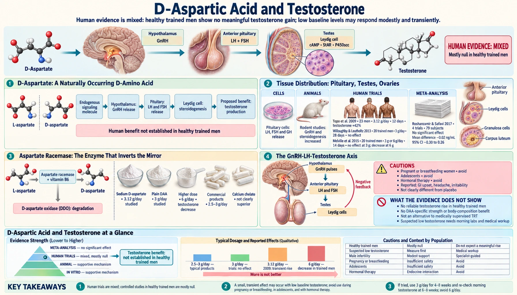
D-aspartic acid (DAA) is the mirror-image stereoisomer of the ordinary L-aspartic acid found in dietary protein — and unlike most D-amino acids, the body makes and uses it. In animal studies D-aspartate concentrates in the pituitary gland and the testes, where it is involved in the release of luteinizing hormone (LH), prolactin, growth hormone and testosterone. A 2009 Italian study by Topo, D'Aniello and colleagues reported that 3.12 g/day of sodium D-aspartate for 12 days raised serum testosterone by 42% in 23 men — the paper behind a popular supplement category. Later placebo-controlled trials in resistance-trained men did not reproduce it: Willoughby and Leutholtz 2013 found no change in testosterone, and Melville and colleagues 2015 found that 6 g/day lowered total and free testosterone. This page lays out the underlying neuroendocrinology, walks through both the supportive and contradictory clinical literature, and summarizes what the current evidence shows.
Table of Contents
- D-Aspartate: A Naturally Occurring D-Amino Acid
- Tissue Distribution: Pituitary, Testes, Ovaries
- Aspartate Racemase: The Enzyme That Inverts the Mirror
- The GnRH-LH-Testosterone Axis
- The D'Aniello / Topo 2009 Human Trial
- Willoughby and Leutholtz 2013: The Failed Replication
- Subsequent Trials and the Current Evidence
- Growth Hormone, Sperm Quality, and Female Fertility
- Supplement Products and Safety Data
- What the Evidence Means in Context
- Key Research Papers
- Connections
- Featured Videos
D-Aspartate: A Naturally Occurring D-Amino Acid
Amino acids exist in two stereoisomeric forms — the L and D configurations — that are non-superimposable mirror images. Ribosomal protein synthesis uses only L-amino acids, so nearly all amino acid mass in the human body is L-form. D-amino acids were long assumed to be biologically irrelevant in mammals, occurring mainly in bacterial cell walls and certain peptide antibiotics.
The discovery that mammals contain meaningful concentrations of free D-aspartate (and, separately, D-serine) overturned this assumption. Both are now recognized as physiologically active signaling molecules. D-serine functions in the central nervous system as a co-agonist at NMDA receptors. D-aspartate, the focus of this page, acts in the nervous system as an NMDA-receptor agonist and in the neuroendocrine system — the hypothalamus, pituitary, and gonads — as a modulator of hormone release.
A 2005 review by Furuchi and Homma describes high D-aspartate concentrations in embryos, which disappear from nervous tissue after birth but rise temporarily in endocrine glands — particularly the pituitary, pineal and adrenal glands — at specific stages; testicular D-aspartate rises just before birth and during sexual maturation. These animal findings fueled the original interest in DAA as a hormonal supplement.
Tissue Distribution: Pituitary, Testes, Ovaries
The Italian neuroendocrinologist Antimo D'Aniello and colleagues, and Japanese groups including Homma's, mapped the tissue distribution of D-aspartate, mostly in rats, in a series of papers from the 1990s onward. The pattern:
- Pineal gland — D-aspartate that appears to come from other tissues suppresses melatonin secretion from pineal cells (rat studies, reviewed by Furuchi and Homma 2005)
- Anterior pituitary — found in prolactin-containing cells; in rat studies it stimulates the secretion of prolactin, LH and growth hormone, and a rat pituitary cell line synthesizes it
- Testes — localized in Leydig cells (testosterone-producing) and Sertoli cells; in rats it appears in two periods, before birth and at sexual maturity, in parallel with testosterone synthesis
- Ovaries — present in human pre-ovulatory follicular fluid, at lower concentrations in women aged 35-40 than in women aged 22-34
- Adrenal gland — a transient rise at specific developmental stages in animals
- Hypothalamus — in the rat hypothalamus it enhances gonadotropin-releasing hormone (GnRH) release
The presence of D-aspartate in several steroid-producing tissues suggests a role in steroidogenic regulation. The proposed mechanism, based on animal and in vitro work, involves D-aspartate produced inside the seminiferous tubules acting on Leydig cells to enhance testosterone synthesis by increasing expression of StAR (steroidogenic acute regulatory protein), plus stimulation of upstream releasing-hormone secretion.
Aspartate Racemase: The Enzyme That Inverts the Mirror
D-aspartate is generated from L-aspartate by an aspartate racemase. The reaction is a stereoinversion: same carbon skeleton, same functional groups, mirror-image three-dimensional arrangement around the alpha carbon. A mammalian aspartate racemase (named DR) was identified and cloned by Kim and colleagues in 2010; it converts L-aspartate to D-aspartate and colocalizes with D-aspartate in the brain and neuroendocrine tissues.
Breakdown runs through a separate enzyme, D-aspartate oxidase (DDO), which oxidatively deaminates D-aspartate and generates hydrogen peroxide as a byproduct. DDO is the only enzyme known to selectively degrade D-aspartate, and the postnatal onset of DDO activity explains the fall in brain D-aspartate after birth. The balance between synthesis and DDO degradation determines steady-state tissue D-aspartate concentrations.
Mice lacking the Ddo gene have elevated D-aspartate, and these mice, like mice given D-aspartate, show changes in hippocampal NMDA-receptor-dependent long-term potentiation and spatial memory — an effect attributed to D-aspartate's action as an NMDA-receptor agonist, distinct from its endocrine role.
Whether vitamin B6 status affects D-aspartate synthesis in humans has not been studied in dietary contexts.
The GnRH-LH-Testosterone Axis
To understand how D-aspartate is proposed to affect testosterone, the hypothalamic-pituitary-gonadal (HPG) axis needs a brief review:
- Hypothalamus: GnRH neurons in the medial preoptic area and the infundibular nucleus release gonadotropin-releasing hormone in pulses into the hypophyseal portal circulation. The pulse frequency is critical — slow pulses favor FSH, fast pulses favor LH.
- Pituitary: GnRH binds GnRH receptors on anterior pituitary gonadotrope cells, triggering release of LH and FSH into systemic circulation.
- Testes (males): LH binds LH receptors on Leydig cells in the testicular interstitium, activating cholesterol mobilization via StAR protein, cholesterol delivery to mitochondria, side-chain cleavage by P450scc to produce pregnenolone, and subsequent steroidogenic enzymes to produce testosterone. FSH acts on Sertoli cells supporting spermatogenesis.
- Ovaries (females): LH and FSH coordinate follicular development, estrogen production, ovulation, and corpus luteum maintenance.
- Negative feedback: Testosterone (and estradiol in females) feeds back to suppress GnRH and gonadotropin release, closing the loop.
D-aspartate is proposed to act at three levels in this axis, based on rat and in vitro studies. At the hypothalamus, it enhances GnRH release. At the pituitary, it increases LH release and synthesis, with cGMP as the second messenger. At the testes, it accumulates in Leydig cells and increases testosterone synthesis and release, with cAMP as the second messenger. The cumulative effect, in principle, would be elevated testosterone production at every step of the axis.
The D'Aniello / Topo 2009 Human Trial
The single most consequential paper in the DAA story is Topo, Soricelli, D'Aniello et al. (2009), published in Reproductive Biology and Endocrinology, titled "The role and molecular mechanism of D-aspartic acid in the release and synthesis of LH and testosterone in humans and rats." The Italian group studied healthy volunteers at a hospital IVF unit:
- Subjects: 23 men aged 27-37 years
- Intervention: 3.12 g sodium D-aspartate per day (10 ml of a 2.0 M solution, which also contained vitamin B6, folic acid and vitamin B12), diluted in water or fruit juice, for 12 days
- Comparison: 20 men of the same age given a placebo solution of sodium chloride with the same vitamins
- Outcomes measured: morning serum LH and testosterone before treatment, at day 6, at day 12, and 3 days after treatment stopped
The reported results: in the D-aspartate group, mean serum LH rose 33% (from 4.2 to 5.6 mIU/mL at day 12) and mean serum testosterone rose 42% (from 4.5 to 6.4 ng/mL at day 12); 20 of the 23 men had increased testosterone. The day-6 increases were not statistically significant. Three days after treatment stopped, LH was 1.14-fold above baseline (not significant) and testosterone 1.28-fold above baseline (significant). The placebo group showed no change.
The paper also reported rat work: rats drinking 20 mM D-aspartate for 12 days had higher serum LH and testosterone, and in isolated rat tissue D-aspartate increased LH release and synthesis in the pituitary (via cGMP) and testosterone synthesis in Leydig cells (via cAMP). The combination of a positive human result plus a plausible animal mechanism made the paper highly cited and drove the commercialization of D-aspartic-acid supplements through bodybuilding and "T-booster" channels.
The methodological limits of the 2009 paper are notable in retrospect: small sample size, a treatment period of only 12 days, measurements at only four time points, and no description of randomization or blinding in the paper. The supplement used also contained B vitamins, although the placebo carried the same vitamins.
Willoughby and Leutholtz 2013: The Failed Replication
Willoughby and Leutholtz (2013), Nutrition Research, tested DAA in a different population. The design:
- Subjects: Resistance-trained men
- Intervention: 3 g/day D-aspartic acid for 28 days, alongside resistance training 4 times per week
- Comparison: 3 g/day placebo
- Outcomes: Body composition, muscle strength, serum gonadal hormones, and serum D-aspartate and D-aspartate oxidase, before and after the intervention
Total and free testosterone, LH, GnRH and estradiol were unchanged with DAA supplementation. Body composition and muscle strength increased in both groups (reflecting the training stimulus), with no difference between them. Serum D-aspartate oxidase, the enzyme that breaks D-aspartate down, was significantly higher in the DAA group than with placebo. The authors concluded that, at the dose provided, DAA did not up-regulate the HPG axis and had no anabolic or ergogenic effect.
Unlike the 2009 paper, the Willoughby study was placebo-controlled in resistance-trained men — the population to which the supplement is most commonly marketed — and ran longer. Two later trials in resistance-trained men (below) also found no rise in testosterone.
Explanations proposed for the difference between the 2009 and 2013 results include training status (Melville and colleagues 2015 contrasted the 2009 participants with resistance-trained men and hypothesized that experienced lifters might need a higher dose), the short 12-day duration of the 2009 study, and the limits of a single small trial without described blinding.
Subsequent Trials and the Current Evidence
Later placebo-controlled trials in resistance-trained men have not found a testosterone rise:
- Melville et al. (2015), Journal of the International Society of Sports Nutrition. 24 resistance-trained men randomized to placebo (6 g/day plain flour), 3 g/day or 6 g/day DAA for 14 days. The 3 g arm showed no significant effect on testosterone. In the 6 g arm, total testosterone fell significantly and free testosterone was significantly lower than with placebo. Initial testosterone level did not predict responsiveness to DAA.
- Melville et al. (2017), PLoS One. 22 resistance-trained men randomized to 6 g/day DAA or placebo for 12 weeks of supervised training (double-blind). No change in total or free testosterone; estradiol fell 16% in the DAA group; strength and muscle size rose similarly in both groups.
- Roshanzamir and Safavi (2017), systematic review. Included 23 animal studies and 4 human studies published to October 2015. Animal studies showed DAA raising testosterone in males; the human studies gave inconsistent results, and the authors judged the human evidence sparse because of the limited number and poor quality of studies, calling for larger, longer, well-designed trials.
The pattern across the post-2009 trials is no effect in young resistance-trained men with normal baseline testosterone, and a decrease at 6 g/day in one 14-day trial. The hypothesis that men with low baseline testosterone respond better has not been confirmed: the 2015 trial found no correlation between initial testosterone and response, and no trial has been run specifically in men with low testosterone.
The 2017 systematic review concluded that the human evidence remains sparse. No trial has compared DAA with testosterone replacement therapy, which is the treatment that endocrine guidelines describe for confirmed hypogonadism.
Growth Hormone, Sperm Quality, and Female Fertility
Beyond the testosterone question, several other DAA effects have been studied:
Growth hormone. In rat studies reviewed by D'Aniello (2007), D-aspartate stimulates growth hormone secretion from the pituitary. No human trial of DAA and growth hormone was found in PubMed, and the supplement market has focused on testosterone rather than growth hormone.
Sperm quality. D-aspartate concentrations in seminal plasma and spermatozoa were significantly lower in oligoasthenoteratospermic and azoospermic men than in normospermic men (D'Aniello et al. 2005, 30 men). In a follow-up study (D'Aniello et al. 2012, Advances in Sexual Medicine), 60 sub-fertile men (30 with oligo-asthenozoospermia, 30 with asthenozoospermia) took sodium D-aspartate (2.66 g D-aspartic acid daily) for 90 days; the authors reported increased sperm concentration and motility and a higher pregnancy rate among their partners. That study had no placebo group, and independent replication is lacking.
Female fertility. Limited human data. In 20 women undergoing IVF, D-aspartate concentration in pre-ovulatory follicular fluid was higher in women aged 22-34 than in those aged 35-40 and was related to the proportion of good-quality oocytes and the fertilization rate (D'Aniello et al. 2007). This is an observational finding; no trials of DAA supplementation in female infertility were found. The supplement is generally not marketed to women.
Postmenopausal women. No studies were found.
Supplement Products and Safety Data
D-aspartic acid supplements remain on the market despite the failed replications. Common product types:
- Standalone DAA — often around 3 g per serving, marketed for "natural testosterone support"
- DAA in "T-booster" blends — combined with zinc, magnesium, vitamin D, fenugreek, ashwagandha, and other ingredients with varying evidence; a blend as a whole may have effects independent of DAA itself
- "Chelated" DAA — D-aspartate calcium chelate or other mineral salts marketed as more bioavailable; no trial has compared them with sodium D-aspartate
- Fertility formulas — positioned for male infertility, often combined with CoQ10, vitamin E, zinc, selenium, and L-carnitine
The human trials lasted 12 days to 12 weeks and were not designed to assess safety; long-term safety data in humans are lacking. In rats, ingested D-aspartate accumulated in the pituitary and testes and declined after treatment stopped (Topo 2009). In the one 14-day trial that tested 6 g/day, total and free testosterone fell.
What the Evidence Means in Context
How the DAA research fits with clinical practice for low testosterone:
- Diagnosis of low testosterone — The Endocrine Society's 2018 guideline recommends diagnosing hypogonadism only in men with symptoms and signs of testosterone deficiency and unequivocally and consistently low serum testosterone, using fasting morning total testosterone confirmed by a repeat measurement, with free testosterone in some cases. No DAA trial enrolled men diagnosed this way.
- Causes — The same guideline recommends further evaluation to find the cause in men with androgen deficiency. Recognized causes include pituitary disease, hemochromatosis, obesity, opioid use and past anabolic steroid use. The guideline recommends testosterone therapy for men with symptomatic, confirmed testosterone deficiency.
- Doses studied — Human trials used 2.66 g to 6 g/day for 12 days to 12 weeks. The positive testosterone result came from a single 12-day study at 3.12 g/day; 3 g/day for 14 or 28 days in trained men showed no effect; 6 g/day lowered testosterone over 14 days and left it unchanged over 12 weeks.
- Male infertility — The fertility evidence rests on observational measurements and one uncontrolled 60-man study from the same research group.
- Populations with no data — The trials enrolled adult men only. There are no safety data for pregnancy, breastfeeding, adolescents, or people on hormonal therapy.
Decisions about supplements and hormone testing belong with a clinician.
Key Research Papers
- Topo E, Soricelli A, D'Aniello A, Ronsini S, D'Aniello G (2009). The role and molecular mechanism of D-aspartic acid in the release and synthesis of LH and testosterone in humans and rats. Reproductive Biology and Endocrinology 7:120. The foundational positive trial. — PubMed PMID: 19860889
- Willoughby DS, Leutholtz B (2013). D-aspartic acid supplementation combined with 28 days of heavy resistance training has no effect on body composition, muscle strength, and serum hormones associated with the hypothalamo-pituitary-gonadal axis in resistance-trained men. Nutrition Research 33(10):803-10. The pivotal failed replication. — PubMed PMID: 24074738
- Melville GW, Siegler JC, Marshall PW (2015). Three and six grams supplementation of d-aspartic acid in resistance trained men. Journal of the International Society of Sports Nutrition 12:15. — PubMed PMID: 25844073
- Melville GW, Siegler JC, Marshall PWM (2017). The effects of d-aspartic acid supplementation in resistance-trained men over a three month training period: A randomised controlled trial. PLoS One 12(8):e0182630. — PubMed PMID: 28841667
- D'Aniello G, Ronsini S, Notari T, Grieco N, Infante V, D'Angel N, Mascia F, Di Fiore MM, Fisher G, D'Aniello A (2012). D-Aspartate, a key element for the improvement of sperm quality. Advances in Sexual Medicine 2:45-53. — doi:10.4236/asm.2012.24008
- D'Aniello G, Ronsini S, Guida F, Spinelli P, D'Aniello A (2005). Occurrence of D-aspartic acid in human seminal plasma and spermatozoa: possible role in reproduction. Fertility and Sterility 84(5):1444-9. — PubMed PMID: 16275242
- D'Aniello A, Di Cosmo A, Di Cristo C, Annunziato L, Petrucelli L, Fisher G (1996). Involvement of D-aspartic acid in the synthesis of testosterone in rat testes. Life Sciences 59(2):97-104. The original rat testosterone mechanism work. — PubMed PMID: 8699926
- Roshanzamir F, Safavi SM (2017). The putative effects of D-Aspartic acid on blood testosterone levels: A systematic review. International Journal of Reproductive BioMedicine 15(1):1-10. Systematic review: human evidence inconsistent and sparse. — PubMed PMID: 28280794
- D'Aniello A (2007). D-Aspartic acid: an endogenous amino acid with an important neuroendocrine role. Brain Research Reviews 53(2):215-34. Review of the broader neuroendocrine biology. — PubMed PMID: 17118457
- Furuchi T, Homma H (2005). Free D-aspartate in mammals. Biological and Pharmaceutical Bulletin 28(9):1566-70. — PubMed PMID: 16141517
- Long Z, Lee JA, Okamoto T, Nimura N, Imai K, Homma H (2000). d-Aspartate in a prolactin-secreting clonal strain of rat pituitary tumor cells (GH(3)). Biochemical and Biophysical Research Communications 276(3):1143-7. — PubMed PMID: 11027602
- Errico F, Napolitano F, Nisticò R, Usiello A (2012). New insights on the role of free D-aspartate in the mammalian brain. Amino Acids 43(5):1861-71. — PubMed PMID: 22851050
- Kim PM, Duan X, Huang AS, Liu CY, Ming GL, Song H, Snyder SH (2010). Aspartate racemase, generating neuronal D-aspartate, regulates adult neurogenesis. Proceedings of the National Academy of Sciences of the USA 107(7):3175-9. — PubMed PMID: 20133766
- D'Aniello G, Grieco N, Di Filippo MA, Cappiello F, Topo E, D'Aniello E, Ronsini S (2007). Reproductive implication of D-aspartic acid in human pre-ovulatory follicular fluid. Human Reproduction 22(12):3178-83. — PubMed PMID: 17951582
- Bhasin S, Brito JP, Cunningham GR, Hayes FJ, Hodis HN, Matsumoto AM, Snyder PJ, Swerdloff RS, Wu FC, Yialamas MA (2018). Testosterone Therapy in Men With Hypogonadism: An Endocrine Society Clinical Practice Guideline. Journal of Clinical Endocrinology and Metabolism 103(5):1715-1744. — PubMed PMID: 29562364
PubMed Topic Searches
Connections
- All Amino Acids
- Aspartic Acid Benefits Hub
- Aspartic Acid Overview
- Neurotransmission & NMDA
- Aspartate in the Urea Cycle
- Aspartate and Energy Production
- Testosterone Lab Test
- Luteinizing Hormone (LH)
- Follicle-Stimulating Hormone (FSH)
- Hypogonadism
- Male Infertility
- Ashwagandha (Adaptogen for T)
- Tongkat Ali
- Zinc (T Synthesis Cofactor)
- Vitamin D3 (T Modulator)
- Vitamin B6 (Racemase Cofactor)
- History
- Sources