Porphyria Cutanea Tarda (PCT)
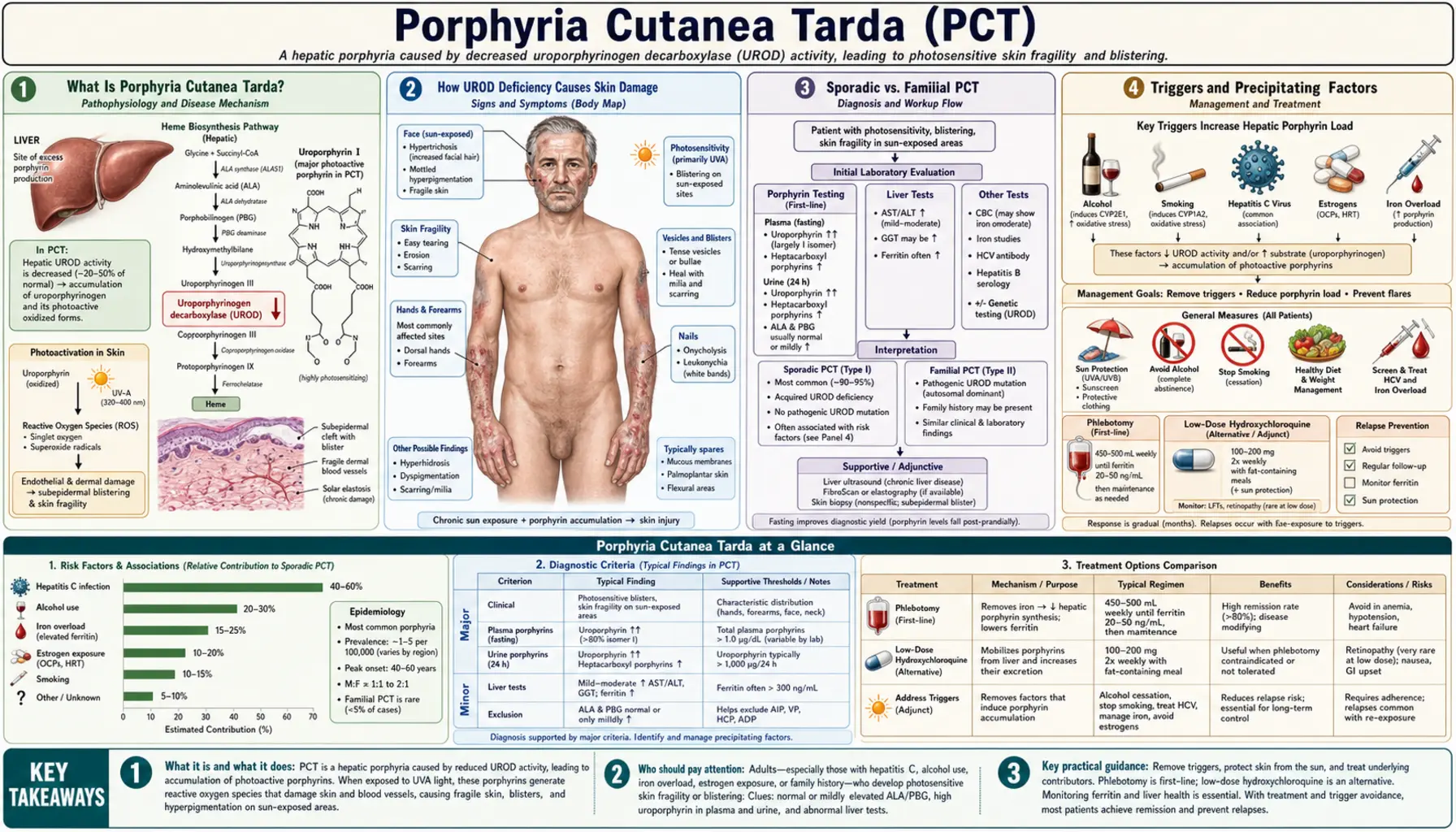
Porphyria Cutanea Tarda is the most common of all the porphyrias — a family of metabolic disorders affecting the heme biosynthesis pathway. In PCT, a deficiency of the enzyme uroporphyrinogen decarboxylase (UROD) causes photoreactive porphyrins to build up in the liver, spill into the bloodstream, and deposit in the skin. When sunlight hits these porphyrins, it triggers a damaging photochemical reaction that produces blistering, scarring, and disfiguring skin changes on sun-exposed areas. The disease is deeply intertwined with treatable triggers — alcohol, hepatitis C, iron overload, and estrogens — meaning that in most patients, identifying and removing the cause can lead to full remission without the need for complex medications.
- Porphyria Cutanea Tarda Explained
- How UROD Deficiency Causes Skin Damage
- Sporadic vs. Familial PCT
- Triggers and Precipitating Factors
- Skin Findings and Symptoms
- Diagnosing PCT
- Treatment: Removing Triggers and Clearing Porphyrins
- Phlebotomy and Hydroxychloroquine in Detail
- Hepatitis C, HFE Mutations, and PCT
- Prognosis and Long-Term Outlook
- Key Research Papers
- Connections
Porphyria Cutanea Tarda Explained
Porphyrias are a group of eight distinct disorders, each caused by a deficiency of one of the eight enzymes in the heme biosynthesis pathway. Heme — the iron-containing core of hemoglobin and of cytochrome enzymes throughout the body — is assembled from simpler molecules through this sequential eight-step pathway in the liver and bone marrow. When any one enzyme is deficient, the upstream precursors (porphyrins and their precursors) accumulate, overflow into the blood, and cause disease in specific organs depending on which enzyme is affected and how toxic the accumulating compound is.
PCT affects the fifth enzyme in the pathway: uroporphyrinogen decarboxylase (UROD). When UROD activity falls below approximately 20% of normal in the liver, the enzyme's substrates — uroporphyrins and heptacarboxylporphyrins — back up dramatically and leak into the plasma. These specific porphyrins are photosensitizers: they absorb ultraviolet (UV) and visible light at approximately 400–410 nanometers (the Soret band) and, when excited by light, transfer that energy to surrounding molecules in a way that generates reactive oxygen species and destroys nearby tissue. In skin, this photochemical reaction severs dermal-epidermal connections, causing the characteristic blistering.
PCT is the most common porphyria worldwide, with an estimated prevalence of approximately 1 in 10,000, though many mild cases go unrecognized. It is an acquired condition in the majority of patients (sporadic PCT) — the enzyme deficiency is not inherited but rather induced by specific environmental and metabolic triggers in genetically predisposed individuals. A minority (about 20%) carry a germline UROD mutation that halves their baseline enzyme activity, making them far more susceptible to these triggers. PCT is not a cancer, not contagious, and not a rapidly fatal disease — but untreated, it causes progressive skin disfigurement and liver damage, and it signals the presence of serious underlying conditions that require their own management.
How UROD Deficiency Causes Skin Damage
The connection between a liver enzyme deficiency and blistering skin rashes is not immediately obvious — it requires understanding the specific photochemical properties of the porphyrins that accumulate and the route by which they reach the skin.
The UROD Enzyme and Its Substrates
In normal heme synthesis, UROD converts uroporphyrinogen III to coproporphyrinogen III through a series of four successive decarboxylation steps, producing a cascade of partially decarboxylated intermediates: heptacarboxylporphyrinogen → hexacarboxylporphyrinogen → pentacarboxylporphyrinogen → coproporphyrinogen. When UROD activity in the liver is suppressed, these intermediates — particularly uroporphyrinogen and heptacarboxylporphyrinogen — accumulate. These compounds are readily auto-oxidized in the presence of iron and oxygen to their porphyrin (oxidized) forms: uroporphyrin I and III, and heptacarboxylporphyrin.
Iron as the Critical Cofactor
Iron is central to PCT pathogenesis and is the reason that iron overload is a consistent finding and phlebotomy is the most effective treatment. Hepatic iron, particularly in the ferric (Fe³⁺) state, appears to drive the formation of an oxidized inhibitor of UROD — uroporphomethene, identified in a 2007 study of mouse models of PCT, with an inhibitor also detected in liver biopsy samples from people with PCT — that competitively blocks the enzyme rather than destroying it (an iron-dependent oxidation). This means iron overload and elevated hepatic iron do not simply co-exist with PCT; they actively drive the enzyme deficiency. Reducing hepatic iron through phlebotomy removes the inhibitory compound, restores UROD activity, and allows porphyrins to normalize.
From Liver to Skin: The Photosensitizer Route
Uroporphyrins and heptacarboxylporphyrins accumulating in the hepatocyte cytoplasm are released into bile and plasma. Plasma porphyrins circulate freely and deposit in sun-exposed skin, particularly in the dermis where small blood vessels carry them close to the surface. When ultraviolet or visible light (particularly the 400–410 nm Soret band, which penetrates window glass) strikes these porphyrin-laden skin cells, a Type I and Type II photochemical reaction occurs: singlet oxygen and free radicals are generated, which oxidize proteins, lipids, and nucleic acids in the dermal-epidermal junction. This destroys the anchoring proteins connecting the epidermis to the dermis, producing subepidermal blistering — mechanically similar to, but pathologically distinct from, the autoimmune blistering of bullous pemphigoid.
Liver Damage and Fibrosis
The accumulated porphyrins are not simply cosmetically problematic — they are hepatotoxic. Chronic porphyrin accumulation in the liver drives oxidative stress, inflammation, and hepatic fibrosis. The same triggers (alcohol, hepatitis C) that precipitate PCT are independently hepatotoxic, compounding the liver injury. Long-term uncontrolled PCT substantially increases the risk of hepatocellular carcinoma, and liver surveillance is a component of PCT management in patients with established liver disease.
Sporadic vs. Familial PCT
PCT is classified into two forms based on whether the UROD deficiency is inherited or acquired. This distinction influences counseling, family screening, and the threshold at which disease manifests.
Type I — Sporadic PCT (80% of Cases)
The majority of PCT patients have no inherited UROD mutation. Their germline UROD gene is normal — they have two functional copies — and the enzyme deficiency is entirely acquired, localized to the liver, and caused by the action of environmental and metabolic triggers (alcohol, iron, hepatitis C virus (HCV), estrogens, aromatic hydrocarbons) on the hepatic UROD enzyme. The enzyme inhibitor formed in this process is present only in the liver, not in red blood cell precursors in the bone marrow. When triggers are removed and iron is depleted, UROD activity in these patients fully recovers and porphyrin levels normalize. Sporadic PCT patients have no increased baseline risk of the disease in the absence of triggers, and their children do not inherit an elevated risk (unless the child independently acquires the same triggers and the gene variants that increase susceptibility).
Type II — Familial PCT (20% of Cases)
In familial PCT, one copy of the UROD gene carries a loss-of-function mutation, inherited in an autosomal dominant pattern. This baseline heterozygosity means UROD enzyme activity starts at approximately 50% of normal in all tissues — liver, bone marrow, red blood cells, everywhere. With this head start toward the critical threshold of enzyme insufficiency, the same environmental triggers that might be harmless in a person with two normal UROD copies push the familial PCT patient below the disease threshold more easily. Type II PCT patients have lower thresholds for disease activation, tend to manifest earlier in life, and — importantly — have measurably reduced UROD activity in their red blood cells (a reliable diagnostic marker, unlike sporadic PCT where red blood cell UROD is normal). Their children have a 50% chance of inheriting the UROD mutation, though inheriting the mutation does not guarantee developing PCT (full clinical penetrance still requires exposure to a precipitating trigger). Genetic counseling and family screening are part of care when Type II is diagnosed.
Measured shares of the inherited form are close to one in five: in two US series, 17% of 143 patients (Jalil 2010) and 19% of 39 patients (Egger 2002) had low red-blood-cell UROD activity, the marker of familial PCT. A 2003 review of 50 studies found hepatitis C in 57% of people with sporadic PCT but in 26% of those with the familial form (Gisbert 2003).
Type III — Familial Without UROD Mutation
A rare subgroup of familial PCT (Type III) shows familial clustering of PCT without a detectable UROD germline mutation. These families likely share genetic variants that increase susceptibility to trigger-induced UROD inhibition — perhaps HFE hemochromatosis mutations, CYP1A2 polymorphisms, or other modifier genes — without a structural UROD defect. Type III is poorly characterized.
Triggers and Precipitating Factors
PCT is almost never a spontaneous disease. In virtually all patients, the biochemical deficiency is unmasked or worsened by one or more identifiable environmental, metabolic, or infectious triggers. Identifying and removing triggers is the first and most important step in management — and in many patients, trigger removal alone is sufficient to achieve remission.
Most people with PCT carry several triggers at once. In a US study of 143 patients, the most common risk factors were alcohol use (87%), smoking (81%), hepatitis C (69%) and changes in the HFE iron gene (53%); 13% had human immunodeficiency virus (HIV) infection, 66% of the women took estrogen, and 70% of patients had three or more risk factors together — PCT is rarely caused by a single trigger (Jalil 2010).
Alcohol — The Most Common Trigger
Alcohol is the most frequent precipitant of PCT in developed countries. Multiple mechanisms converge: alcohol promotes hepatic iron accumulation by upregulating iron absorption and impairing iron export; alcohol generates hepatic oxidative stress through acetaldehyde and reactive oxygen species; and alcohol suppresses UROD enzyme activity directly. The relationship is dose-dependent — heavier, longer alcohol consumption causes more severe porphyrin accumulation. Stopping alcohol completely is a standard part of PCT management. In patients who abstain completely and have no other triggers, some achieve partial or complete remission from abstinence alone, though phlebotomy is usually still necessary to clear the accumulated hepatic iron.
Hepatitis C Virus (HCV) — A Strikingly Strong Association
Hepatitis C virus infection is found in approximately 50% of PCT patients in European and North American series — a prevalence far higher than in the general population. The mechanism is not fully elucidated but likely involves hepatitis C virus-induced hepatic iron dysregulation (hepatitis C virus suppresses hepcidin, the master regulator of iron homeostasis, leading to iron loading even without HFE mutations), combined with hepatitis C virus-driven liver oxidative stress and inflammation. Strikingly, treating hepatitis C virus with modern direct-acting antivirals (DAAs) — agents like sofosbuvir/velpatasvir, glecaprevir/pibrentasvir — achieves sustained virological response in nearly 95–99% of patients and in many cases leads to spontaneous PCT remission without phlebotomy, as the liver normalizes iron handling and UROD inhibitor production falls. A 1998 North American study concluded that all PCT patients should be tested for hepatitis C infection and for HFE gene mutations; of all the triggers, hepatitis C is the one that modern medicines can cure.
Iron Overload and HFE Hemochromatosis Mutations
Elevated hepatic iron and elevated serum ferritin are almost universal in PCT at the time of diagnosis. HFE gene mutations — particularly C282Y homozygosity and C282Y/H63D compound heterozygosity, which cause hereditary hemochromatosis — are found at substantially increased frequency in PCT patients compared to the general population. These mutations amplify iron loading, lowering the threshold at which triggers can inhibit UROD sufficiently to cause disease. People with two copies of C282Y usually need more phlebotomies, and because hereditary hemochromatosis is inherited, their relatives share the genetic risk.
Estrogens
Combined oral contraceptive pills and hormone replacement therapy containing estrogen are recognized PCT triggers, particularly in women with other predisposing factors. Estrogens increase hepatic iron mobilization and alter porphyrin metabolism. PCT brought on by oral contraceptive pills typically resolves when the pill is stopped, though iron depletion (phlebotomy) is still required to complete remission. Progestin-only and non-hormonal contraception contain no estrogen, which is why women with PCT are commonly switched to them; estrogen hormone replacement therapy (HRT) in menopausal women with PCT is commonly stopped or reconsidered for the same reason.
Human Immunodeficiency Virus Infection
Human immunodeficiency virus (HIV) co-infection is found at increased frequency in PCT patients, likely through mechanisms similar to hepatitis C virus: immune dysregulation, chronic hepatic inflammation, and alteration of iron metabolism. The effect of antiretroviral therapy on PCT activity is less clear than that of hepatitis C virus treatment with DAAs.
Chlorinated Hydrocarbons
The first large epidemic of PCT ever documented was caused by environmental hexachlorobenzene contamination of wheat grain in Turkey between 1955 and 1959, affecting an estimated 4,000 people. This catastrophic exposure demonstrated that exogenous aromatic hydrocarbons can directly inhibit UROD and trigger epidemic PCT. Occupational exposure to certain chlorinated solvents, dioxins, and polychlorinated biphenyls has been implicated in sporadic cases. Modern PCT in developed countries rarely has this cause, but it established the fundamental principle that exogenous chemicals can precipitate PCT.
Smoking
Smoking is an independent risk factor for PCT, possibly through the pro-oxidant and aryl hydrocarbon receptor-activating compounds in tobacco smoke. The effect is additive with alcohol and other triggers.
Skin Findings and Symptoms
PCT is a purely cutaneous porphyria. Unlike the acute hepatic porphyrias (acute intermittent porphyria, variegate porphyria, hereditary coproporphyria), PCT does not cause neurovisceral attacks — no abdominal pain crises, no neurological manifestations, no psychiatric attacks. All of its disease expression is in the skin, on sun-exposed areas, developing insidiously over months to years rather than suddenly. Patients typically have had subtle skin changes for years before recognizing them as a disease.
Blistering on Sun-Exposed Skin
The hallmark of PCT is tense, fluid-filled blisters (bullae) appearing on sun-exposed areas — most prominently the dorsal hands and fingers, forearms, face, ears, and the V-area of the chest when a shirt collar exposes it. The blisters typically develop after minor sun exposure or trivial trauma (pressure, friction) and are not immediately painful — patients often report finding blisters that they did not feel forming. They rupture to leave shallow erosions, which heal slowly over weeks, leaving behind scars and milia (small, firm, white keratin-filled cysts at healed blister sites — a highly characteristic finding).
Skin Fragility
Skin fragility — the tendency to develop erosions from even trivial mechanical injury — is a prominent early feature. Patients notice skin tears from picking up objects, gardening, or simply putting on gloves. The dorsal hands are particularly fragile. This fragility predates visible blistering in some patients and may be the first clue. The mechanism is photodamage to the dermal-epidermal junction proteins (type IV collagen, laminin) making the skin structurally weak even when blisters have not yet visibly formed.
Hyperpigmentation
Diffuse hyperpigmentation on sun-exposed areas — particularly the face and periorbital regions — develops in established PCT. The pigmentation resembles a tan but is irregular and does not fully fade in winter. Periorbital darkening can be striking, giving a characteristic panda-bear appearance. The mechanism involves porphyrin-induced melanocyte stimulation and post-inflammatory hyperpigmentation from chronic low-grade skin injury.
Hypertrichosis — Increased Facial Hair
One of the most distinctive and diagnostically useful features of PCT is hypertrichosis — abnormal, excessive hair growth — particularly over the temples, cheeks, and sideburn areas in women, and generalized facial hypertrichosis in both sexes. This is not virilization (no clitoromegaly, no voice change, no breast atrophy) — it is a porphyrin-induced stimulation of vellus hair follicles to produce longer, darker, terminal-type hairs. In women, who do not normally have coarse facial hair, this finding is cosmetically distressing and is often the presenting complaint that brings them to medical attention. The hypertrichosis reverses with PCT remission, though slowly.
Sclerodermoid Changes
In chronic, inadequately treated PCT, progressive skin thickening and induration develop over sun-exposed areas — a sclerodermoid pattern that mimics localized scleroderma. The skin becomes waxy, thickened, and tight, particularly over the face, forehead, and hands. Calcinosis (calcium deposits palpable under the skin) can develop within sclerodermoid areas. Unlike scleroderma, PCT's skin thickening is driven by porphyrin-induced collagen stimulation from chronic photodamage rather than by autoimmune fibrosis; it affects only sun-exposed areas and regresses (partially) with treatment. Distinguishing PCT from scleroderma requires porphyrin testing — the clinical appearance alone can be confusing.
What PCT Does NOT Cause
PCT does not cause abdominal pain, peripheral neuropathy, psychiatric symptoms, or cardiovascular complications. These features, if present, suggest a different diagnosis — particularly the acute hepatic porphyrias (acute intermittent porphyria, variegate porphyria) which cause neurovisceral attacks and share some biochemical overlap. PCT also does not cause photosensitivity of the immediate burning/stinging type (as in erythropoietic protoporphyria, EPP, which is agonizing and immediate); PCT's skin damage builds gradually over hours to days after cumulative sun exposure, not within minutes.
Diagnosing PCT
The diagnosis of PCT rests on a combination of characteristic clinical features — blistering, fragility, hypertrichosis, hyperpigmentation on sun-exposed areas — and laboratory confirmation of the specific porphyrin pattern. PCT is suspected whenever a patient presents with these features, particularly in the context of alcohol use, hepatitis C virus infection, or iron overload.
Urine Porphyrin Testing — The Diagnostic Cornerstone
A 24-hour urine collection or a spot urine sample sent for quantitative porphyrin fractionation is the most important diagnostic test. In PCT, there is dramatic elevation of uroporphyrins (primarily uroporphyrin I) and heptacarboxylporphyrins, with uroporphyrins substantially exceeding coproporphyrins — a ratio that distinguishes PCT from other porphyrias. In active PCT, 24-hour urine uroporphyrin may be 10–50 times the upper limit of normal. The pink or dark brown discoloration of PCT urine under ordinary light, and its orange-red fluorescence under a Wood's lamp (365 nm ultraviolet), reflect this massive porphyrinuria and are useful bedside clues — though formal quantification is always required for diagnosis.
Plasma Porphyrin Fluorescence Spectroscopy
Plasma porphyrin measurement with fluorescence scanning is a highly sensitive and specific test for PCT. The emission peak of PCT plasma at 615–620 nanometers, obtained by scanning plasma fluorescence after excitation at 405 nm, is characteristic and distinguishes PCT from variegate porphyria (peak at 625–627 nm) and erythropoietic protoporphyria (peak at 634 nm). Plasma scanning is increasingly available at reference laboratories and is particularly useful when the clinical picture suggests PCT but urine results are equivocal.
Stool Porphyrins
Stool porphyrin analysis shows elevation of isocoproporphyrins (a PCT-specific finding — isocoproporphyrins are generated because UROD's partial block produces an atypical porphyrin side-product specific to this enzyme deficiency). Elevated fecal isocoproporphyrin is pathognomonic for PCT or hepatoerythropoietic porphyria (HEP, the rare homozygous form of PCT). Stool testing is not routinely required when urine and plasma results are clear but is used in difficult diagnostic cases.
Laboratory Workup for Triggers and Organ Involvement
- Iron studies: serum iron, total iron-binding capacity (TIBC), transferrin saturation, serum ferritin — ferritin is usually elevated (often 300–1000 ng/mL); elevated transferrin saturation suggests hereditary hemochromatosis
- HFE gene testing: C282Y and H63D mutations; importantly, compound heterozygosity and C282Y homozygosity both substantially increase PCT risk and iron loading severity
- Hepatitis C serology: anti-HCV antibody and HCV RNA (viral load) — tested in every PCT patient, because hepatitis C is a common and curable trigger
- Human immunodeficiency virus serology
- Liver function tests: alanine aminotransferase (ALT), aspartate aminotransferase (AST), gamma-glutamyl transferase (GGT) (usually elevated in PCT, reflecting liver porphyrin toxicity and co-existing trigger-related liver disease)
- Liver imaging: ultrasound to assess for hepatic steatosis, fibrosis, or cirrhosis; transient elastography (a liver-stiffness scan) or liver magnetic resonance imaging (MRI) in patients with established liver disease
- Red blood cell UROD enzyme activity: normal in sporadic PCT, reduced to approximately 50% in familial PCT — useful for distinguishing Types I and II
- UROD gene sequencing: if familial PCT suspected; identifies the specific mutation for family counseling
Skin Biopsy
Skin biopsy is not required for diagnosis of PCT when porphyrin testing is diagnostic, but it may be performed when the clinical picture is atypical or when another blistering disorder must be excluded. Biopsy shows subepidermal blisters (the blister is below the epidermis), a festooning pattern of dermal papillae protruding into the blister base (characteristic but not specific to PCT), and periodic acid–Schiff (PAS)-positive hyaline material deposited around superficial dermal blood vessel walls (reflecting porphyrin-induced vessel wall damage). Direct immunofluorescence is negative for immunoglobulins (distinguishing PCT from bullous pemphigoid and dermatitis herpetiformis, which show immunoglobulin G (IgG) and immunoglobulin A (IgA) deposits respectively).
Key Differential Diagnoses
- Bullous pemphigoid: autoimmune subepidermal blistering; tense bullae on normal and urticarial skin; immunoglobulin G antibodies against the basement membrane zone (BMZ) on direct immunofluorescence; no porphyrinuria; no hypertrichosis; responds to steroids
- Dermatitis herpetiformis: intensely pruritic papulovesicles on extensor surfaces; immunoglobulin A deposits on direct immunofluorescence; associated with celiac disease; no porphyrinuria
- Epidermolysis bullosa acquisita: acquired autoimmune form of epidermolysis bullosa; anti-collagen VII antibodies; distinguished by immunofluorescence and porphyrin testing
- Pseudoporphyria: drug-induced PCT-like blistering (nonsteroidal anti-inflammatory drugs (NSAIDs), tetracyclines, furosemide, nalidixic acid, voriconazole) with normal porphyrin levels; clinical features identical to PCT; drug history is key; it clears once the causative drug is stopped
- Variegate porphyria: shares photocutaneous features with PCT plus causes acute neurovisceral attacks; distinguished by plasma porphyrin fluorescence peak (625–627 nm vs. 615–620 nm in PCT) and stool protoporphyrin elevation
Treatment: Removing Triggers and Clearing Porphyrins
The treatment strategy for PCT follows a logical sequence: first, identify and eliminate all precipitating triggers; second, deplete the accumulated hepatic iron that is perpetuating UROD inhibition; third, if phlebotomy is not tolerated or effective enough, add low-dose hydroxychloroquine to mobilize porphyrins. In the modern era, treating hepatitis C virus where present has become an additional powerful treatment lever. With appropriate management, the vast majority of PCT patients achieve complete biochemical and clinical remission.
Trigger Removal as the First Step
Treatment works poorly while the precipitating factors remain, so removing them comes first:
- Alcohol: complete abstinence is the standard, not reduction. Continuing alcohol consumption makes phlebotomy far less effective and may prevent remission entirely.
- Estrogens: oral contraceptives are commonly stopped (progestin-only or non-hormonal contraception contain no estrogen), and so is estrogen hormone replacement therapy, after weighing whether the estrogen is truly needed and what alternatives exist.
- Iron supplements: iron supplements and multivitamins containing iron add to the iron load that drives PCT.
- Hepatotoxic drugs: the medication list is reviewed for other potential liver stressors.
Sun Protection — Immediate Symptom Control
While treatments work to clear porphyrins from the body (a process taking months), sun protection is the only immediate measure that prevents new blister formation. Because uroporphyrins absorb at ~400 nm, standard ultraviolet-blocking chemical sunscreens (which protect against 280–320 nm ultraviolet B (UVB) and partially against ultraviolet A (UVA)) do not adequately block the phototoxic wavelengths in PCT. Physical sunscreens containing zinc oxide or titanium dioxide, which reflect and scatter the full visible and ultraviolet spectrum, give that protection when applied thickly to all sun-exposed areas each day. Broad-brimmed hats, ultraviolet-protective long sleeves and staying out of the midday sun add to it. Standard window glass does not block the 400 nm Soret band — blistering can occur from sitting near a sunny window for extended periods.
Hepatitis C Treatment
Hepatitis C in people with PCT is treated with antiviral medicines. Modern direct-acting antivirals (DAAs) — sofosbuvir-based regimens (sofosbuvir/velpatasvir, ledipasvir/sofosbuvir, sofosbuvir/velpatasvir/voxilaprevir) or glecaprevir/pibrentasvir — achieve sustained virological response (SVR12, undetectable HCV RNA 12 weeks after treatment) in 95–99% of patients regardless of genotype. Following SVR, many hepatitis C virus-positive PCT patients experience progressive PCT remission as liver iron metabolism normalizes and hepatic UROD inhibition recedes. In some patients with HCV-PCT and minimal iron overload, hepatitis C virus treatment alone achieves full PCT remission without phlebotomy.
Phlebotomy and Hydroxychloroquine in Detail
The two active treatments for PCT — phlebotomy and low-dose hydroxychloroquine — work by different mechanisms and have different risk profiles. Phlebotomy is the first-line treatment for most patients; hydroxychloroquine is used when phlebotomy is not feasible or insufficient.
Phlebotomy — First-Line Treatment
Phlebotomy (therapeutic venesection) removes iron from the body by removing iron-containing red blood cells. Each unit (450 mL) of blood removed contains approximately 200–250 mg of iron. By serially depleting the body's iron stores, phlebotomy reduces hepatic iron to the level at which it can no longer generate the oxidized UROD inhibitor — UROD activity recovers, porphyrin production falls, plasma and urine porphyrins normalize, and skin blistering stops.
Protocol: 450 mL phlebotomy every 2–4 weeks. The target endpoint is a serum ferritin of 15–20 ng/mL — a level of relative iron depletion that is safe but below the threshold that perpetuates UROD inhibition. Most patients require 4–8 phlebotomies to reach this target, though heavily iron-loaded patients (C282Y homozygotes, heavy drinkers with alcoholic liver disease) may need 12 or more. Phlebotomy is not painful beyond the needle stick and can be done at blood donation centers or outpatient clinical settings. Iron studies are typically checked every 1–2 phlebotomies to track progress.
Response: urine porphyrins begin falling within 1–2 months of starting phlebotomy. Clinical response (cessation of new blistering) typically lags biochemical improvement by 1–3 months. Full skin healing takes 3–6 months after porphyrin normalization. Scars and milia persist after remission; hypertrichosis gradually recedes over 6–12 months.
Contraindications and cautions: phlebotomy is limited by baseline anemia. Hemoglobin is checked before each session, and phlebotomy is paused if hemoglobin falls below approximately 11–12 g/dL. People with significant heart or lung disease usually have phlebotomies at a slower pace and in monitored settings.
Low-Dose Hydroxychloroquine — Alternative or Adjunct
Hydroxychloroquine (HCQ) — a 4-aminoquinoline antimalarial — works in PCT by a fundamentally different mechanism from phlebotomy. HCQ forms complexes with uroporphyrins in the liver, dramatically increasing their solubility and mobilizing them from hepatic storage into the plasma, from which they are excreted in the urine. The result is a sharp initial spike in urinary porphyrin excretion (which can temporarily worsen photosensitivity and is distinct from disease worsening), followed by progressive porphyrin normalization as hepatic porphyrin stores are depleted.
Critical dosing caveat: in PCT, HCQ is used at significantly lower doses than in its standard clinical uses. Standard HCQ doses for lupus or rheumatoid arthritis (200–400 mg daily) cause acute toxic hepatitis in PCT by releasing too much porphyrin too rapidly from the liver — a dangerous and potentially fatal complication. Low-dose regimens in PCT are 100 mg or 200 mg twice weekly — far below lupus dosing. The 2012 head-to-head trial tested 100 mg twice weekly, and its authors concluded that higher-dose regimens of hydroxychloroquine, which have more side effects, do not seem justified. At these low doses, HCQ is safe and effective, achieving PCT remission over 6–12 months. When given at the low dose, the risk of HCQ retinal toxicity (which is dose- and duration-dependent) is minimal, but annual eye (ophthalmological) screening is commonly done in patients on long-term therapy.
Head-to-head results: in the 2012 trial, 17 patients treated with phlebotomy (450 mL every 2 weeks until serum ferritin reached 20 ng/mL) and 13 treated with hydroxychloroquine 100 mg twice weekly reached normal plasma porphyrin levels in a median 6.9 and 6.1 months respectively — a difference that was not significant. Compliance was substantially better and projected costs were lower with hydroxychloroquine, and there were no significant side effects with either treatment. What is not known: the study was too small to prove that hydroxychloroquine is no worse than phlebotomy (noninferiority was not established), and how long remission lasts with each treatment was not compared — the authors called for long-term studies (Singal 2012).
When to use HCQ over phlebotomy: HCQ is preferred when phlebotomy is contraindicated (baseline anemia, poor venous access, severe cardiac disease) or when phlebotomy has been completed to target ferritin but porphyrins remain elevated. It can also be combined with phlebotomy in severe or refractory cases. Some centers use low-dose HCQ as maintenance therapy after remission to prevent relapse if triggers cannot be fully eliminated.
Hepatitis C, HFE Mutations, and PCT
The intersection of PCT with hepatitis C and iron metabolism disorders deserves dedicated discussion because these associations profoundly affect management, prognosis, and counseling.
The HCV–PCT Relationship
The association between hepatitis C virus and PCT is so strong that hepatitis C virus testing is part of the standard work-up of every newly diagnosed PCT patient. A 2003 meta-analysis of 50 studies (2,167 patients) found a mean hepatitis C prevalence of 47% by antibody test and 50% by polymerase chain reaction (PCR, a test for the virus itself), with large differences between countries (Gisbert 2003). The mechanisms by which hepatitis C virus promotes PCT are multifactorial: hepatitis C virus directly suppresses hepcidin (the liver-derived hormone that governs systemic iron homeostasis) by inhibiting hepcidin gene transcription, leading to iron accumulation even in patients without HFE mutations; hepatitis C virus induces chronic hepatic inflammation and oxidative stress, promoting UROD inhibitor formation; and hepatitis C virus may directly impair porphyrin metabolism. The hepcidin suppression pathway means that HCV-PCT patients often have moderately elevated ferritin and elevated transferrin saturation despite lacking the HFE mutations traditionally associated with iron overload — hepatitis C virus itself is loading them with iron.
The therapeutic implication is powerful: treating and curing hepatitis C virus with DAAs removes the major driver of hepcidin suppression. As hepcidin levels normalize post-SVR, the liver begins appropriately restricting iron absorption, hepatic iron falls, UROD inhibitor formation drops, and PCT remits — sometimes completely, without any phlebotomy. In a 2023 study, 15 people with both PCT and hepatitis C were treated with ledipasvir/sofosbuvir alone; of the 13 who stayed in the study, 12 were cured of hepatitis C, and all 12 reached lasting PCT remission (no new blisters, urine porphyrins down to the study's remission level) without phlebotomy or low-dose hydroxychloroquine (Bonkovsky 2023). In a Spanish series of 13 people with hepatitis C-related PCT, all remaining skin lesions resolved after hepatitis C was cured with direct-acting antivirals (García-Fraile 2022). In practice, most specialists treat hepatitis C virus first, then reassess whether phlebotomy is still needed 6–12 months after SVR.
HFE Hemochromatosis and PCT
HFE mutations — particularly C282Y, which in homozygous form causes hereditary hemochromatosis — were found in about half to three-quarters of PCT patients in US series (73% in Bonkovsky 1998, 53% in Jalil 2010), with two copies of C282Y in about 6–15%; among 190 German patients with sporadic PCT, 39% carried C282Y and 12% had two copies (Tannapfel 2001) — substantially exceeding the general population frequency (C282Y homozygosity occurs in approximately 0.3–0.5% of Northern Europeans). HFE mutations amplify iron loading: C282Y homozygotes absorb iron far more aggressively from the gut because of chronically suppressed hepcidin, leading to severe hepatic iron overload. When this iron load is combined with alcohol, hepatitis C virus, or estrogen exposure, PCT threshold is crossed far more readily. C282Y homozygotes with PCT typically require many more phlebotomies to deplete iron (often 15–30 units over 12–24 months) and are evaluated and managed under hereditary hemochromatosis guidelines, which include screening of first-degree relatives for HFE mutations.
Hepatocellular Carcinoma Risk
Long-term PCT patients with chronic hepatitis C virus infection, significant hepatic fibrosis or cirrhosis, or hemochromatosis have substantially elevated risk of hepatocellular carcinoma (HCC). Liver surveillance with ultrasound every 6 months, with or without AFP (alpha-fetoprotein) measurement, is recommended for PCT patients with cirrhosis or advanced fibrosis — the same surveillance protocol used for any chronic liver disease at HCC risk. Much of this risk comes from the underlying liver diseases (hepatitis C virus, alcohol, hemochromatosis) that drive PCT, but PCT itself may add to it. A Norwegian study of all adults in the country from 2000 to 2016, including 612 people with PCT, found that people with PCT were about 20 times more likely than the general population to be diagnosed with liver cancer (adjusted hazard ratio 19.7) and about 3 times more likely than people with a history of chronic alcohol abuse (3.1); they also had a 1.5-fold higher risk of dying early. The authors judged that most of these findings are likely explained by common lifestyle risk factors, but that something inherent in PCT may contribute to liver cancer (Baravelli 2019).
Prognosis and Long-Term Outlook
PCT has an excellent prognosis for skin disease when properly diagnosed and treated. Most patients achieve complete clinical and biochemical remission within 6–18 months of starting appropriate management. The skin blistering stops, hypertrichosis recedes, hyperpigmentation fades over years, and sclerodermoid thickening partially resolves. For most patients, PCT does not shorten life expectancy directly — the skin disease is manageable, reversible, and does not progress to systemic involvement.
Relapse
PCT can relapse if triggers are reintroduced after remission. Patients who resume alcohol, restart estrogen therapy, or develop new hepatitis C virus infection (or experience failure of hepatitis C virus treatment) are at risk of biochemical and clinical relapse. Annual monitoring of urine porphyrins and iron studies allows early detection of relapse before clinical blistering recurs. Some patients benefit from continued sun protection and trigger avoidance monitoring indefinitely.
The Prognostic Driver — Liver Disease
The factor that most influences long-term survival in PCT patients is not the porphyria itself but the underlying liver diseases — hepatitis C virus cirrhosis, alcoholic cirrhosis, and hemochromatosis-related liver fibrosis — that triggered PCT. Patients who achieve hepatitis C virus cure and stop alcohol have dramatically better liver outcomes than those who do not. The relationship between PCT management and liver disease management is thus synergistic: the steps that eliminate PCT triggers (curing hepatitis C virus, stopping alcohol, depleting iron) are simultaneously the most effective hepatoprotective interventions available.
Quality of Life in Remission
After remission, most PCT patients can live normal lives with appropriate sun protection as an ongoing precaution. Permanent scarring from past blistering, residual milia, and sometimes residual skin fragility on the hands remain. Patients who achieved PCT remission before developing sclerodermoid changes or significant scarring have the best cosmetic outcomes. The psychological burden of PCT — particularly the hypertrichosis in women, the visible hand scarring, and the lifestyle restrictions imposed by strict sun protection — is substantial; dermatology follow-up and patient support resources are part of comprehensive PCT care.
Key Research Papers
- Bonkovsky HL, Poh-Fitzpatrick M, Pimstone N, et al. Porphyria cutanea tarda, hepatitis C, and HFE gene mutations in North America. Hepatology. 1998;27(6):1661–1669. — PubMed PMID: 9620340
- Phillips JD, Bergonia HA, Reilly CA, et al. A porphomethene inhibitor of uroporphyrinogen decarboxylase causes porphyria cutanea tarda. Proc Natl Acad Sci USA. 2007;104(12):5079–5084. — PubMed PMID: 17360334
- Singal AK, Kormos-Hallberg C, Lee C, et al. Low-dose hydroxychloroquine is as effective as phlebotomy in treatment of patients with porphyria cutanea tarda. Clin Gastroenterol Hepatol. 2012;10(12):1402–1409. — PubMed PMID: 22985607
- Jalil S, Grady JJ, Lee C, et al. Associations among behavior-related susceptibility factors in porphyria cutanea tarda. Clin Gastroenterol Hepatol. 2010;8(3):297–302. — PubMed PMID: 19948245
- Ryan Caballes F, Sendi H, Bonkovsky HL. Hepatitis C, porphyria cutanea tarda and liver iron: an update. Liver Int. 2012;32(6):880–893. — PubMed PMID: 22510500
- Méndez M, Poblete-Gutiérrez P, García-Bravo M, et al. Molecular heterogeneity of familial porphyria cutanea tarda in Spain: characterization of 10 novel mutations in the UROD gene. Br J Dermatol. 2007;157(3):501–507. — PubMed PMID: 17627795
- Andant C, Puy H, Bogard C, et al. Hepatocellular carcinoma in patients with acute hepatic porphyria: frequency of occurrence and related factors. J Hepatol. 2000;32(6):933–939. — PubMed PMID: 10898313
- Gisbert JP, García-Buey L, Pajares JM, et al. Prevalence of hepatitis C virus infection in porphyria cutanea tarda: systematic review and meta-analysis. J Hepatol. 2003;39(4):620–627. — PubMed PMID: 12971974
- Egger NG, Goeger DE, Payne DA, et al. Porphyria cutanea tarda: multiplicity of risk factors including HFE mutations, hepatitis C, and inherited uroporphyrinogen decarboxylase deficiency. Dig Dis Sci. 2002;47(2):419–426. — PubMed PMID: 11855561
- Tannapfel A, Stölzel U, Köstler E, et al. C282Y and H63D mutation of the hemochromatosis gene in German porphyria cutanea tarda patients. Virchows Arch. 2001;439(1):1–5. — PubMed PMID: 11499833
- Bonkovsky HL, Barnard GF. Diagnosis of porphyric syndromes: a practical approach in the era of molecular biology. Semin Liver Dis. 1998;18(1):57–65. — PubMed PMID: 9516679
- Bonkovsky HL, Rudnick SP, Ma CD, et al. Ledipasvir/Sofosbuvir is effective as sole treatment of porphyria cutanea tarda with chronic hepatitis C. Dig Dis Sci. 2023;68(6):2738–2746. — PubMed PMID: 36811718
- García-Fraile LJ, García-Buey L, Alonso Cerezo C, et al. Evolution of HCV associated porphyria cutanea tarda after HCV sustained virologic response by direct acting antivirals. Gastroenterol Hepatol. 2022;45(4):249–255. — PubMed PMID: 34562521
- Baravelli CM, Sandberg S, Aarsand AK, Tollånes MC. Porphyria cutanea tarda increases risk of hepatocellular carcinoma and premature death: a nationwide cohort study. Orphanet J Rare Dis. 2019;14(1):77. — PubMed PMID: 30944007
Connections
- Dermatology
- Epidermolysis Bullosa — another blistering skin disorder, but caused by inherited structural protein defects rather than porphyrin accumulation; classified by anatomical level of skin split rather than biochemistry
- Bullous Pemphigoid — autoimmune blistering disease of the elderly; shares tense blisters and subepidermal split with PCT but is driven by immunoglobulin G autoantibodies against BP180/BP230, not porphyrins; direct immunofluorescence distinguishes the two
- Dermatitis Herpetiformis — gluten-triggered immunoglobulin A-mediated blistering skin disorder; pruritic vesicles on elbows/knees; distinguished by immunoglobulin A deposits on direct immunofluorescence (DIF) and association with celiac disease
- Lichen Sclerosus — chronic inflammatory skin disease causing skin thickening and sclerosis; shares sclerodermoid appearance with advanced PCT but does not blister and has no porphyrin biochemistry
- Morphea — localized scleroderma with skin induration mimicking the sclerodermoid changes of chronic PCT; differentiated by histology and normal porphyrin levels
- Iron — iron overload is the central metabolic driver of PCT; depletion of excess hepatic iron through phlebotomy is the primary treatment mechanism; understanding iron homeostasis is essential to understanding PCT
- Lab Tests — porphyrin fractionation (urine and plasma) and iron studies are the cornerstone diagnostic tests for PCT; fluorescence spectroscopy distinguishes PCT from variegate porphyria